Full Length Research Paper

ABSTRACT
The study was conducted to evaluate the antibacterial, antioxidant, and wound healing properties of the stem bark of Khaya grandifoliola (Welw) CDC (Meliaceae). A preliminary phytochemical screening conducted showed the presence of tannins, alkaloids, saponins, reducing sugars, flavonoids, terpenoids, and phenols in the stem bark (KG) as well as its ethanol extract (KGE). The antibacterial activity of KGE was evaluated using agar well diffusion method against Pseudomonas aeruginosa, Staphylococcus aureus, Escherichia coli, and Klebsiella pneumoniae. In-vitro antioxidant activity of KGE was also evaluated using the total phenolic content, DPPH radical scavenging activity, and total antioxidant capacity assays. In the wound healing activity test, topical formulations of varying concentrations of KGE (1-15% w/w) with Emulsifying Ointment BP were used in an excision wound model involving Wistar albino rats. KGE demonstrated in-vitro antibacterial activity against all test microorganisms in a dose-dependent manner. It also showed good antioxidant effects, with a strong correlation between the antioxidant capacity and phenol content (r = 0.9898); indicating that the observed effects may be due to the phenolic compounds initially detected. KGE showed significant wound healing effects as compared to the untreated group (p < 0.05). Additionally, the observed wound healing effects from the adopted doses were showed to be comparable (p > 0.05). In effect, the smallest dose was as effective as the highest dose. These outcomes showed that KG was effective as an antibacterial and antioxidant agent, and a wound healing promoter, justifying its reported traditional uses for infections and wound management.
Key words: Wound healing activity, antibacterial agent, antioxidant effects, tannic acid, medicinal plant, African mahogany.
INTRODUCTION
A wound is defined as the loss or breaking of cellular and anatomical or functional continuity of the skin, mucous membrane or tissue surface (Agyare et al., 2013; Kokane et al., 2009)which can be caused by physical, chemical and microbiological sources, and/or immunological mechanisms (Raina et al., 2008). There are several types and severities of wounds (Builders and Builders, 2016). Those that heal slowly tend to be infected if not taken care of efficiently and can even proceed to amputations, which will in turn affect the social and economic life of an affected individual (Deufert and Graml, 2017). Acute wounds heal normally in an orderly and efficient manner, and within an expected time-frame (Shedoeva et al., 2019). Chronic wounds on the other hand occur when there is an interruption in the healing process. Consequently, healing time is protracted and the healing outcome is distorted (Shedoeva et al., 2019)and often poses a risk to the health and well-being of the individual (Deufert and Graml, 2017).
Wounds are thought to constitute among the major causes of patient visits to health facilities in the African region (30% - 42%) and account for 9% of death every year (Builders and Builders, 2016). Due to poor access to hospitals in many parts of the region, especially for those living in rural communities, the majority have resorted to the use of herbal medicines for their wound care (Agyare et al., 2016).
During the natural wound healing process, colonization may occur from pathogenic aerobic and anaerobic microorganisms originating mostly from mucosal surfaces like those of the oral cavity and gut (Bowler et al., 2001). Infections from these organisms, including Pseudomonas aeruginosa, Staphylococcus aureus, Bacillus sp. and Escherichia coli tend to delay the healing process, by protracting the inflammatory phase, disrupting the normal clotting mechanism, hence ultimately delaying angiogenesis (Bowler et al., 2001; Shedoeva et al., 2019). Delay in the inflammatory phase is also shown to lead to the generation of reactive oxygen species (ROS), which due to their detrimental effects on cells and tissue, are harmful to the wound healing process (Adly, 2010). The increase in free radical production and diminished antioxidant activity may worsen the condition and account for the delay in healing (Adly, 2010).
It is, therefore, necessary that treatment options should target holistically all the facets of the condition and not just one. For example, the treatment should focus on getting rid of infections, taking care of ROS, facilitating wound closure among others. In recent times, a broad range of antibiotics have been used to manage wound infections but are now limited by their associated side effects, cost of treatment, and ultimately, antimicrobial resistance (Singh and Gupta, 2017). In place of this, medicinal plants have attracted attention, especially because of their easy access and additional beneficial effects to the whole wound management process (Raina et al., 2008). Many of these medicinal plants have been reported to possess wound healing as well as antimicrobial and antioxidant activities, deemed essential in wound healing (Agyare et al., 2016; Firdous and Sautya, 2018; Shedoeva et al., 2019).
Khaya grandifoliola (Welw.) CDC of the family Meliaceae, also known as African mahogany (Ojokuku et al., 2010)is a tree found in most parts of Africa, including Benin, the Democratic Republic of Congo, Ivory Coast, Ghana, Guinea, Nigeria, Sudan, Togo, and Uganda (Ojokuku et al., 2010). The tree has valuable use in the timber industry. It is usually grown in plantation within its natural habitat. In Benin, Nigeria, Congo, and Senegal, it has also been used for a wide range of medicinal purposes, including the treatment of convulsion, cough, stomach ache, fever, threatened abortion, rheumatism, dermatomycosis, malaria fever (Ojokuku et al., 2010), lumbago, gastric pains, worm infestation (Stephen et al., 2009)among others. Extracts of the bark have shown antiplasmodial activity in mice infected with Plasmodium berghei (Agbedahunsi et al., 1998), hypoglycemic, hypoproteinemic and hypocholesterolemic effects (Bumah et al., 2005), as well as cytoprotective effects against gastric ulcerations (Sandrine et al., 2016). Although some studies have demonstrated the antioxidant (Njayou et al., 2013; Sandrine et al., 2016; Yunga et al., 2018)and antimicrobial effects (Stephen et al., 2009)of the plant, very limited evidence exists on its wound-healing effects. In Ghana, the people of Bimbilla in the Northern region use the plant for treating wounds. The same has been reported in the Oyo State of Nigeria (Rafiu and Sonibare, 2017). The current study was thus premised on the afore-mentioned medicinal benefits of the plant, and it was proposed that K. grandifoliola (KG) may possess wound healing effects in addition to antioxidant and antibacterial effects.
MATERIALS AND METHODS
Chemicals, reagents, reference compounds, and equipment
All the chemicals and reagents used were of analytical grade. Chemicals used included ethanol (98%v/v, GPR, BDH, Poole, UK.), methanol (≥ 99.9%, VWR Chemicals, UK), chloroform (99.9%v/v, AR, Marek, UK), normal saline (N/S) infusion (0.9%w/v, Sanbao (GH) Pharmaceuticals Ltd, Ghana), Fehling’s solution A and B (GPR, BDH, Poole, UK), sulphuric acid (98.5%v/v, GPR, BDH, Poole, UK), hydrochloric acid (36%v/v, GPR, BDH, Poole, UK), Sodium hydroxide 96%v/v (GPR, BDH, Poole, UK), Dragendorff’s reagent (50%v/v, AR, Marek, UK), ammonia solution (30%v/v, AR, Marek, UK) and iron (ΙΙ) chloride (97%v/v, GPR, BDH, Poole, UK). The reference compounds used included Azithromycin (Ernest Chemist Limited, Ghana), 2, 2-Diphenyl-1-picryhydrazyl (90%w/w, Sigma Aldrich, Darmstadt, Germany), Ascorbic acid (Ernest Chemist Limited, Ghana) and Tannic acid (Sigma Aldrich, Darmstadt, Germany). Drez® ointment (Indus Life Sciences Pvt. Ltd, India) was used as a positive control drug. Equipment used included Rotary evaporator-R 10 (Buchi, Germany) and UV-spectrophotometer (Cecil 2000 series, Basildon Ltd., UK).
Plant material collection and processing
The stem bark of KG was collected in Bimbilla in the Northern Region of Ghana (8°51’33.444’’N 0°3’29.772’’E) by a local herbalist in January 2019. The plant material was then authenticated at the Department of Medicinal Chemistry and Pharmacognosy in the School of Pharmacy at Central University, Ghana. A voucher specimen (CU/PD/2019/SB003) was deposited in the school’s herbarium. The stem bark was cleaned of foreign matter, chopped into smaller pieces, and sun-dried for a week. The dried material was then coarsely powdered using a mechanical grinder.
Extract preparation
The extract was prepared following described methods (Singh, 2008). 500 g of pulverized KG was cold macerated with 1000 mL of ethanol with regular shaking for 72 h. The mixture was decanted, pre-filtered with cotton wool, and finally with Whatman No. 1 filter paper. The filtrate was concentrated in-vacuo using Rotary evaporator-R 10 and then air-dried to obtain the final extract (KGE; %yield = 6.16% w/w). KGE was then stored in a desiccator for future use.
Phytochemical screening
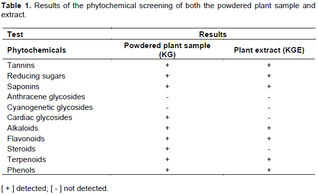
KG and KGE were subjected to preliminary phytochemical screening (Orman et al., 2015; Mireku-Gyimah et al., 2018). Phytochemicals tested included tannins, saponins, reducing sugars, anthracene glycosides, cardiac glycosides, cyanogenetic glycosides, steroids, phenols, terpenoids, alkaloids and flavonoids.
In-vitro antibacterial activity
Microorganisms
Reference strains of the microorganisms, Pseudomonas aeruginosa ATCC 27853, Staphylococcus aureus ATCC 25923, Escherichia coli ATCC 25922 and Klebsiella pneumoniae ATCC 8308 were employed for the studies. These organisms were obtained from the Department of Microbiology, Korle-Bu Teaching Hospital, Accra, Ghana. The isolates were identified by biochemical tests (Kar., 2008). Stock cultures of the organisms were maintained in nutrient agar plates at 4 oC and sub-cultured in nutrient broth at 37°C for 24 h before the experiment.
Antibacterial activity test
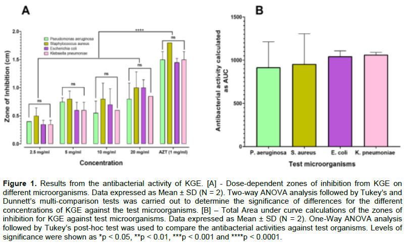
Agar well diffusion method as described by Srinivasan et al. (2001)was used to evaluate the antibacterial activity of KGE (at concentrations, 20, 10, 5, and 2.5 mg/ml) against the test organisms. Azithromycin (AZT, at 1 mg/mL) was used as a positive control. Plates of nutrient agar were seeded with cultures and wells were cut with a sterile cork borer (size = 6 mm). 0.5 ml each of KGE solutions (dissolved in distilled water) were pipetted into the wells, allowed to diffuse at room temperature for 1 h, and incubated at 37°C for 24 h. The activity of KGE was evaluated by measuring the zones of inhibition from each well. The experiment was carried out in duplicate and the mean diameter of the zones of inhibition was calculated.
In-vitro antioxidant assays
The antioxidant activity of KGE was assessed using the DPPH free radical scavenging, total antioxidant capacity, and total phenol content in-vitro models.
DPPH radical scavenging assay
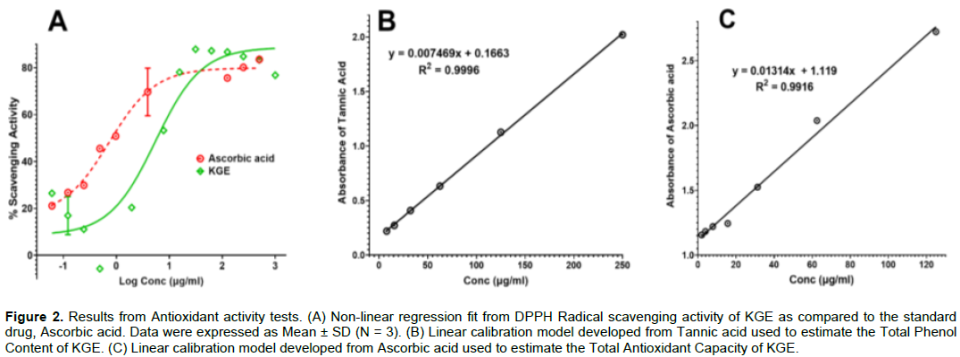
The procedure adopted involved the use of the stable radical, 2,2-diphenyl-1-picrylhydrazyl (DPPH), and ascorbic acid as the standard (Amponsah et al., 2016; Ayensu and Quartey, 2015). A solution of DPPH (20 mg/L), as well as serial concentrations of KGE (0.0610- 1000 µg/ml) and ascorbic acid (0.0610- 500 µg/ml), were prepared in methanol. 1 ml each of KGE solutions was added to 3 mL of the DPPH solution and incubated for 30 min at 37°C in the dark. Similarly, 1mL of ascorbic acid solutions was also added to 3 mL of the DPPH solution and incubated for 30 min at 37°C in the dark. The absorbances for resultant mixtures were taken at 517 nm with a UV-spectrophotometer using methanol as blank. Triplicate determinations were carried out. Radical scavenging activity for each test solution was determined using the formula:

A non-linear regression fit of the scavenging activities of KGE and ascorbic acid was adopted to estimate the EC50 of tested agents.
Total phenol content
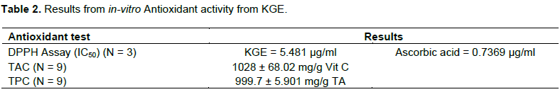
The method adopted was as described in the literature (de Oliveira et al., 2012). The procedure involved the use of 0.5 N Folin – Ciocalteu reagent and tannic acid as a reference standard. Serial concentrations of tannic acid (7.8125 - 250 µg/ml) were prepared and 0.5 ml of each was added to 0.1 mL of Folin – Ciocalteu reagent and incubated at room temperature for 15 min. 2.5 ml of saturated sodium carbonate was then added and re-incubated for a further 30 min at room temperature. The absorbances for the resultant solutions were taken at 760 nm using a UV-spectrophotometer. Triplicate determinations were carried out. Selected concentrations of KGE (32.25, 62.50, and 250 µg/ml) were prepared and taken through the same procedure, to also record triplicate absorbances. A linear regression model (Figure 2b) was developed from the outcomes of the reference standard and this was used to estimate the total phenol content of KGE as expressed in terms of tannic acid equivalent (mg/g TA) (Table 2). All tests were done in triplicates.
Total antioxidant capacity
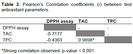
The procedure adopted was as described in the literature (Prieto et al., 1999)and involved the use of ascorbic acid as a reference standard and a standard reagent mixture (SRM) prepared from 0.6 M sulphuric acid, 28 mM disodium phosphate and 4mM ammonium molybdate. Serial concentrations of ascorbic acid (1.953 – 125 µg/ml) were prepared and 1 mL of each was added to 3 ml of SRM, incubated at 95°C for 24 h, cooled at room temperature and centrifuged for 10 min. The absorbance of the supernatant liquid of the various concentrations was measured at 695 nm with a UV-spectrophotometer. Triplicate determinations were carried out. Similarly, selected concentrations of KGE (31.25, 62.50, and 125 µg/ml) were prepared and taken through the same procedure to also record triplicate absorbances. A linear regression model (Figure 2c) was developed from the outcomes of the reference standard and this was used to estimate the total antioxidant capacity of KGE as expressed in terms of ascorbic acid equivalent (mg/g Vit C) (Table 2)
Wound healing activity
Animals
The wound healing studies were performed according to internationally accepted principles for laboratory animal use and care (NIH, 2011)and with due permission from Animal Ethics Committee (FPPS-AEC/CA02/19) in the Department of Pharmacology, Faculty of Pharmacy and Pharmaceutical Sciences, Kwame Nkrumah University of Science and Technology, Kumasi, Ghana. Healthy Wistar albino rats of both sexes, weighing 80-150 g, were procured from the Noguchi Memorial Institute for Medical Research (NMIMR), University of Ghana and were individually housed and maintained under standard environmental conditions of temperature (24.5 ± 0.50°C) and humidity (50 ± 5%) and 12 h light/dark cycle. They were fed with commercial pellet diet (GAFCO, Ghana) and clean autoclaved water ad libitum. Animals were allowed to acclimatize for a week before the test was initiated. They were then randomized into 6 groups of fives with labels, A, B, C, D, E, and F, and weighed. Groups A, B, C, and D were dedicated to four concentrations of KGE (that is, 15, 10, 5, and 1% respectively). Groups E and F were designated for negative and positive control groups, with the negative control group being untreated whiles the positive control group received treatment with Drez® ointment.
Sample preparation
Topical aqueous creams (200 g each) of varying concentrations of KGE (that is, 1, 5, 10, and 15% w/w) were prepared using Emulsifying Ointment BP (BPC, 2012)as a base.
Excision wound model
The excision wound model as described by Boakye et al. (2018) was adopted. The dorsal fur of the animals was gently shaved to expose the skin and cleaned with 70% v/v ethanol. The animals were anesthetized with chloroform and excision wounds of about 20 mm in diameter carefully created under aseptic conditions. The wounds were cleaned with a wad of cotton and N/S. The animals were closely monitored for any infection and those showing signs of infection were excluded from the study and replaced accordingly. The wound diameters were recorded after 24 h and wounds were subsequently treated topically, with daily applications of KGE (15, 10, 5 and 1%) and Drez® ointment after cleaning with N/S, in the test groups as earlier described. Wound contractions were calculated from the wound diameters recorded every 72 h for the next 15 days.
where n = number of days 3rd, 5th, 7th, 9th, 11th, 13th, and 15th day.
Data analysis
Data analysis was performed using GraphPad Prism for Windows (version 8.0.2, GraphPad Software Inc, San Diego, USA, 2019). Data were described using the descriptive parameters, means, standard deviations, and standard error of means. Analysis of variance (ANOVA) followed by Tukey’s and Dunnett's post-hoc tests was carried out to compare the means of outcomes between and within test groups at a 95% confidence level. Data from reference compounds in the total antioxidant capacity and phenol content tests were analyzed using linear regression by the least-squares method to develop linear regression models used to estimate the antioxidant capacity and phenol content of KGE. Data from the radical scavenging test were fitted to a non-linear regression model (Equation 3), to determine concentrations responsible for 50% of the maximum antioxidant activity (EC50) for both KGE and ascorbic acid. The correlation between total antioxidant capacity and phenol content was evaluated by determining the Pearson’s correlation coefficient for the mean absorbances obtained from the optimized concentration ranges for the above-mentioned tests.
Where ‘y’ is the response and ‘x’ is the logarithm of concentration of the KGE.
RESULTS AND DISCUSSION
The phytochemical screening showed the presence of tannins, alkaloids, saponins, reducing sugars, flavonoids, terpenoids, and phenols in both KG and KGE (Table 1). Anthracene and cyanogenetic glycosides were however absent in both. The presence of tannins, saponins, reducing sugars, alkaloids confirmed the outcomes from previous phytochemical studies on KG (Falodun et al., 2009; Ibrahim et al., 2006; Ojokuku et al., 2010; Stephen et al., 2009). On the contrary, whereas anthracene and cardiac glycosides were not detected in the current study, they were shown to be present in previous studies (Ibrahim et al., 2006; Ojokuku et al., 2010; Stephen et al., 2009). Also, while flavonoids were present in the current study, Ojokuku et al. (2010)and Ibrahim et al. (2006)reported their absence. These differences have been argued to be as a result of differences in geographical locations of the plants (Liu et al., 2016)as well as effects from the different modes of handling and preparing the plant samples (Tiwari and Cummins, 2013). Comparatively, KG, and KGE contained similar phytoconstituents except for cardiac glycosides and steroids, which were detected in KG but not in KGE. This obviously could be attributed to the ethanol solvent used; which has a high affinity for polar compounds than non-polar compounds (Dhawan and Gupta, 2016; Truong et al., 2019).
Most of these phytochemicals have been shown to possess antibacterial activity. For example, in a review conducted by Barbieri et al. (2017), the authors enumerated several phytochemicals classified under alkaloids, sulfur-containing phytocompounds, terpenoids, carotenoids, and polyphenolic compounds, possessing antibacterial effects; with some exerting effects against resistant and multi-resistant microorganisms (Barbieri et al., 2017). It could thus be inferred that the presence of these compounds confers antimicrobial properties on KGE, as documented in the literature (Galani et al., 2016; Okpe et al., 2019; Stephen et al., 2009). In the current study, KGE demonstrated dose-dependent antibacterial activities against the test organisms, S. aureus, E. coli, and K. pneumoniae. In other words, as the concentration of KGE increased, the zones of inhibition became larger (Figure 1a). The inhibitions produced from similar test concentrations of KGE against the test organisms were shown to be comparable (F(12,20) = 0.4254; p = 0.9345, Figure 1). This was further confirmed from their Area under Curve calculations (AUCs), which were comparable for all the test organisms (F(3,4) = 0.1858; p = 0.9009; Figure 1b). However, KGE’s activities against the test organisms were lower than that of the standard drug AZT (1 mg/ml) (p < 0.0001; Figure 1a).
KGE also demonstrated good antioxidant effects (Figure 2 and Table 2). The DPPH scavenging test assessed the ability of KGE to terminate free radical initiated reactions, which occur during oxidative stress conditions. The total antioxidant capacity measured the phytochemical constituents with antioxidant properties while the total phenolic content measured all phenolic and polyphenolic phytochemicals in the plant extract. The higher the values, the higher the antioxidant effect (Barku et al., 2013). Thus, KGE was shown to have a relatively higher antioxidant capacity and phenol content when compared to other types of extracts from the same plant reported elsewhere (Njayou et al., 2013). The high correlation between the total phenolic content and the antioxidant capacity (Table 3) could mean that the antioxidant activity of KGE was mainly due to the phenolic compounds like flavonoids, terpenoids and tannins present (Gülçin et al., 2010; Liu et al., 2009; Pietta, 2000; Villaño et al., 2007). Most of these phytochemicals possess aromatic rings in their structures and are thought to react with free radicals, resulting in the delocalization of the gained electron over the phenolic structure, leading to stabilization by resonance effect. This phenomenon disrupts the free radical reaction chain. Thus, a high amount of these phyto-chemicals in a plant shows a high antioxidant activity (Lee et al., 2017).
KGE was established to possess wound healing effects, as most of the test doses of KGE produced % contractions significantly higher than that of the untreated or negative control group (Figure 3a and 3b). It is thought that the observed antioxidant and antibacterial activities as earlier discussed could contribute to and/or promote KGE’s wound-healing effects. The phytochemicals present in the plant may be responsible for the observed wound-healing effect. Saponins have been shown to possess anti-inflammatory effects on the body thereby reducing inflammation during the early phase of the wound, reducing edema, and promoting re-epithelisation of the wound (Kim et al., 2011). They also promote angiogenesis by stimulating the production of vascular endothelial growth factor, through the increase of HIF-1α expression in keratinocytes, and elevation of IL-1β. These result from macrophage accumulation in the wound (Kimura et al., 2006). Tannins are also known to improve wound healing due to their astringent and antimicrobial activities. They also increase epithelisation and the rate of wound contraction and act as free radical scavengers (Okoli et al., 2009). Flavonoids which are known for their antioxidant properties also prevent and slow the onset of cell death and improve vascularity. This improves wound healing by providing nutrients to the affected area. Antioxidants are known to play an important role in wound repair (Okoli et al., 2009). KGE was further shown to be as effective as and a potential alternative wound healing therapy to the positive control, as they were shown to be comparable (p > 0.05; Figure 2b).

Even though KGE demonstrated a dose-dependent effect (Figure 3a), the % contractions from adopted doses were comparable (p > 0.05; Figure 3b). In effect, the smallest dose (1% w/w) was as effective as the highest dose (15% w/w). This may be thought of as welcoming especially when the administration of high doses of plant medicines, just as in allopathic medicines, are associated with side and/or adverse effects due to the matrix of compounds in the plant parts adopted (Fatima and Nayeem, 2016).
CONCLUSIONS
The outcomes of the study conducted showed that KGE contains some key phytochemicals and these are thought to be responsible for the observed antibacterial, antioxidant, and wound-healing effects. These observations may thus confirm the local uses of K. grandifoliola stem bark for the treatment of various wounds, microbial infections, and other ailments associated with oxidative stress in traditional medicine.
CONFLICT OF INTERESTS
The authors have not declared any conflict of interests.
ACKNOWLEDGMENTS
The authors are grateful to the technical staff of the Department of Pharmaceutical Sciences in the School of Pharmacy, Central University, and the Department of Pharmacology, Faculty of Pharmacy and Pharmaceutical Sciences, KNUST, Ghana for their technical support in the conduct of the studies.
REFERENCES
|
Adly AAM (2010). Oxidative Stress and Disease: An Update Review. Research Journal of Immunology 3(2):129-145. |
|
|
Agbedahunsi JM, Elujoba AA, Makinde JM, Oduda AMJ (1998). Antimalarial activity of Khaya grandifoliola stem-bark. Pharmaceutical Biology 36(1):8-12. |
|
|
Agyare C, Dwobeng AS, Agyepong N, Boakye YD, Mensah KB, Patrick George Ayande A, Adarkwa-Yiadom M (2013). Antimicrobial, Antioxidant, and Wound Healing Properties of Kigelia africana. Advances in Pharmacological Sciences 2013:10. |
|
|
Agyare C, Kisseih E, Hensel A, Appiah T (2016). Review: African medicinal plants with wound healing properties. Journal of Ethnopharmacology 177:85-100. |
|
|
Amponsah IK, Orman E, Mensah AY, Sarpong FM, Armah FA, Sarpong LM (2016). Development and validation of a radical scavenging antioxidant assay using potassium permanganate. Journal of Scientific and Innovative Research 5(2):36-42. |
|
|
Ayensu I, Quartey AK (2015). Phytochemical Screening and in-Vitro Antioxidant Properties of the Stem Bark of Trichilia Tessemannii. World Journal of Pharmacy and Pharmaceutical Sciences 4(3):76-90. |
|
|
Barbieri R, Coppo E, Marchese A, Daglia M, Sobarzo-Sánchez E, Nabavi SF, Nabavi SM (2017). Phytochemicals for human disease: An update on plant-derived compounds antibacterial activity. Microbiological Research 196:44-68. |
|
|
Barku VYA, Opoku-Boahen Y, Owusu-Ansah E, Mensah EF (2013). Antioxidant activity and the estimation of total phenolic and flavonoid contents of the root extract of Amaranthus spinosus. Asian Journal of Plant Science and Research 3(1):69-74. |
|
|
Boakye YD, Agyare C, Ayande GP, Titiloye N, Asiamah EA, Danquah KO (2018). Assessment of wound-healing properties of medicinal plants: The case of Phyllanthus muellerianus. Frontiers in Pharmacology 9(AUG):1-12. |
|
|
Bowler PG, Duerden BI, Armstrong DG (2001). Wound Microbiology and Associated Approaches to Wound Management. Clinical Microbiology Reviews 14(2):245-269. |
|
|
British Pharmacopoeial Commission (BPC) (2012). British Pharmacopoeia (Vol 1 & 2), 2013th ed. The Stationery Office on behalf of the Medicines and Healthcare products Regulatory Agency (MHRA), London. |
|
|
Builders PF, Builders MI (2016). Wound Care: Traditional African Medicine Approach. In: Vicente da Fonseca CJ, ed. Worldwide Wound Healing: Innovation in Natural and Conventional Methods. InTech, Rijeka, Croatia. pp. 1-24. |
|
|
Bumah VV, Essien EU, Agbedahunsi JM, Ekah OU (2005). Effects of Khaya grandifoliola (Meliaceae) on some biochemical parameters in rats. Journal of Ethnopharmacology 102(3):446-449. |
|
|
de Oliveira AMF, Pinheiro LS, Pereira CKS, Matias WN, Gomes RA, Chaves OS, de Souza M de FV, de Almeida RN, de Assis TS (2012). Total phenolic content and antioxidant activity of some malvaceae family species. Antioxidants 1(1):33-43. |
|
|
Deufert D, Graml R (2017). Disease-specific, health-related quality of life (HRQoL) of people with chronic wounds - A descriptive cross-sectional study using the Wound-QoL. Wound Medicine 16:29-33. |
|
|
Dhawan D, Gupta J (2016). Comparison of Different Solvents for Phytochemical Extraction Potential from Datura metel Plant Leaves. International Journal of Biological Chemistry 11(1):17-22. |
|
|
Falodun A, Poh CF, Adelusi SA, Emmanuel O (2009). Phytochemical and anti-inflammatory evaluation of Khaya grandifoliola stem bark extract. International Journal of PharmTech Research 1(4):1061-1064. |
|
|
Fatima N, Nayeem N (2016). Toxic Effects as a Result of Herbal Medicine Intake. Toxicology-New Aspects to This Scientific Conundrum. London, UK: InTech Open, pp 193-207. |
|
|
Firdous SM, Sautya D (2018). Medicinal plants with wound healing potential. Bangladesh Journal of Pharmacology 13(1):41-52. |
|
|
Galani BRT, Sahuc ME, Sass G, Njayou FN, Loscher C, Mkounga P, Deloison G, Brodin P, Rouillé Y, Tiegs G, Séron K, Moundipa PF (2016). Khaya grandifoliola C.DC: a potential source of active ingredients against hepatitis C virus in vitro. Archives of Virology 161(5):1169-1181. |
|
|
Gülçin Ä°, Huyut Z, ElmastaÅŸ M, Aboul-Enein HY (2010). Radical scavenging and antioxidant activity of tannic acid. Arabian Journal of Chemistry 3(1):43-53. |
|
|
Ibrahim JA, Ayodele EA, Jegede AI, Kunle YF (2006). Comparative studies on Khaya. A. Juss (Meliaceae) in Nigeria. African Journal of Biotechnology 5(11):1154-1160. |
|
|
Kar A (2008). Identification of Microorganisms. In: Pharmaceutical Microbiology. New Age International Publishers, New Delhi (India). pp. 112-145. |
|
|
Kim YS, Cho IH, Jeong MJ, Jeong SJ, Nah SY, Cho YS, Kim SH, Go A, Kim SE, Kang SS, Moon CJ, Kim JC, Kim SH, Bae CS (2011). Therapeutic efect of total ginseng saponin on skin wound healing. Journal of Ginseng Research 35(3):360-367. |
|
|
Kimura Y, Sumiyoshi M, Kawahira K, Sakanaka M (2006). Effects of ginseng saponins isolated from Red Ginseng roots on burn wound healing in mice. British Journal of Pharmacology 148(6):860-870. |
|
|
Kokane DD, More RY, Kale MB, Nehete MN, Mehendale PC, Gadgoli CH (2009). Evaluation of wound healing activity of root of Mimosa pudica. Journal of Ethnopharmacology 124(2):311-315. |
|
|
Lee MT, Lin WC, Yu B, Lee TT (2017). Antioxidant capacity of phytochemicals and their potential effects on oxidative status in animals - A review. Asian-Australasian Journal of Animal Sciences 30(3):299-308. |
|
|
Liu L, Sun Y, Laura T, Liang X, Ye H, Zeng X (2009). Determination of polyphenolic content and antioxidant activity of kudingcha made from Ilex kudingcha C.J. Tseng. Food Chemistry 112(1):35-41. |
|
|
Liu W, Yin D, Li N, Hou X, Wang D, Li D, Liu J (2016). Influence of environmental factors on the active substance production and antioxidant activity in Potentilla fruticosa L. and its quality assessment. Scientific Reports 6(28591):1-18. |
|
|
Mireku-Gyimah NA, Sarpong K, Amponsah IK, Mensah AY, Dickson RA (2018). Comparative Pharmacognostic Studies of Two Ghanaian Medicinal Plants: Saba Senegalensis and Saba Thompsonii. International Journal of Pharmaceutical Sciences and Research 9(4):1451-1461. |
|
|
National Institutes of Health (NIH) (2011). Guide for the Care and Use of Laboratory Animals, Eighth Edi. Washington: The National Academies Press. |
|
|
Njayou FN, Njayou FN, Tandjang MK, Tchana AK, Ngadjui BT, Moundipa PF (2013). Hepatoprotective and antioxidant activities of stem bark extract of Khaya grandifoliola (Welw) CDC and Entada africana Guill. et Perr. Journal of Natural Products 6:73-80. |
|
|
Ojokuku SA, Okunowo WO, Apena A (2010). Evaluation of the chemical composition of Khaya grandifoliola and Ficus capensis. Journal of Medicinal Plants Research 4(12):1126-1129. |
|
|
Okoli CO, Ezike AC, Akah PA, Udegbunam, SO, Okoye TC, Mbanu TP, Ugwu E (2009). Studies on wound healing and antiulcer activities of extract of aerial parts of Phyllanthus niruri L. (Euphorbiaceae). American Journal of Pharmacology and Toxicology 4(4):118-126. |
|
|
Okpe O, Ndidi US, Ojowu J, Maifada SR, Etim EE, Awen DA, Ovur CE (2019). GC-MS Profiling and Antimalarial Activity of Khaya grandifoliola on Plasmodium berghei-infected mice. Journal of Herbs, Spices and Medicinal Plants 25(1):21-32. |
|
|
Orman E, Addo P, Ofori M, Adosraku R (2015). Investigating the In-vivo Antiplasmodial Properties of Aqueous Extract of Moringa oleifera Lam (Moringaceae) Leaves. British Journal of Pharmaceutical Research 5(6):419-430. |
|
|
Pietta PG (2000). Flavonoids as antioxidants. Journal of Natural Products 63(7):1035-1042. |
|
|
Prieto P, Pineda M, Aguilar M (1999). Spectrophotometric quantitation of antioxidant capacity through the formation of a phosphomolybdenum complex: Specific application to the determination of Vitam E. Analytical Biochemistry 269:337-341. |
|
|
Rafiu B, Sonibare MA (2017). Ethnobotanical survey of tree species used for wound healing in Ibadan, southwest Nigeria. Nigerian Journal of Natural Products and Medicine 20:96-104. |
|
|
Raina R, Prawez S, Verma PK, Pankaj NK (2008). Medicinal Plants and their Role in Wound Healing. Vetscan 3(1):1-7. |
|
|
Sandrine E, Christophe M, Ernestine N, George E-O, Vernyuy T, Barthelemy N (2016). Cytoprotective and Antioxidant Properties of the Stem Bark Aqueous extract of Khaya grandifoliola (Meliaceae) in Rats. British Journal of Pharmaceutical Research 9(2):1-11. |
|
|
Shedoeva A, Leavesley D, Upton Z, Fan C (2019). Wound healing and the use of medicinal plants. Evidence-based Complementary and Alternative Medicine 2019:30 pp. |
|
|
Singh J (2008). Maceration, Percolation and Infusion Techniques for the Extraction of Me- dicinal and Aromatic Plants. In: Handa SS, Khanuja SPS, Longo G, Rakesh DD, eds. Extraction Technologies for Medicinal and Aromatic Plants. United Nations Industrial Development Organization and International Centre for Science and High Technology, Trieste, pp 67-82. |
|
|
Singh SK, Gupta B (2017). Choices and Challenges of Antibiotics Therapy in Diabetic Foot Infection. Indian Journal of Endocrinology and Metabolism 21(5):647. |
|
|
Srinivasan D, Nathan S, Suresh T, Lakshmana PP (2001). Antimicrobial activity of certain Indian medicinal plants used in folkloric medicine. Journal of Ethnopharmacology 74(3):217-220. |
|
|
Stephen UA, Abiodun F, Osahon O, Ewaen E (2009). Phytochemical Analysis and Antibacterial Activity of Khaya gradifoliola Stem Bark. Journal of Biological Sciences 9(1):63-67. |
|
|
Tiwari U, Cummins E (2013). Factors influencing levels of phytochemicals in selected fruit and vegetables during pre- and post- harvest food processing operations. Food Research International 50(2):497-506. |
|
|
Truong DH, Nguyen DH, Ta NTA, Bui AV, Do TH, Nguyen HC (2019). Evaluation of the use of different solvents for phytochemical constituents, antioxidants, and in vitro anti-inflammatory activities of severinia buxifolia. Journal of Food Quality 2019. |
|
|
Villaño D, Fernández-Pachón MS, Moyá ML, Troncoso AM, García-Parrilla MC (2007). Radical scavenging ability of polyphenolic compounds towards DPPH free radical. Talanta 71(1):230-235. |
|
|
Yunga ST, Health O, Mbacham W, Sipping M (2018). Evaluation of in vitro antioxidant and immunomodulatory activities of polysaccharide fractions of Khaya grandifoliola C . D . C ( Welw ) stem bark and Cryptolepis sanguinolenta ( Lindl .) Schltr leaves. African Journal of Biotechnology. |
|
Copyright © 2024 Author(s) retain the copyright of this article.
This article is published under the terms of the Creative Commons Attribution License 4.0


