Full Length Research Paper

ABSTRACT
Bauhinia ungulata L. species, belongs to the genus Bauhinia, popularly known as pata de vaca, is among the species of medicinal employ in Brazil, used to treat diabetes. The aim of this study was to characterize the chemical composition, antimicrobial activity, toxicity on Artemia salina and acetylcholinesterase enzyme inhibition by essential oil from B. ungulata L. The chromatographic analysis revealed 18 components, the majority were the β-caryophyllene (15.9%), caryophyllene oxide (9.2%), α-humulene (8.1%), epi-γ-eudesmol (7.5%), α-bisabolol (4.7%), copaene (3.5%), nerolidol (3.3%), α-bisabolol oxide B (2.5%), spathulenol (2.1%). The essential oil showed high toxicity compared to the tests with Artemia salina and inhibited 95.96%±0.62 of the acetylcholinesterase enzyme. The microorganisms show antimicrobial inhibition with Candida albicans (85%), Bacillus cereus (65.5%) and Staphylococcus aureus (66.4%), Salmonella typhimurium (68.7%) and Citrobacter freundii (46.1%). The oil showed great potential when tested in bioassays.
Key words: Volatile oil, cytotoxicity on microcrustacean, antifungal, antibacterial, neurodegenerative disease.
INTRODUCTION
Among many medicinal plants, there is the Bauhinia ungulata L. species, belonging to the genus Bauhinia belonging of Fabaceae family (Caesalpinioideae subfamily) one of the largest of angiosperms, which can be found mainly in tropical areas, with about 250-300 species (Souza and Lorenzi, 2008; Joly, 1993, 1998).
In Brazil this species is popularly known pata de vaca, unha de boi, pé de boi, escada de macaco, unha de jaboti and mororó (Silva and Cechinel, 2002; Lorenzi and Matos, 2002). It is applied in the treatment of diabetes mellitus, cholesterol control as diuretic and expectorant (Correa, 1998). It was also reported the use of B. ungulata by Tapebas Indians in Ceará state, Brazil for the treatment of diabetes, and this species most frequently studied for its hypoglycemic action (Silva et al., 2002; Pepato et al., 2002; Morais et al., 2005).
Species of this genus have shown molluscicidal activity (Singh et al., 2012), larvicidal activity against Aedes aegypti L. (Gois et al., 2011), antiviral against Arbovirus mayaro MAYV (Santos et al., 2014), antioxidants (Santos et al., 2014; Port's et al., 2013; Paula et al., 2014), inhibition of the acetylcholinesterase enzyme (Santos et al., 2011), antibacterial action (Cechinel-Filho, 2000, 2009), anti-helminth, in B. variegata against Ferentima posthuma and Ascardia galli (Bairagi et al., 2012) and antitumor activity tested in rats (Rajkapoor et al., 2003).
In addition to the genus activities, aforementioned, there are the major compounds and compounds highlighted, folow. The essential oil from the leaves of another species, B. acurana, has as main constituents spathulenol, sesquiterpenes, epi-α-cardinol and caryophyllene oxide (Gois et al., 2011). The major constituents of essential oil from B. ungulata are spathulenol and caryophyllene oxide (Gramosa et al., 2009).
Essential oils are a lipophilic moiety of the chemical composition of a plant. Generally, consisting of sesquiterpenes, monoterpenes and phenylpropanoids (Cunha et al., 2004). Aromatic drugs are frequently used to destroy infection causing agents such as bacteria and pathogenic fungi (Costa, 2002). They are also used in industries, food and cosmetics (Bizzo, 2009).
This study aims to characterize the main volatile chemical constituents using gas chromatography and biological activities by essential oil from B. ungulata leaves.
MATERIALS AND METHODS
Plant material and essential oil extraction
The leaves of B. ungulata were collected along the Água Boa River, near the BR 174, at 11 Km in Boa Vista, Roraima, Brazil, in January 2016 (dry season). The plant material was identified by José Ferreira Ramos (National Institute for Research in the Amazon, INPA), and a voucher specimen (272558) was deposited at the INPA Herbarium.
Fresh leaves (600 g) were cut into smaller pieces with scissors and putting in a Clevenger apparatus to obtain the essential oil by hydrodistillation. Water droplets were removed from the essential oil by anhydrous sodium sulfate and stored at -20°C before analysis (Rubiolo et al., 2010; Sefidkon, 2002).
GC/FID analysis
The essential oil was analyzed on a HP 7820A Gas Chromatograph (GC) equipped with a flame ionization detector (FID) using a capillary column (HP5 30 m × 0.32 mm × 0.25 µ, Agilent). Column temperature: 50°C (0 min) at 3°C min-1 up to 230°C. Gun: 250°C Split (1:30). FID Detector: 250°C. Carrier gas: hydrogen at 3 mL min-1. Vol injection: 1 μL mL-1. Essential oil was diluted at 1% in chloroform. Data acquisition software used was Compact EZChrom Elite (Agilent). The quantitative analysis was accomplished using standard areas from the chromatograms obtained by GC-FID.
Gas chromatography/mass spectrometry analysis
A GCMS-QP2010 ULTRA (Shimadzu) was used. Column: Rxi-1MS, 30 m × 0.25 mm × 0.25µ (Restek). Column Temperature: 50°C (3 min), 3°C min-1 to 250°C. Injector: 250°C Split (1:10), GC-MS interface at 250°C. MS detector (electron impact at 70 eV) temperature was 250°C. Carrier gas: helium at 1.5 mL min-1. Vol injection: 1 μL. Essential oil was diluted at 0.1% in chloroform. Data acquisition software used was GC-MS Solution (Shimadzu) together with NIST11 library. Identification of peaks was made by comparison of the mass spectra obtained by GC-MS spectra with the NIST11 library and also by comparing the Kovats indices calculated by GC-FID and literature data.
Determination of toxicity on Artemia salina
The essential oil was solubilized in Tween 20 (1%) and saline supplemented with water to give concentrations (1000, 500, 250 e 125 μL mL-1). They were transferred to tubes (3 mL) and added 10 organisms (nauplii Artemia salina). The tests were performed in triplicate for each concentration. Saline without extract was used as negative control also in triplicate and was subjected to the same experimental procedure. This system was incubated at room temperature for 24 h, with aeration and other tubes kept under illumination. After 24 h, the number of dead and live larvae in each tube was counted. Thereafter, the probability of mortality was calculated according to the formula:

Where: r = number of dead nauplii; n = total number of A. salina in each tube.
It was given the lethal concentration 50% LC50, using the statistical program Microsoft Excel 2010 (Meyer et al., 1982; Mclaughlin et al., 1993).
Acetylcholinesterase (AChE) inhibition assay
Aliquots of a working solution (25 μL) (sample in Tween/DMSO/30%) was added to microplate wells, positive and negative controls were also prepared. To the first five wells of a column (positive control), 25 μL of an eserine solution prepared at 10 mg mL-1 (in Tris/HCl at pH 8.0) was added. Thereafter, 25 μL of acetylthiocholine iodide (ATChI, Sigma A5751); the reaction mixture, 125 μL of 5'.5-dithio-bis (2-nitrobenzoate) (DTNB, Sigma D8130) and 50 μL of Tris/HCl (pH 8) containing 0.1% (m/v) bovine serum albumin were added to each well. Absorbance was measured at 405 nm every 1 min for 8 times. Then 25 μL (0.226 U mL-1) of Electric eel AChE (type VI-S) provided by Sigma (C3389-500UN) in Tris/HCl were added to each well. Absorbance was measured at 405 nm by 9 times (Frank and Gupta, 2005; Ellman et al., 1961). Percentual inhibition was calculated using the formula:
Where PCA = (absorbance of the sample with enzyme - absorbance of the sample without enzyme); PSA = (absorbance of negative control with enzyme - absorbance of negative control without enzyme).
Antibacterial and yeast assay
Two Gram-negative: Salmonella typhimurium (ATCC 13311) e Citobacter Freundii (ATCC8090), two bacterium Gram-positive: Staphylococcus aureus (ATCC 25923), and Bacillus cereus (ATCC 11778) and one fungus (yeast) Candida albicans (ATCC 18804) yeast were used in the assay. Concentrations assayed were 250, 125, 62.5, 31.25, 15.6, 3.9 and 1.95 μg mL-1 (Zacchino and Gupta, 2007). Samples were weighed and dissolved in DMSO to 500 mg mL-1. 124 μL of this solution was added to a flask containing 2976 μL of BHI (Brain Heart Infusion) broth (working solution) for bacterium and 2976 μL of Sabouraud for yeasts. A pre-inoculum was prepared in which the bacteria and the yeast, stored under refrigeration, were transferred with a platinum loop to test tubes containing 3 mL of freshly made BHI broth. The tubes were incubated at 37°C for 24 h. Then, the pre-inoculum (500 μL) was transferred to tubes containing 4.5 mL of sterile distilled water. The tubes were homogenized and the concentration adjusted to 0.5 of McFarland turbidity standard (108 CFU mL-1), thereby obtaining the inocula used in the bioassays.
Assays were performed in 96-microwell plates in triplicate. 100 μL of BHI broth was added to each well. In the first well, 200 μL of working solution were also added. The solution was homogenized and 100 μL transferred to the next well and so on until the last well, from where 100 μL was discarded. Then, 100 μL of microorganism inocula were added to wells. Eight different concentrations of each sample were tested. A positive control devoid of the working solution allowed us to examine microorganism growth. A negative control, which lacked the inoculum made it possible to discount the color coming from the working solution. A control plate containing 100 μL of BHI culture medium and 100 μL of sterile distilled water were added to the experiment as a control of BHI broth sterility.
Another control was also prepared, containing the standard antibiotics Ampicillin (antibacterial), miconazole and nystatin (antifungals) to observe the activity of these antibiotics over the microorganisms. Microorganism growth was measured in ELISA plate reader (492 nm) immediately after ending the experiment (0 h). They were incubated at 37°C and read again after 24 h of experiments, ending the test. Results were calculated as percentual inhibition using the formula:
AC1 = absorbance of the sample; AC2 = absorbance of control sample; AH = absorbance of microorganisms in the control control and AM = absorbance of culture medium control.
RESULTS AND DISCUSSION
The yield of essential oils of B. ungulata in this study was 0.066%, higher than that obtained by Gramosa et al. (2009) who in their study developed this species in the Northeast, which was 0.007%. The yields of essential oils obtained from the genus Bauhinia varies between bauhinia rufa with 0.37% (Silva and Camara, 2014) and B. acuruana with 0.01% (Gois et al., 2011).
Table 1 shows the chemical components identified by chromatographic analysis GC-FID and GC-MS with their Kovats index and mass and Figure 1 shows chemical structures of the major constituents identified in the essential oil in B. ungulata.
.png)
.png)
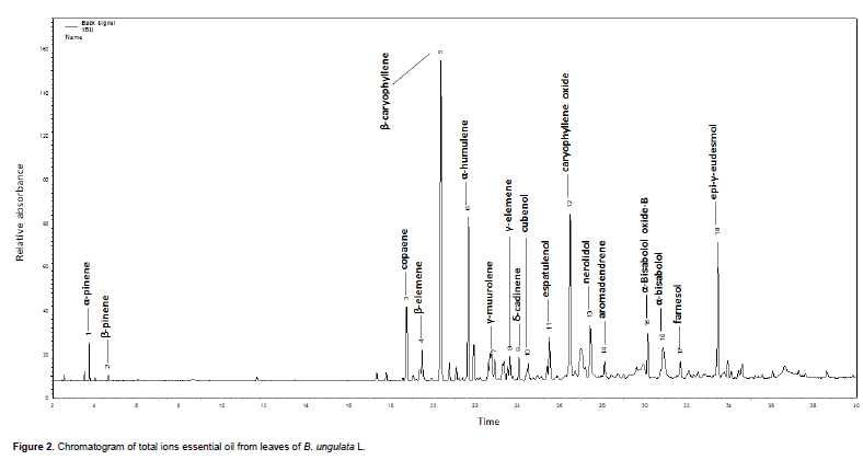
The nine major components identified in this study are β-caryophyllene (15.9%), caryophyllene oxide (9.2%), α-humulene (8.1%), epi-γ-eudesmol (7.5%), α-bisabolol (4.7%), copaene (3.5%), nerolidol (3.3%), α-bisabolol oxide B (2.5%), spathulenol (2.1%). The structures are shown in Figure 1.
Figure 2 shows the chromatographic profile for B. ungulata oil. 18 components corresponding to 65.5% of essential oil composition were identified. Among the compounds identified, nine are majority corresponding to 61.1%.
In Boa Vista, Roraima, Brazil compared to studies Gramosa et al. (2009), the composition varies mainly in the major constituents. Secondly, according to Oliveira et al. (1998), plant species, in general, may present variations in relation to the yield and chemical composition of essential oils according to the part of the plant studied as well as their interactions with the environment, climate, micro-organisms and also genetic factors.
Gramosa et al. (2009) identified 13 compounds, which represent 95.9% of the essential oil content, the majority were spathulenol (47.7%), caryophyllene oxide (18.3%), humulene epoxide II (5.2%), β-caryophyllene (4.2%), α-humulene (3.5%) and α-copaene (2.9%).
Duó-Bartolomeu et al. (2014) identified: germacrene-D, biciclogermacrene, β-elemene, trans-cariofilene, α-humulene, espatulenol, trans-nerolidol, β-ionone, e β- elemene.
It is observed so spathulenol presented itself as the main constituents for the essential oil of B. ungulata in studies of Gramosa et al., (2009), with a percentage of 47.7. However in the present study this compound presents a lesser amount (2.1%).
According to Figueiredo et al. (2008) and Simões et al. (2004), depending on the part of the plant, type of collection, climatic conditions and extraction methods, essential oils, may suffer variations in their income.
In this study it was found that the highest percentage constituent was the β-caryophyllene (15.9%), a fact equivalent to the study of Neto (2006), where this substance was also the majority with 25.65%.
Oils with high concentrations of β-caryophyllene showed good correlation with the free radical scavenging by dpph, and acetylcholinesterase inhibition (Alcântara et al., 2010); in vivo tests reduced cell death in neurodesgenerativas diseases such as Parkinson's and Alzheimer (Ferreira, 2014).
The α-bisabolol (4.7%) was not identified in other regions for the species B. ungulate, indicating that it may have been the formation of a new chemotype for this species in Boa Vista, RR.
The α-bisabolol compound has correlation with activity against acetylcholinesterase. According to Nurulain et al. (2015), the compound binds directly to the receptor a7-nAChRs, leading to inhibition of acetylcholine.
It also has anti-inflammatory action (Kim et al., 2011) and biological activity against bacteria and fungi, as well as, Aedes aegypt, S. aureus, Pseudomonas aeruginosa, Microsporum gypseum, Trichophyton mentagrophytes and Trichophyton rubrum (Vila et al., 2010). The Figure 3 shows the bioactivity of essential oil from B. ungulata leaves for A. salina.
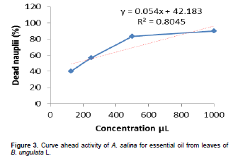
Interpretations of the results of toxicity were carried out taking into account the above literature, which can be classified as highly toxic LC50 values between 0-500 µg mL-1; moderate toxicity between 500-1000 µg mL-1 and low toxicity or nontoxic values above 1000 µg mL-1 (Meyer et al., 1982; Lopes et al., 2002; Rodriguezet al., 2004).
Through the straight equation formula Y = A + BX, we can calculate the CL50. Considering Y = 50, A = 42.183 e B = 0.054, is the value of X is equal to 144.75 µg mL-1. It can be considered that the essential oil of B. ungulata has high toxicity, based on the LC50 value found to be less than 500 µg mL-1.
According to Amarante et al. (2011), the plant extracts and derivatives that have a high toxicity against A. salina are high potential indications for biological activities. This finding reinforces the importance of the method as it is very useful to use this bioassay, when you want to develop biological studies. The results of the inhibition tests for acetylcholinesterase essential oil B. ungulata are as shown in Table 2.
.png)
The oil showed a good inhibitory potential, corresponding to 95.96%, and can see that the standards used for Eserine and galantamine showed inhibition of 91.93 and 94.36%, respectively, which were lower than the inhibition of essential oil. Savalev et al. (2003) observed synergistic interactions among the components (1.8-cineole, camphor, α-pinene, β-pinene, borneol, caryophyllene oxide, linalool and bornyl acetate) which come from the species Salvia lavandulaefolia. Among those components mentioned above was found the essential oil of B. ungulata in the presence of α-pinene, β-pinene and caryophyllene oxide; certainly there was synergism of compounds present in the oil of the species studied by high inhibition of the enzyme, highlighting the chemical components β-caryophyllene and α-bisabolol, which has a close relationship with neurodegenerative diseases and reported by Nurulain et al. (2015), Ferreira (2014), Alcantara et al. (2010) and Santos et al. (2015).
Another biological activity of essential oil of B. ungulata leaves deserves attention as well as the bioactivity against the pathogenic microorganisms (Table 3).
.png)
Figure 4. Activity curve for essential oil from leaves of B. ungulata (L) front of the A) Candida albicans; B) Bacillus cereus; C) Staphylococcus aureus; D) Salmonella typhimurium Graphs were plotted with the aid of software origin 8.0, as can be seen in Figure 4 and using equation Y = A2 + (A1-A2) / (1 + (x/x0) ^p) was calculated from IC50.
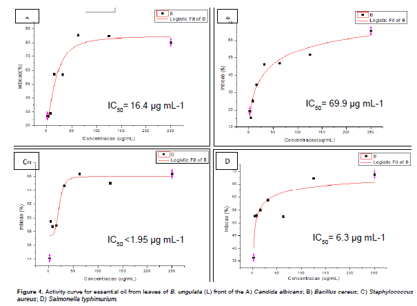
By analyzing the percentages of inhibition and IC50, it can be seen that there has been satisfactory inhibition of four of the tested microorganisms, whereas inhibition was greater than 50%. Checking greater emphasis on the S. aureus which was inhibited in all eight concentrations tested and showed a IC50 < 1.95 µg mL-1 as well as C. albicans to inhibit the microorganism concentration 15.62 μg mL-1 of essential oil, this value was found to be in agreement with the IC50 (16.4 µg mL-1). It is noteworthy that the three highest concentrations were those that had greater inhibition ranging from 80-85%.
The Figure 4 shows graphs of the minimum inhibitory concentrations of the essential oil to the front microorganisms studied. The bacterium C. freundii showed an inhibition of 46.16%, thus less than 50%, not showing satisfactory results.
Species of this genus Bauhinia action also demonstrated inhibition of acetylcholinesterase (Santos et al., 2011), antibacterial action (Cechinel-Filho, 2000, 2009), anthelmintic (Bairagi et al., 2012) and antitumor activity (tested in rats) (Rajkapoor et al., 2003).
CONFLICT OF INTERESTS
The authors have not declared any conflict of interests.
ACKNOWLEDGMENT
The authors are grateful to CAPES for a fellowship and to the staff of the Herbarium of the INPA, especially Mr. José Ramos for identification.
REFERENCES
|
Alcantara JM, Yamaguchi KKL, Silva JRA, Veiga Junior VF (2010). Chemical composition and biological activity of essential oils from the leaves and stems of Rhodostemono daphne parvifolia Madri-án (Lauraceae). Acta Amaz. 40(3):567-572. |
|
|
Amarante CB (2011). Phytochemical study biomonitored by toxicity tests front of Artemia salina and antiplasmodial activity aninga stem (Montrichardia linifera). Acta Amaz. 41(3):431-434. |
|
|
Bairagi SM, Abhijeet AA, Knimase P (2012). In vitro Anthelmintic Activity of Bauhinia variegata Bark (Leguminoseae). |
|
|
Bizzo RH (2009). Essential oils in Brazil: General Aspects, development and research. Química Nova. 3(32):588-594. |
|
|
Cechinel-Filho V (2000). Major Advances and Prospects on Natural Products Area Assets: developed studies in Niqfar/Univale. Química Nova 5:680-685. |
|
|
Cechinel-Filho V (2009). Natural and Synthetic Products with Therapeutic Potential: 15 years Studies at the core of Chemical investigations - Pharmacist at Niqfar/Univale. Rev. Fitos. 4(2):6-23. |
|
|
Costa FA (2002). Pharmacognosy, Lisboa: Fundação calouste gulbenkian. pp. 1-1031. |
|
|
Cunha AP (2004). Medicinal plants and plant products with ethics, Lisboa: Fundação calouste gulbenkian. pp. 9-310. |
|
|
Duó-Bartolomeu AC, Fiori GML, Bastos DV, França SC, Pereira MAS, Taleb-Contini SH (2014). Compostos voláteis de Bauhinia forficata L. 2014. (Apresentação de Trabalho/Simpósio). |
|
|
Ferreira DAS (2014). The protective effect evaluation of β-caryophyllene in cellular models of neurodegenerative diseases. College science pharmaceutical in Ribeirão Preto. Doctoral thesis pp. 1-71. |
|
|
Figueiredo AC, Barroso JG, Pedro LG, Scheffer JC (2008). Factors Affecting Secondary Metabolite Production in Plants: VolatileComponents and Essential Oil. A Review. Flavour Fragr. J. 23:213-226. |
|
|
Gois RWS, Sousa LM, Lemos TLG, Arriaga AMC, Andrade-Neto M, Santiago GMP, Ferreira YS Alves, PB, Jesus HCR (2011). Chemical Composition and Larvicidal Effects of Essential oil from Bauhinia acuruana (Moric.) against Aedes aegypti. J. Essent. Oil Res. 23:59-62. |
|
|
Gramosa NV, Freitas JVB, Neto MND (2009). Volatile components of the essential oil from Bauhinia ungulata L. J. Essent. Oil Res. 21(6):495-496. |
|
|
Joly AB (1993). Botany: Introduction to plant taxonomy.11ª. Ed., Nacional: São Paulo. |
|
|
Joly AB (1998). Botany: introduction to vegetal taxonomy. 12ª. Ed., Nacional editor: São Paulo. |
|
|
Kim S, Jung E, Kim JH, Park YH, Lee J, Park D (2011). Inhibitory effects of (-) α- bisabolol on LPS- induced inflamatory responde in RAW264 macrophages. Food Chem. Toxicol. 49(10):2580-2585. |
|
|
Lopes WB (2002). Development of an alternative method to the use of laboratory animals for toxicity evaluation of plant extracts. Rev. Eletrôn Horizonte Científico. 1:1-11. |
|
|
Lorenzi H, Matos FJA (2002). Medicinal plants in Brazil: native and exotic grown, Nova Odessa editor: São Paulo. |
|
|
Mclaughlin JL, Chang CJ, Smith DL (1993). Simple bench-top bioassays (BS & PD) for discovery of plant antitumor compounds-review of recent progress in human medicinal agents from plants. Nova York: Kinghorn & Balandrini. pp. 112-137. |
|
|
Meyer BN, Ferrigni NR, Putnam LB, Jacobsen DE, Nichols and McLaughlin JL (1982). A convenient general bioassay for active plant constituents. Planta Méd. 45:31-34. |
|
|
Morais SM, Dantas JDP, Silva ARA, Magalhães EF (2005). Medicinal plants used by the Tapebas Indians of Ceara. Bras. J. Pharmacogn. 15(2):169-177. |
|
|
Neto MM (2006). Contribution to the knowledge of chemical plant in northeastern Brazil. Dissertation (Master in Chemistry) – Federal University of Ceará. Fortaleza. pp. 1-170. |
|
|
Nurulain S, Prytkova T, Sultan AM, Ievglvskyi O, Lokke D, Yang KHS, Petroianu G, Howarth FC, Kabbani N, Oz M (2015). Inhibitory actions of bisabolol on α 7- nicotinec acetylcholine receptors. Rev. Neurosci. 306:91-99. |
|
|
Oliveira F, Akisue G, Akisue KM (1998). Pharmacognosy. São Paulo: Atheneu editor. 1-412. |
|
|
Paula SC, Canteli VCD, Hirota BCK, Campos R, Oliveira VB, Kalegari M, Silva CB, Silva GM, Miguel OG, Miguel MD (2014). Antioxidant potential of Bauhinia Ungulata. Rev. Ciênc. Farm. Básic. Apl. 35(2):217-222. |
|
|
Pepato MT, Keller EH, Baviera AM, Kettelhut IC, Vendramini RC, Brunetti IL (2002). Anti-diabetic activity of Bauhinia forticata decoction in Streptozotocin-diabetic rats. J. Ethopharmacol. 81:191-197. |
|
|
Port's S P, Chisté RC, Godoy HT, Prado MA (2013). The phenolic compounds and the antioxidant potential of infusion of herbs from the Brazilian Amazonian region. Food Res. Int. 53:875-881. |
|
|
Rajkapoor B, Jayakak B, Murugesh N (2003). Antitumour Activity of Bauhinia variegata on Dalto's Asiatic lymphoma. J. Ethnopharmacol. 89:107-109. |
|
|
Rodriguez AG, Texeira JRA, Salles FG, Vital JP, Peixoto DS. (2009). Artemia salina bioassay for detection of toxins in foods. Estudos 36(5):795-808. |
|
|
Rubiolo P, Sgorbini B, Liberto E, Cordero C, Bicchi C (2010). Essential oils and volatiles: sample preparation and analysis. A review. Flavour Fragr. J. 25:282-290. |
|
|
Santos AE, Kuster RM, Yamamoto KA, Salles TS, Campos R, Meneses MDF, Soares MR, Ferreira D (2014). Quercetin and quercetin 3-glicosides from Bauhinia longifolia (Bong) steud. Show anti-Mayaro virus activity. Parasit. Vectors 7:130. |
|
|
Santos RC, Melo Filho AA, Chagas EA, Takahashi JA, Ferraz VP, Montero IF, Ribeiro PRE, Melo ACGR, Holanda LC (2015). Chemical composition, antimicrobial and antiacetylcholinesterase activities of essential oil from Lantana camara (Verbenaceae) flowers. J. Med. Plants Res. 9(35):922-928. |
|
|
Santos KM, Gonçalves OS, Paiva MJN, Lacerda GA (2011). Acetylcholinesterase Inhibition from Startin extracts of Bauhinia variegata L, Bauhinia Var. Candida (Aiton) Buch – ham and Bauhinia ungulata L. Rev. Soc. Bras. Med. Trop. 44(6):741-783. |
|
|
Santos MP, Limab ES, Moraesc MO, Costac PM, Meirac AS, Pessoac CÓ, Valented LMM, Juniora VFVN (2014). Profile of flavonoids and evaluation of the antioxidant potential and of cytotoxic Bauhinia purpurea (Fabaceae) the Amazon Region. Química Nova 37(1):89-94. |
|
|
Savalev S, Okello E, Perry NSL, Wilkins RM, Perry EK (2003). Synergistic and antagonistic interactions of anticholinesterase terpenoides in Salvia lavandulaefolia essential oil. Pharmacol. Biochem. Behav. 75:661-668. |
|
|
Sefidkon F (2002). Essential oil of Lantana camara L. occurring in Iran. Flavour Fragr. J. 17:78-80. |
|
|
Silva FRMB, Szpoganicz B, Pizzolatti MG, Willrich MAV, Sousa E (2002). Acute effect of Bauhinia forticata on serum glucose levels in normal and alloxan- induced diabetic rats. J. Ethnopharmacol. 83:33-37. |
|
|
Silva KLC, Camara CAG (2014). Chemical composition and evaluation of acaricide potential of the essential oil from the leaves of bauhinia rufa species Steud against the mite Rajad the tetranichus urticae koch. in: 54º comgresso brasileiro de quimica cbq, natal. Resumo, Natal:cbq. pp. 1-54. |
|
|
Silva LK, Cechinel-Filho V (2000). Plants Bauhinia Gender: Chemical Composition and Potential Pharmacological. Química Nova 3:449-454. |
|
|
Simões CMO, Spitzer V (2004). Volatele oils. In: Simões CMO, Schenkel EP, Gosmann, G, Mello JCP, Mentz LA, Petrovick PR (Org.). Farmacognosia: da planta ao medicamento. 5ª ed. Porto Alegre: Editora UFRGS/ Florianópolis: Editora UFSC. |
|
|
Singh KL, Singh DK, Singh VK (2012). Characterization of the molluscicidal activity Bauhinia Variegata and mimusops clengi plant extracts against the fasciola vector Limnaea acuminate. Rev. Inst. Med. Trop. 53(3):135-140. |
|
|
Souza VC, Lorenzi H (2008). Systematic Botany: Illustrated Guide to the identification of the families of native and exotic Phanerogams in Brazil, based on APG II. 2ª. ed.; Instituto Plantarum: Nova Odessa, São Paulo. |
|
|
Vila R, Santana AI, Rosés RP, Valderrama A, Castelli MV, Mendonça S, Zacchio S, Gupta MP, Canigueral S (2010). Composition and biológical activity of essential oil from leaves of Plinia cerrocampanensis a new souce of α- bisabolol. Bioresouce Technol. 101:2510-2514. |
|
Copyright © 2024 Author(s) retain the copyright of this article.
This article is published under the terms of the Creative Commons Attribution License 4.0


