Full Length Research Paper

ABSTRACT
Interest is renewed in herbal medicine since it is believed it has less side effects and is safer. In addition, there has been continued demand to obtain more drugs from plant sources to alleviate various ailments of mankind. This study is aimed at investigating the antimalarial activity in an herbal cocktail and individual plant extracts contained on Plasmodium berghei in infected Wistar rats. Thirty five Wistar rats randomly assigned into seven groups of five were used. The cocktail and individual aqueous extracts of Azadirachta indica, Mangifera indica, Carica papaya, and Citrus limon were orally administered to the infected Wistar rats weighing an average of 200 g at standard doses of 100 mg/kg/day for seven days, with the exception of aqueous leaf extract of A. indica which was administered at a dose of 10 mg/kg/day. The therapeutic effects of the cocktail and the individual extracts against P. berghei were investigated and the effects on the liver were histologically assessed. Biochemical assays for liver markers aspartate aminotransferase (AST), alanine aminotransferase (ALT), total protein (TP) and albumin (ALB) were also assessed. The results showed that the cocktail and individual extracts possess antimalarial activity, by reducing the degree of parasitaemia, inducing recovery of hepatic cells and reduction of malaria associated liver pathology. Administration of the extracts did not significantly alter the level of albumin and total protein, no increase was observed in AST activity (P > 0.05). Significant increase was observed in the ALT activities among the rats administered with the cocktail or that which contained extract (P < 0.0001). In conclusion, the cocktail and the individual extracts possess antimalarial activity, thus justifying their usage in traditional medical practice. Extensive studies for validation of various medicinal plants used in treating malaria should be further conducted
Key words: Plasmodium berghei, cocktail, aspartate aminotransferase, alanine aminotransferase, total protein, albumin.
INTRODUCTION
Phytotherapy has been a backbone of medicine since way back in which various herbs and their extracts containing active ingredients of therapeutic significance is used (Tiwari et al., 2014). Natural plant extracts contain a variety of phenolic compounds which are assigned various biological activities (Makni et al., 2018). Malaria remains a public health problem worldwide and a leading cause of death and disease in many developing countries especially the tropical and subtropical regions of the world where young children and pregnant women are mostly affected (Vineet and Bagai, 2014; Iyamah and Idu, 2015).
Malaria is one of the most common vector-borne diseases recognized as a crucial parasitic disease of humans, it is a life-threatening blood disease caused by parasites transmitted to human through female anopheles mosquitoes bite (Arzoo and Kumari, 2017). Until now, malaria is still a health problem worldwide, especially in tropical countries like Africa and Asia and about 3.3 billion people worldwide are at risk of malaria (Taek et al., 2018). Variety of Nigerian medicinal plants has demonstrated antimalarial potential and could serve as possible sources of antiplasmodial compound (Ibrahim et al., 2012). The World Health Organization defines herbal medicines to include herbs, herbal materials, herbal preparations and finished herbal products; containing active ingredients plants parts, or other plant materials, or combinations of both (WHO, 2005). In Nigeria, malaria remains a major public health problem and the high cost of the effective antimalarial drugs, poor quality drugs and increased emergence of Plasmodial resistance necessitates the need for alternative source of medicine in malaria treatment and prevention (Okere et al., 2014). Malaria is responsible for approximately 60% of outpatient visits and 30% of admissions in Nigeria and it is also believed to contribute up to 11% of maternal mortality, 25% of infant mortality, and 30% of under-5 mortality (FMOH, 2015). It is estimated that about 110 million clinically diagnosed cases of malaria and nearly 300,000 malaria-related childhood deaths occur each year (FMOH, 2015). Children under age five and pregnant women are mostly affected and Africa still bears over 80% of the global malaria burden which Nigeria accounts for about 29% of this burden. Moreover, in combination with the Democratic Republic of Congo, Nigeria contributes up to 40% of the global burden (WHO, 2014).
Malaria infection has been reported to induce acute injuries to vital organs and the most pronounced changes inflicted due to this disease involve the blood, spleen, liver and kidney of the infected host (Vineet and Bagai, 2014). The greatest impact of the disease is on the poor people of the world and most of these populations are found in the rural settings, especially in African communities where the people have poor nutritional status and also lack access to good health facilities (Ajala et al., 2011). Thus, the rural dwellers depend more on herbs and other forms of traditional medicines for cure (Idowu et al., 2010). Various medicinal properties have been attributed to natural herbs (Vaghasiya et al., 2011) and plants constitute the main source of new pharmaceuticals and healthcare products (Ivanova et al., 2005).
WHO (2016), indicated malaria to be endemic in 91 countries as at the beginning of 2016 and a total funding for malaria control and elimination in 2015 was estimated at US$2.9 billion. About 212 million cases of malaria occurred worldwide, where 429,000 deaths were recorded globally and 303,000 malaria deaths are estimated to have occurred in children aged under 5, which is equivalent of to 70% of the global total (WHO, 2016).
Plants provide many advantages such as ornament, oxygen, food, beverages, clothing, perfumes and building materials and they are source of an enormous number of compounds, known as ‘secondary metabolites’ (Laudicina et al., 2013). In Nigeria, various plants are used for the management of malaria and these vary from one locality to another (Aiyeloja and Bello, 2006; Odugbemi et al., 2007). Within the context of traditional practice, malaria is commonly treated with decoctions or infusions from bitter plants (Randrianarivelojosia et al., 2003), and the popularity of herbs in traditional medicine has been linked to their higher likelihood of containing pharmacologically active compounds compared to woody plant forms (Thomas et al., 2009).
In the last decade, there has been resurgence in search for new lead compounds from plants to treat malaria (Noronha et al., 2018). A number of traditional herbs have been tested and used in the prevention and treatment of malaria including, leaves of Carica papaya, Azadirachta indica popularly called Dongoyaro in Nigeria, Mangifera indica and Citrus limon (Idowu et al., 2010; Gbolahan et al., 2014). Chloroform extract of C. papaya have been found to be active against the malarial parasites (Abass et al., 2017).
Etuk et al. (2010) and Ene and Atawodi (2012) reported the use of M. indica leaf decoctions in the treatment of malaria. Various parts of M. indica tree have been used in traditional medicine for the treatment of different ailments and a number of bioactive phytochemical constituents such as polyphenols, terpenes, sterols, carotenoids, vitamins, and amino acids, and so forth have been reported with several studies proving the pharmacological potential of different parts of mango trees such as leaves, bark, fruit peel and flesh, roots, and flowers as anticancer, anti-inflammatory, anti-diabetic, antioxidant, antibacterial, antifungal, anthelmintic, gastroprotective, hepatoprotective, immunomodulatory, antiplasmodial, and antihyperlipemic (Ediriweera et al., 2017).
A large variety of chemical compounds have been reported in M. indica which includes mangiferin, gallic acid, catechins, quercetin, kaempferol, protocatechuic acid, ellagic acids, propyl and methyl gallate, rhamnetin, and anthocyanins as the major polyphenolic compounds found in M. indica (Nayan et al., 2017). C. papaya belongs to the family of Caricaceae. It is commonly called paw-paw and it is known for its food and nutritional values worldwide (Melariri et al., 2012).
The properties of papaya fruit and other parts of the plant are also well known in traditional system of medicine and the medicinal application of papaya makes it a valuable nutraceutical fruit plant. The different parts of the C. papaya plant includes leaves, seeds, latex and fruit exhibited to have medicinal value and have been
used in the treatment of various ailments. Young leaves are rich in flavonoids (kaempferol and myricetin), alkaloids (carpaine, pseudocarpaine, dehydrocarpaine I and II), phenolic compounds (ferulic acid, caffeic acid, chlorogenic acid), and cynogenetic compounds (benzyl glucosinolate). Both the leaf and fruit of C. papaya Linn. possess carotenoids namely β - carotene, lycopene, anthraquinones glycoside, as compared to matured leaves; hence, possess medicinal properties like anti-inflammatory hypoglycaemic, anti-fertility, abortifacient, hepatoprotective, wound healing. Recently, its antihypertensive and antitumor activities have also been established (Anjum et al., 2013).
The latex from unripe papaya fruit contains enzymes papain and chymopapain, while vitamin C and E are also constituents of the leaves (Yogiraj et al., 2014). Variety of substances are present in citrus fruits and they include carbohydrates, fibre, vitamin C, potassium, folate, calcium, thiamine, niacin, vitamin B6, vitamin A, phosphorus, magnesium, copper, riboflavin, pantothenic acid and a variety of phytochemicals which are necessary for proper functioning of the body, although some confer additional protection against chronic disease over basic nutrition. In addition, phytochemicals including essential oils, alkaloids, flavonoids, coumarins, psoralens and carotenoids are also present with previous pharma-cological studies reporting the antimicrobial, anthelmintic, insect repellent, antioxidant, anticancer, cardiovascular, central nervous, anti-inflammatory, analgesic, antidiabetic, reproductive, gastrointestinal, immunological, respiratory and many other pharmacological effects of the C. limon (Al-Snaf, 2016).
Titanji et al. (2008) reported the use of C. papaya, M. indica as part of the medicinal plants used in the treatment on malaria in Cameroon in the form of decoction, dried powder or ground material prepared from plants for consumption as teas, steam bath or enema which are sold as herbal remedies. Water extracts of pawpaw and mango leaves have been confirmed to show potencies against malaria parasites and they compare favorably with an established long acting orthodox anti-malarial drug, sulphadoxine/pyrimethamine (Gbolahan et al., 2014). Indigenous dwellers also claimed that the usage of selected forest plants leaves and parts are effective in the management of malaria. This study evaluates the efficacy of a herbal cocktail which contains fruit extract of C. limon, aqueous leaf extract of C. papaya, A. indica and M. indica commonly used in the treatment of malaria in some communities in Nigeria, as well as to determine the biochemical or histomor-phological changes that could occur in the liver following treatment with the cocktail or extracts contained therein.
MATERIALS AND METHODS
Experimental animals
This experiment was conducted in strict compliance with the humane animal care standards of the University of Benin, Benin City, Nigeria. Six weeks old inbred Wistar rats, weighing an average of 200 g obtained from the Anatomy Department of the University of Benin animal house were used as experimental animals for this study. All animals purchased were kept in ventilated cages in the Animal House at the Anatomy Department, University of Benin, Benin city, Nigeria. They were fed with growers mash obtained from Edo Feeds and Flour Mill Limited, Ewu, Edo State, Nigeria throughout the duration of this study.
Parasite and infection
Chloroquine-sensitive Plasmodium berghei (NK 65 strain) obtained from the Institute of Advance Medical Research and Training (IMRAT), University of Ibadan, Nigeria was used for this study. Thirty-five (35) adult Wistar rats weighing an average of 200 g were divided into seven groups labeled I, II, III, IV, V, VI and VII. Each of the groups consisted of 5 rats, which were allowed to attain the requisite weights before commencement of the experiment.
Group I: Non-infected control group (Negative control) (Distilled water was administered only).
Group II: Infected untreated group (P. berghei + Distilled water was administered).
Group III: P. berghei + Herbal cocktail was administered.
Group IV: P. berghei + A. indica extract was administered.
Group V: P. berghei + M. indica extract was administered.
Group VI: P. berghei + C. papaya extract was administered.
Group VII: P. berghei + C. limon extract was administered.
Inoculation procedure
Three donor mice with rising parasitemia of 25% were sacrificed and blood was collected into ethelyene diaminetetraacetic (EDTA) bottle and diluted with phosphate buffer saline to ![]() parasitized erythrocytes/mL. Healthy Wistar rats were inoculated intraperitoneally with preparation of the infected blood (Peter and Anatoli, 1998).
parasitized erythrocytes/mL. Healthy Wistar rats were inoculated intraperitoneally with preparation of the infected blood (Peter and Anatoli, 1998).
Blood cytology evaluation
A small drop of blood from the tail of each infected rats was collected on clean grease free slide. Thin blood films were stained with Giemsa and were allowed to air-dry and viewed with oil immersion objectives (Akin-Osaniye et al., 2013). The percentage parasitaemia evaluation was determined (total number of pRBC/total number of RBC × 100) for each infected rat (Innocent et al., 2017). Rats with parasitaemia, at least equal to 25%, were either treated with the cocktail or each constituent extract on the seventh post-infection day.
Plant
Cocktail and individual extract
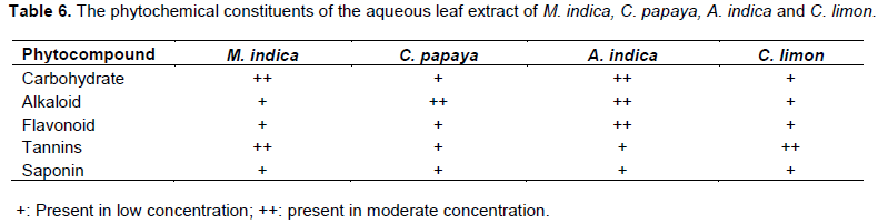
The plant samples collected were identified by a plant Taxonomist from Plant Biology and Biotechnology Department of the University of Benin, Benin City, Nigeria as A. indica (family Meliaceae), M. indica (family Anacardiaceae), C. papaya (family Caricaceae), and C. limon (family Rutaceae) (Arzoo and Parina Kumari, 2017). The cocktail, C. limon juice extract and aqueous leaf extracts of M. indica and C. papya were administered to respective groups at a dose of 100 mg/kg/body weight, while aqueous leaf extract of A. indica was administered at 10 mg/kg/body. The chemotherapeutic effect of the extract against P. berghei was investigated after seven days of administration. Phytochemical studies were carried out at the Pharmacognosy Department of the University of Benin, Benin City, Nigeria to determine the presence of carbohydrate, alkaloid, flavonoid, tannins and saponin in aqueous leaf extract of A. indica, M. indica, C. papaya and fruit extract of C. limon, using standard procedures proposed by Sofowora (1982) and Evans (2002).
Histopathology studies
The liver was excised and immediately transferred into 10% neutral buffered formalin and processed for light microscopic study, using an automatic tissue processor machine (Shandon 2000, Leica, Frankfurt, Germany). Tissues were dehydrated in various grades of alcohol then cleared in two changes of xylene, infiltrated in two changes of wax bath and finally embedded in paraffin wax. Five microns thick paraffin sections were obtained, which were finally stained using the Hematoxylin and Eosin staining procedure and the sections mounted with DPX and examined microscopically by means of ×10 and ×40 objective lenses (Avwioro, 2014).
Biochemical studies
Biochemical markers associated with liver functions were determined using the plasma obtained from the Wistar rats upon necropsy after seven days of treatment. Aspartate amino-transferase (AST), alanine aminotransferase (ALT), total protein and albumin were analyzed using commercial diagnostic kits from Randox laboratory, United Kingdom. The kits employed the procedure of Reitman and Frankel (1957) for the analysis of AST and ALT, while total protein was estimated using the procedure of Tiez (1995). Albumin was estimated using the method of Grant (1987).
Statistical analysis
Data analysis was performed using statistical package for social sciences (SPSS) version 20.0. Data were expressed as mean ± standard deviation (SD). Test of significance was calculated using paired Student’s t-test. p<0.05 was considered to be significant.
RESULTS
Histopathological findings
Histopathological findings indicated that extracts of C. papaya and C. limon were the most efficacious as there was significant reduction in the malaria associated liver pathology; while the groups administered with aqueous leaf extract of M. indica was observed to be least potent. Sections of uninfected rats administered with distilled water only showed normal histological features composed only of hepatocytes, portal vein and sinusoids, the morphology of the hepatocytes appeared normal and the sinusoids were not infiltrated; and no pathological lesion observed (Figure 1). Liver sections of the infected untreated group showed very poor architecture. There is severe portal triaditis and the portal tracts showed periportal infiltration of inflammatory cells with mild congestion of the portal vein, some of the hepatocytes showed foamy cytoplasms and some showed cellular debris engulfed by Kupffer cells. Heavy infiltrates of inflammatory cells and mild vascular congestion was observed. Parasites were engulfed in Kupffer cells and malaria pigment (haemozoin) was also present (Figure 2). Liver sections of infected rats administered with the cocktail showed sinusoids mildly packed with parasite-laden Kupffer cells and malaria pigment, mild inflammatory cellular infiltrates were also observed to be present around the portal zone, the hepatic portal vein was not congested (Figure 3). Liver sections of infected rats treated with aqueous leaf extract of A. indica showed decreased inflammatory cellular infiltrates indicating resolving malaria infection; however, the sinusoids were mildly packed with Kupffer cells which have engulfed haemozoin pigment and cellular debris and mild congestion was observed within the portal vein (Figure 4). Comparison with the untreated infected group administered with distilled water only showed no observable congestion in the blood vessels and there was significant reduction in the polymorphornuclear cellular infiltrates (Figure 4). Liver sections of infected rats treated with the aqueous extract of M. indica showed sinusoids moderately packed with parasite-laden Kupffer cells and cellular debris (Figure 5). There was a mild observable congestion in the portal vein with severe periportal infiltration by inflammatory cells, and various regions of the liver were packed with haemozoin. The morphology of the hepatocytes showed micro vesicular steatosis and the cytoplasms are vacuolated and infiltrated with fat. Few cells with engulfed parasites are also seen (Figure 5). Infected group administered with aqueous leaf extract of C. papaya showed normal central venules without congestion. There is no observable congestion of the hepatic portal vein and the hepatocytes appeared normal. Mild sinusoidal Kupffer cell activation is present; however, the central venules is not congested and the sinusoids appeared normal and not infiltrated, no pathological lesion seen (Figure 6) Liver sections of infected group treated with fruit extract of C. limon showed normal morphology of most of the hepatocytes, however, with a few engulfed parasites. There was no portal vein congestion, liver sinusoids were mildly packed with Kupffer cells and haemozoin pigment was seen in various regions (Figure 7).


Biochemical studies
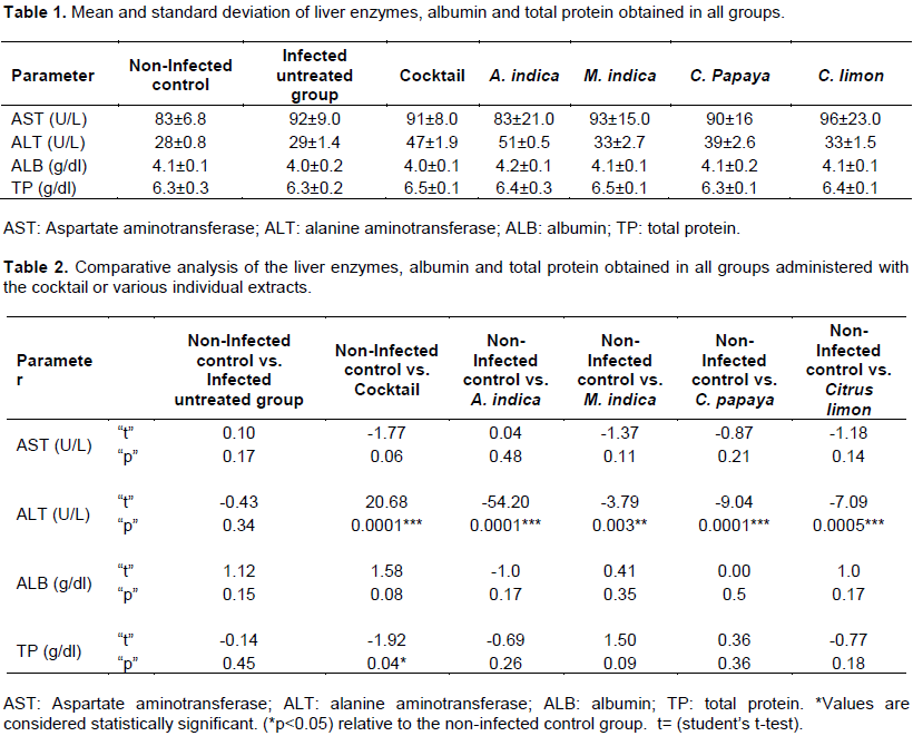
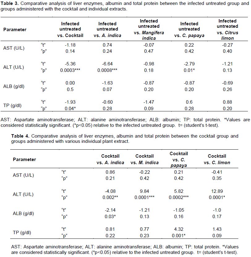
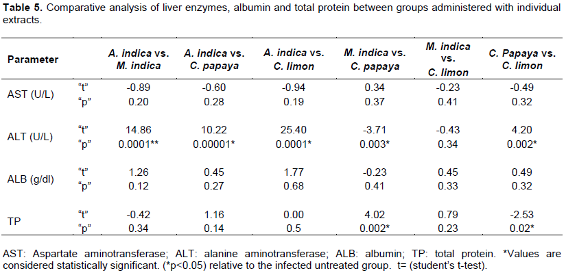
Table 1 shows the mean and standard deviation of liver enzymes, albumin and total protein obtained in all groups. Table 2 shows the comparative analysis of liver enzymes, albumin and total protein obtained in all groups administered with the cocktail or either extract contained therein. There were statistical significant variations in ALT activity (P < 0.0001) when all groups administered with the cocktail or either of the constituent extract were compared with the non-infected control group administered with distilled water only. AST activities and albumin values were observed to be normal among all groups (P > 0.05), while rats administered with the cocktail showed increase in total protein values (P < 0.04). Table 3 shows the comparative analysis of liver enzymes, albumin and total protein between the infected untreated groups and groups administered with the cocktail or either extracts showed no increase in albumin values and AST activity was observed to be normal (P > 0.05). However, there was a significant increase in ALT activities in rats administered with either of the cocktail (P < 0.0003), A. indica (P < 0.0008) or C. papaya (P < 0.01). Significant increase was observed in the total protein values among rats administered with the cocktail (P < 0.04) when compared with the infected untreated group administered with distilled water only. Comparative analysis of liver enzymes, albumin and total protein between the cocktail group and groups administered with various individual plant extract showed no significant increase in AST activity (P > 0.05). However, significant increase in ALT activity (P < 0.0001) was observed in other groups. Significant increase was observed in the groups administered with A. indica (P < 0.03) and in the total protein values of groups administered with aqueous leaf extract of C. papaya (P < 0.001) shown in Table 4. In addition, comparative analysis of liver enzymes, albumin and total protein between groups administered with individual extracts showed no statistical significant increase in the albumin values and AST activities; however, there was variations in the ALT activities (P < 0.00001) with the exception of rats administered with aqueous leaf extract of M. indica or C. limon (P > 0.05) while increase in total protein values was observed in rats administered with either M. indica or C. papaya <0.002 and groups administered with C. papaya or C. limon (P < 0.02) (Table 5).
Phytochemical studies
The results obtained from the phytochemical analysis of M. indica, C. papaya, A. indica and C. limon extract showed the presence of some secondary metabolite like carbohydrate, alkaloid, flavonoid, tannins and saponin. The presence of these secondary metabolites in this extract may be responsible for the antiplasmodial activity of the various extract (Table 6).
DISCUSSION
This study indicated that the herbal cocktail and individual extract contained therein has antiplasmodial effect on P. berghei. Histologcal studies of the infected rats showed recovery of the hepatocytes from congested black pigmentation (haemozoin pigment) as induced by these plant extracts; thus highlighting their importance in traditional treatment of malaria. Examination of the liver section clearly indicate that the administration of aqueous leaf extract of C. papaya and fruit extract of C. limon significantly reduced malaria associated with liver pathology when compared with the infected untreated group administered with distilled water only. Rats administered with the cocktail and plants extracts of A. indica also showed ameliorative effect on malaria associated liver pathology; while the group administered with aqueous leaf extract of M. indica was observed to be least potent. Photomicrographs of the liver of the uninfected control group administered with distilled water appeared normal as no endemic inflammation of hepatocytes was observed. Liver changes in severe malaria reported by Whitten et al. (2011) were observed in the untreated infected group administered with distilled water only. Sinusoids was observed to be densely packed with malaria pigments (haemozoin) and cellular debris engulfed by Kupffer cells. Heavy infiltrates of inflammatory cells was also confirmed to be present as observed by Olayode et al. (2015), mild vascular congestion of the hepatic portal vein was observed. The sinusiods of the infected rats treated with the cocktail was observed to be mildly packed with parasite laden Kupffer cells and haemozoin, a mild inflammation was also observed around the portal zone. The hepatic portal vein was not congested. Infected rats treated with aqueous leaf extract of A. indica showed decreased inflammatory cellular infiltrates in the liver sections; indicating that infection is resolving, which is in line with the observation of Anyaechie (2009) who documented that an active ingredient Irodin A isolated from Neem leaves is toxic to causative strains of malaria. Comparison with the infected untreated group administered with distilled water only showed no observable congestion in the blood vessels, and there was significant reduction in the polymorphor nuclear cellular infiltrates.
The sinusoids of infected rats administered with aqueous extract of M. indica were moderately packed with parasite-laden Kupffer cells and cellular debris; mild observable congestion in the portal vein and various regions of the liver were packed with haemozoin. The infected group treated with aqueous leaf extract of C. papaya had mild sinusoidal Kupffer cell activation and haemozoin pigments were observed in various regions of the liver section. Relatively few cellular infiltrates was observed to be present, indicating a resolving plasmodial infection. There was no observable congestion of the hepatic portal vein. This observation is in line with Longdet and Adoga (2017) who observed that extract of C. papaya leaf confers a dose dependent decrease on the level of parasitaemia when compared to the non-infected control group. This study corroborates Arise et al. (2012) who reported that extract of C. papaya gave significant suppression (P < 0.05) of parasitemia, following five days administration in established infection. Fatmawaty et al. (2017) further buttressed the antiplasmodial activity of C. papaya in P. berghei infected mice and histological examination of liver tissues of treated and untreated mice; which further supports the potential antimalaria of this plant, hence validating its traditional use in the treatment of malaria. Thomas et al. (2004), Fatmawaty (2013) and Okpe et al. (2016) also reported the antilarvicidal and antimalarial potential of fruit and leaf extract of C. papaya, both in vitro and in vivo. Liver sections of the group treated with C. limon showed reduced inflammatory cellular infiltrates, and the sinusoids were mildly packed with Kupffer cells and haemozoin pigment was seen in various regions. There was no portal vein congestion observed.
Observation in this study further corroborates Saganuwan et al. (2014) who indicated that many Nigerian plants can be used for the treatment of malaria as the groups administered with the cocktail or either extracts of A. indica, M. indica, C. papaya or C. limon and they showed reduction in the liver pathology associated with malaria infection. The leaves used in this study (C. papaya, A. indica, M. indica) showed the presence of phytochemical constituents, including flavonoid, alkaloid and tannin which compounds are potentially used for various treatments such as herbal remedies (Longdet and Adoga, 2017). This study corroborates the observation of some studies (Dhar et al., 1996; Mulla and Su, 1999; Nathan et al., 2005) that showed that azadirachtin and other limonoids available in neem extracts are active on malaria vectors. Furthermore, observation in these study are in line with Osanaiya et al. (2013) who confirmed that antimalarial activity of A. indica leaf extract in P. berghei infected albino mice.
Biochemical studies indicated that the cocktail and individual extracts do not alter the AST activity in the infected rats administered with the cocktail or that of the constituent extracts contained therein when compared with the non-infected group administered with distilled water only. However, there was an observable increase in the activity of ALT in the rats treated with the cocktail or that of the extract. Rats administered with aqueous leaf extract of M. indica showed a reduction in ALT activity compared to those administered with the cocktail or either of the extracts of A. indica, C. papaya or C. limon. Thus, the result obtained in this study agrees with the findings of Olayode et al. (2017) who observed that M. indica has an ameliorative effect against hepatocellular injury. There was variation in the AST among the rats in the different group but no significant increase was observed in the enzyme activity (p > 0.05). Also, there was no significant change in albumin levels P > 0.05 and total protein values among groups administered with A. indica, M. indica, C. papaya and C. limon were observed to be normal, while rats administered with the cocktail showed a significantly raised albumin level. AST activity in the infected groups administered with cocktail or that of the extracts remained normal when compared with the infected group administered with distilled water only; there was also no significant difference in the ALT activity among rats in the groups treated with M. indica or C. limon when compared with the infected untreated group administered with distilled water.
Additionally, no difference in the total protein and albumin values were observed in test groups compared to the untreated infected group administered with distilled water; however, the group administered with the cocktail showed an elevated total protein concentration. Comparison of A. indica, M. indica, C. papaya and C. limon extracts with cocktail group showed no significant difference in the AST activity. Notwithstanding, an increase in ALT activity in various groups administered with either of the extract when compared to the group administered with the cocktail was observed. No observable difference in the albumin level between the infected rats administered with the cocktail and groups administered with either of the plant extracts, with the exception of the groups administered with the aqueous leaf extract of A. indica, showed an increased level of albumin when compared to the group administered with cocktail.
Comparative analysis of total protein values among infected rats in the group administered with cocktail and infected rats administered with either extracts of A. indica, M. indica, C. papaya or C. limon showed no significant difference, with the exception of the rats in the group treated with aqueous leaf extract of C. papaya that showed an increase in total protein value when compared with the group administered with the cocktail. No observable difference in the AST activity between various individual extracts when statistically compared with one another. Significant elevation of ALT activity induced by individual extracts was observed.
In addition, increased activity of ALT enzyme in the groups administered with M. indica, C. papaya and C. limon when compared with the groups treated with aqueous leaf extract of A. indica was also observed. Furthermore, there was increase in ALT activity in groups treated with C. papaya upon comparison with the groups administered with M. indica; while the activity in the group treated with C. limon was observed to be insignificant (P > 0.05). Increase in ALT activity occurred in groups administered with C. limon fruit extract when compared with the group administered with aqueous leaf extract of C. papaya; while albumin levels among the various groups treated with various extracts showed no significance. There was increase in the total protein concentration in the group treated with C. papaya compared to the group treated with M. indica. Increase in total protein values in the groups treated with C. limon when compared with the groups administered with aqueous leaf extract of C. papaya was as well observed.
CONCLUSION
This study indicated that the cocktail and individual plant extracts contained therein possess antiplasmodial effect by inducing recovery of hepatic cells from congested black pigmentation (haemozoin pigment) and reduction in malaria associated liver pathology. Thus, observation in this study gave credence to the traditional use of the plants for the treatment of malaria.
CONFLICT OF INTERESTS
The authors have not declared any conflict of interests.
REFERENCES
|
Abass AY, Muhammad I, Bilbis LS, Saidu Y, Onu A (2017). In vitro antimalarial activity of some Nigerianmedicinal plants. Journal of Pharmacognosy and Phytochemistry 6(6):885-888 |
|
|
Aiyeloja AA, Bello OA (2006). Ethnobotanical potentials of common herbs in Nigeria: A case study of Enugu state. Education Research and Reviews 1(1):16-22. |
|
|
Ajala TO, Igwilo CI, Oreagba IA, Oluwatoyin, Odeku OA (2011). The antiplasmodial effect of the extracts and formulated capsules of Phyllanthus amarus on Plasmodium yoelii infection in mice. Asian Pacific Journal of Tropical Medicine 4(4):283-287. |
|
|
Akin-Osaniye BC, NokAJ, Ibrahim S, Inuwa HM, Onyike E, AmLabu E, Haruna E (2013). "Antimalarial effect of Neem leaf and Neem stem bark extracts on Plasmodium berghei infected in the pathology and treatment of malaria". International Journal of Research in Biochemistry and Biophysics 3(1):7–14. |
|
|
Al-Snafi AE (2016). Nutritional value and pharmacological importance of citrus species grown in Iraq. IOSR Journal of Pharmacy 6(8):76-108 |
|
|
Anjum V, Ansari SH, Naquvi KJ, Arora P, Ahmad A (2013). Development of quality standards of Carica papaya Linn. Leaves. Scholars Research Library 5(2):370-376. |
|
|
Anyaehie UB (2009). Medicinal properties of fractionated acetone/water neem (A. indica) leaf extract from Nigeria: A review. Nigerian Journal of Physiological Sciences 24:157-159. |
|
|
Arise RO, Malomo SO, Lawal MM (2012). Comparative antimalarial and toxicological effects of artemisinin with methanolic extract of C. papaya leaves and bark of Alstonia broonai in animal models. Advances in Natural and Applied Sciences 6(2):116-123. |
|
|
Arzoo and Parina K (2017). A review on plants having antimalarial activity. International Journal of Universal Pharmacy and Bio Sciences 6(3):142-158. |
|
|
Avwioro OG (2014). Histochemistry and tissue pathology; Principles and Techniques. 3rdEdition. Society for Cellular Pathology Scientist of Nigeria pp. 97-98. |
|
|
Dhar RH, Dawar S, Garg S, Basir F, Talwar GP (1996). "Effect of volatiles from neem and other natural products on gonotrophic cycle and oviposition of anopheles stephensi and An. Culicifacies (Diptera: Culicidae)". Journal of Medical Entomology 33(2):195-201. |
|
|
Ediriweera MK, Tennekoon KH, Samarakoon SR (2017). A review on ethnopharmacological applications, pharmacological activities, and bioactive compounds of Mangifera indica (Mango) Evidence-Based Complementary and Alternative Medicine pp. 1-24. |
|
|
Ene AC, Atawodi SE (2012). Ethnomedicinal survey of plantsused by the Kanuris of North-eastern Nigeria. Indian Journal of Traditional Knowledge 11(4): 640-645. |
|
|
Etuk EU, Bello SO, Isezuo SA, Mohammed BJ (2010). Ethnobotanicalsurvey of medicinal plants used for the treatment ofDiabetes mellitus in the north western region of Nigeria. Asian Journal of Experimental Biological Sciences 1(1):55-59. |
|
|
Evans WC (2002). Trease and Evans Pharmacognosy, 15th Edition. London: W.B. Sanders. pp. 183-393. |
|
|
Fatmawaty F, Astuti H (2013). Antimalarial activity of Delonix regia on mice with Plasmodium berghei. Journal of Natural Products 6: 61-66. |
|
|
Fatmawaty F, Rosmalena R, Amalia A, Syafitri I, Prasasty VD (2017). Antimalarial effect of flamboyant (Delonix regia) bark and papaya (C. papaya L) leaf ethanolic extracts against Plasmodium berghei in mice. Biomedical and Pharmacology Journal 10(3):1081-1089. |
|
|
Federal Ministry of Health (2015). National Malaria Elimination Programme, FMOH. Federal republic of Nigeria. Abuja, Nigeria. |
|
|
Gbolahan WA, Titus AOl, Isiaka AA, Gbolahan-Ayoade EE (2014). Composition of some traditional malaria remedies and their antiplasmodial effects on (Plasmodium berghei). International Journal of Scientific Research Publications 4(3):1-8. |
|
|
Grant GH (1987). Amino Acids and Proteins. In: Fundamentals of Clinical Chemistry, Tietz NW, 3rd edition, Philadephia, USA: WB Saunders Company pp. 328-329. |
|
|
Ibrahim HA, Imam IA, Bello AM, Umar U, Muhammad S, Abdullahi SA (2012). The potential of Nigerian medicinal plant as antimalarial agent: A review. International Journal of Science and Technology 2(8):600-605. |
|
|
Idowu OA, Soniran OT, Ajana O, Aworinde DO (2010). Ethnobotanical survey of antimalarial plants used in Ogun State, Southwest Nigeria. African Journal or Pharmacy and Pharmacology 4(2):55-60. |
|
|
Innocent AE, Aniekan IP, Aquaisua NA (2017). Histopathological effect of Nauclea latifolia ethanolic leaf extract and Artemether/Lumefantrine on the hippocampus of P. berghei-infected mice. International Journal of Brain and Cognitive Sciences 6(1):9-16. |
|
|
Ivanova D, Genova D, Chervenkov T, Yankova T (2005). Polyphenols and antioxidant capacity of Bulgarian medicinal plants. Journal of Ethnopharmacology 96(1-2):145-150. |
|
|
Iyamah PC, Idu M (2015). Ethnomedicinal survey of plants used in the treatment of malaria in Southern Nigeria. Journal of Ethnopharmacology 173:287-302. |
|
|
Laudicina VA, Palazzolo E, Germanà MA (2013). Current and Potential Use of Citrus Essential Oils. Current Organic Chemistry 17:3042-3049 |
|
|
Longdet I, Emmanuel AA (2017). Effect of Methanolic leaf extract of C. papayaon Plasmodium berghei infection in Albino Mice. European Journal of Medicinal Plants 20(1): 1-7. |
|
|
Makni M, Jemai R, Kriaa W, Chtourou Y, Fetoui H (2018). Citrus limon from Tunisia: Phytochemical and physicochemical properties and biological activities. BioMed Research International pp. 1-8. |
|
|
Melariri P, Campbell W, Etusim P, Smith P (2012). In vitro antiplasmodial activities of extracts from five plants used singly and in combination against Plasmodium falciparum parasites. Journal of Medicinal Plants Research 6(47):5770-5779. |
|
|
Mulla MS, Su T (1999). Activity and biological effects of Neem products against arthropods of medical and veterinary importance. Journal of the American Mosquito Control Association 15 (2):133-152. |
|
|
Nathan SS, Kalaivani K, Muruga K (2005). Effects of Neem limonoids on the malaria vector Anopheles stephensi Liston (Diptera: Culicidae). Acta Tropica 96(1):47-55. |
|
|
Nayan V, Onteru SK, Singh D (2017). Mangifera indica flowerextract mediated biogenic green gold nanoparticles: Efficientnanocatalyst for reduction of 4-nitrophenol. Environmental Progress and Sustainable Energy 37(1):283-294. |
|
|
Noronha M, Guleria S, Jani D, George LB, Highland H, Subramanian RB (2018). Ethnobotanical database based screening and identification of potential plant species with antiplasmodial activity against chloroquine-sensitive (3D7) strain of Plasmodium Falciparum. Asian Pacific Journal of Tropical Biomedicine 8(2):92-97. |
|
|
Odugbemi TO, Akinsulire OR, Aibinu IE, Fabeku PO (2007). Medicinal plants useful for malarial therapy in Okeigbo, Ondo State, Southwest Nigeria. African Journal of Traditional. Complementary and Alternative Medicine 4(2):191-198. |
|
|
Okere SO, Janet OS, Eunice O, Moses DA, Mercy OS (2014). Antiplasmodial activity of aqueous leaf extract of Cymbopogon citratus against Plasmodium falciparum infected rats. American Journal of Biomedical and Life Sciences 2(3):60-64 |
|
|
Okpe O, Nathan H, Joseph I, Vicent AU, Stanley IR, Okoduwa, Omiagocho TI (2016). Antimalarial potential of C. papaya and Vernonia amygdalina in mice infected with Plasmodium berghei. Journal of Tropical Medicine pp. 1-6. |
|
|
Olayode AA, Ofusori DA, Ogunniyi TA, Olusola SS, Bejide RA, Adelodun ST (2017). Modulatory role of aqueous leave extracts of M. indica (Linn) on liver function test in plasmodium - infected albino mice. Anatomy Physiology and Biochemistry International Journal 2(2): 001-005. |
|
|
Olayode AA, Ofusori DA, Ogunniyi TAB, Olusola SS (2015). Histomorphological studies of the liver of Plasmodium-infected albino mice after administration of aqueous leaf extract of M. indica (Linn.). Anatomy 9(3). |
|
|
Peter IT, Anatoli VK (1998). The current global malaria situation. Malaria parasite biology, pathogenesis and protection. Washington D.C, USA: ASM Press pp. 11-12. |
|
|
Randrianarivelojosia M, Rasidimanana VT, Rabarison H, Cheplogoi PK, Ratsimbason M, Mulholland DA, Mauclère P (2003). Plants traditionally prescribed to treat tazo (malaria) in the eastern region of Madagascar. Malaria Journal 2:25. |
|
|
Reitman S, Frankel S (1957). A colorimetric method for determination of serum glutamate oxaloacetate and glutamic pyruvate transaminase. American Journal of Clinical Pathology 28:56-58. |
|
|
Saganuwan SA, Onyeyili PA, Etuk EU (2104). Immunomodulatory potentials and histopathological effects of aqueous leaf extracts of Abrus precatorius leaf in mus musculus. Journal of Haematology Research 1(2):54-62. |
|
|
Sofowora A (1982). Medicinal plants and traditional medicine in Africa. John Wiley and sons Ltd. |
|
|
Taek MM, Bambang PEW, Mangestuti A (2018). Ethnomedicinal plants used for the treatment of malaria in Malaka, West Timor. Journal of Young Pharmacists 10(2). |
|
|
Thomas EI, Vandebroek SS, Van Damme P (2009). Cultural significance of medicinal plant families and species among Quechua farmers in Apillapampa, Bolivia. Journal of Ethnopharmacology 122:60-67. |
|
|
Tietz NW (1995). Clinical guide to laboratory tests. 3rd Edition. Philadephia USA: WB Saunders Company pp. 518-519. |
|
|
TitanjiV PK, Zofou D, Ngemenya MN (2008). The antimalarial potential of medicinal plants used for the treatment of malaria in cameroonian folk medicine. African Journal of Traditional Complementary and Alternative Medicine 5(3):302-321. |
|
|
Tiwari R, Amit KV, Sandip C, Kuldeep D, Shoor VS (2014). Neem (Azadirachta. indica) and its potential for safeguarding health of animals and humans: A Review. Journal of Biological Sciences 14(2):110-123. |
|
|
Vaghasiya Y, Dave R, Chanda S (2011). Phytochemical analysis of some medicinal plants from western Region of India. Research Journal of Medicinal Plant 5(5):567-576. |
|
|
Vineet K, Upma B (2014). Structural changes in spleen architecture upon Plasmodium berghei (NK-65) infection in BALB/c mice. Journal of Pharmacy and Biological Sciences 9(4):16-20. |
|
|
Whitten R, Milner DAJr, Yeh MM, Kamiza S, Molyneux ME, Taylor TE (2011). Liver pathology in Malawian children with fatal encephalopathy. Human Pathology 42:1230-1239. |
|
|
World Health Organization (2005). World Malarial Report. Geneva. Malaria prevention and control.ISBN 92 4 159319 9 |
|
|
World Health Organization (2014). World Malaria Report.Geneva. ISBN 978 92 4 156483 0. |
|
|
World Health Organization. (2016). World Malaria Report.Geneva.ISBN 978-92-4-151171-1 |
|
|
Yogiraj V, Goyal PK, Chauhan CS, Goyal A, Vyas B (2014). Carica papaya Linn: an overview. International Journal of Herbal Medicine 2(5): 01-08. |
|
Copyright © 2024 Author(s) retain the copyright of this article.
This article is published under the terms of the Creative Commons Attribution License 4.0


