Full Length Research Paper

ABSTRACT
Hyperlipidemia is one of the major health concerns worldwide. The present study aimed at utilizing clinicopathological tools to investigate the possible improving effect of Avena sativa mucilaginous extract (ASE) on lipid metabolic and liver function profiles in albino rats. The rats were rendered hyperlipidemic by 6-week supplementation of high-fat diet ad libitum. The rats were grouped into seven groups; with different treatment applications. Rats of group-I received normal diet and served as normal control; those of group-II were kept on high-fat (cholesterol 1% + coconut oil 2%) diet for 6 weeks and served as diseased control. Rats in group-III were kept on high-fat diet and received ezetimibe (1 mg/Kg B. Wt, orally, daily) and served as standard. Those in group-IV and V were kept on high-fat diet and received ASE at doses of 25 and 50 mg/Kg B. Wt., orally, daily (small and high doses, SD and HD, respectively) and served as treated-SD and treated-HD, respectively. While the last two groups (VI and VII) were kept on normal diet and received SD and HD of ASE. Blood samples for serum were taken for clinicochemical analysis on days 28 (4 weeks) and 42 (6 weeks) of the experiment and tissue specimens were taken for histopathology. ASE significantly (P<0.05) decreased the elevated serum lipid profile parameters, including total lipids, tri-acylglycerols (TAGs), cholesterol, LDL-C, VLDL-C, but significantly (P<0.05) normalized the serum HDL-C concentrations of rats kept on high-fat diet. Administration of A. sativa extract significantly decreased elevated serum liver enzyme activities in samples taken from animals kept on high-fat diet compared to the diseased untreated ones. Observations from histopathological examination were parallel and explanatory to clinicochemical analytical results. These data may suggest that the aqueous mucilaginous extract of A. sativa seed has a good health impact in cases associated with hyperlipidemia indicated by clinical pathology.
Key words: Avena sativa, antihyperlipidemic, clinical pathology, liver function, phytotherapy, phytomedicine.
INTRODUCTION
Among metabolic disorders, dyslipidemia, particularly hypercholesterolemia is considered as a major contributor in cardiovascular disease, including athero-sclerosis and atherosclerosis- associated conditions as coronary heart disease, ischemic cerebrovascular disease and peripheral vascular disease (Nelson, 2013). The best target for therapy in these conditions is to normalize blood and/or tissue cholesterol and lipid values. The conventional anti-hyperlipidemic chemical drugs are, usually, associated with some common adverse effects such as malaise, gastric irritation, nausea/vomiting/diarrhea, hyperuricemia, muscle aches, red dry skin, and altered liver function; Kumar et al., 2008). Natural products with health improving impacts constitute, therefore, an interesting research area in minds of concerned personnel, including patients, physicians and researchers based on their safety, efficacy and easy access (Ververidis et al., 2007).
Avena sativa (Oat; Kingdom: Plantae; Family: Poaceae) and its constituents were reported to possess variable beneficial health activities like antimicrobial (Maizel et al., 1964), antiparkinsonian (Zhou and Panchuk-Voloshina, 1997), topical anti-inflammatory (Boyer et al., 1998; Capasso, 2003). Also included are wound healing (Aries et al., 1999), antidiabetic (Jenkins et al., 2002), antioxidant (Bratt et al., 2003; Xu et al., 2009), anti-atherogenic (Liu et al., 2004), vasodilating (Nie et al., 2006), anti-asthmatic (Tabak et al., 2006), immunomodulatory (Ramakers et al., 2007; Dhillon and Bhatia, 2008), and anticancer (Anderson et al., 2009) activities.
Although effect of Avena sativa was tried on lipid profile in earlier studies (Karmally et al., 2005; Robitaille et al., 2005), yet, most of them used it as cereal as a whole or a part of the diet; and at least to our information, no studies have been performed using it as mucilaginous aqueous extract with adjusted doses. Moreover, Kerckhoffs et al. (2003) reported that the cholesterol lowering effect of β-glucan from oat bran in mildly hyper-cholesterolemic subjects may decrease when β-glucan is incorporated into bread and cookies. Therefore, the present study was designed to assess, using clinicopathological tools, the hyperlipidemia improving profile of A. sativa mucilaginous aqueous extract as a natural remedy to the control of dyslipidemia in rats prepared as hyperlipidemic model by high-fat diet supplementation.
MATERIALS AND METHODS
A. sativa mucilaginous extract
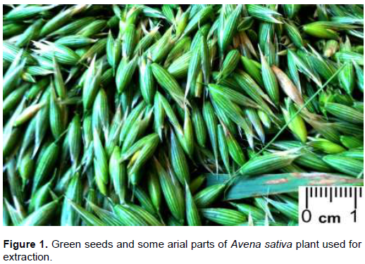
The extract was obtained and standardized according to the classical methodological stepwise described by Harborne (1973) with minor modifications. The green seeds with aerial parts of A. sativa (Figure 1) were collected from the local environment (Qaliuobeya Governorate, 2016) and identified by a plant specialist. Plant parts were refluxed in running tap water and then with bi-distilled water, shade dried at room temperature and chopped. Extract was prepared by macerating a weighed amount of the chopped plant parts (200 g) in a known volume (1.5 L) of water/ethanol (70:30, v/v). Maceration continued for 48 hours in refrigerator with occasional shaking. The hydro-ethanolic extract was then strained through muslin mesh and then concentrated using a shaking water bath at 56°C in a wide-mouthed containers and the mucilage obtained (yield) was then weighed and re-constituted freshly every day by dissolving in measured amount of bi-distilled water. Two stock solutions were prepared, 5 and 10 mg/ml. Avena sativa extract (ASE) was administered to rats at dosage rate of 25 (small dose; SD) and 50 (high dose; HD) mg/Kg B. Wt daily adjusted so that each rat (weighing 200 g) receives 1 ml of ASE orally using a rat gastric tube to the corresponding groups as explained below.
Ezetimibe
EZE, a standard inhibitor of cholesterol absorption, (Lipka, 2003) used in the present study was kindly gifted by SIGMA pharmaceuticals, Quesna Industrial Zone, Egypt. It was obtained as a pure powder. EZE was dissolved in 20% ethanol; where 10 mg of EZE were dissolved firstly in 10 ml absolute alcohol, and then the alcoholic solution of EZE was completed up to 50 ml by bi-distilled water. Each rat within the target group received 1 ml of the prepared solution which is equivalent to a dosage rate of 1 mg/Kg B. Wt., orally, once daily (Patel, 2004).
Experimental animals and protocol
Forty-two white male rats, of age 6-8 weeks and of average weight of 200 g were used in a parallel study design. Rats were divided in separate cages and gained access to clean water and diets ad libitum at room temperature. A week later (for acclimatization), rats were subjected to various experimental treatments, as follows:
Group-I: Rats kept on normal balanced diet and administered no drugs but their vehicles; served as normal control.
Group-II: Rats kept on high-fat diet and administered no drugs but their vehicles; served as diseased control.
Group-III: Rats kept on high-fat diet and administered EZE at a dose rate of 1 mg/Kg B. Wt.; used as a standard anti-hyperlipidemic.
Group-IV: Rats kept on high-fat diet and administered ASE at a dose of 25 mg/Kg.
Group-V: Rats kept on high-fat diet and administered ASE at a dose of 50 mg/Kg.
Group-VI: Rats kept on normal balanced diet and administered ASE at a dose of 25 mg/Kg.
Group-VII: Rats kept on normal balanced diet and administered ASE at a dose of 50 mg/Kg.
Treatments were conducted simultaneously from the first day of the experiment and continued for 6 weeks with continuous observation. All procedures were ethical to animals, and were performed with merciful and humane manner under light ether anesthesia, and adhered to principles published by International Council for Laboratory Animal Science (ICLAS) and those of Benha University Animal Care and Use Committee.
Sampling
Blood for serum was collected on the 28th and the 42th days from the start of the experiment. Blood was harvested into plain sampling tubes from the ocular medial canthus venous plexus using heparinized capillary tubes. The harvested blood was left to coagulate at 37°C for an hour; and then the clot was allowed to shrink by refrigeration for further an hour. Centrifugation at 1000 × g for 10 min were performed to separate clean sera, which were collected into Eppendorf’s tubes using Pasteur pipettes. The obtained samples were at -50°C until analysis of the following clinicochemical parameters: Total lipids (TL), total cholesterol (TC),
tri-acylglycerols (TAGs), high-density lipoprotein-cholesterol (HDL-C), low-density lipoprotein-cholesterol (LDL-C), very low-density lipoprotein-cholesterol (VLDL-C), aspartate amino transferase (AST), and alanine amino transferase (ALT). On the last day of the experiment, and after blood collection, animals were sacrificed by slaughtering using sharp scalpels under ether anesthesia, and tissue specimens from the liver were taken into 10% formalin solution for histopathological examination.
Clinicochemical analysis
The serum total lipids were determined according to the method described by Chabrol and Castellano (1961) using a kit supplied by Spinreact® (Sant Esteve De Bas, Spain); total cholesterol was determined enzymatically according to the method described by Meiattini et al. (1978) using a kit supplied by Spinreact®; HDL-L was determined according to the precipitation method described by Friedewald et al. (1972) using a kit supplied by Spectrum® (Obour city, Egypt); TAGs were measured enzymatically depending on the method explained after Young et al. (1975) using a kit Spinreact®. LDL-C and VLDL-C values were calculated using the formulae described by Friedewald et al. (1972) and Bauer (1982), respectively. Serum AST and ALT were quantitatively determined according to the method described by Murray (1984) using kits supplied by Diamond® (Cairo, Egypt). All laboratory steps were according to the instructions supplied by the kit manufacturers.
Histopathological examination
Liver specimens picked out from dissected rats in all groups were immediately placed in 10% formalin solution; processed for sectioning, staining and microscopical examining as described by Bancroft and Gamble (2008). Samples were allowed to fix for a 24 h period. The fixed samples were then gently washed under slowly running water for over-night. The clean fixed samples were then allowed to dehydrate in a series of increased-concentrations of ethanol starting in 70% and finalizing in absolute alcohol. The fixed, clean, dehydrated samples were placed in xylol for 3 h to clear and then placed in melted paraffin wax tissue boxes. The wax containing tissue specimens was left to solidify and then sections of 4 - 6 µm thickness were obtained using a rotary microtome. Staining procedure started with removal of wax from the microsections by two changes in ethanol (absolute; 5 minutes each). Ethanol was washed away with water. Sections were stained with Haematoxylin and Eosin (HandE) for 10 minutes, and then the extra stains were gently flushed under running water for 15 minutes. The stained microsections were dehydrated by alcohol series as mentioned above, then allowed to clear in xylol and finally covered with Dibutylphthalate Polystyrene Xylene (DBX). The prepared microsection slides were examined microscopically and interpreted by a specialist.
Data analysis and presentation
Data were presented as mean ± S.E of 6 observations. The obtained data were statistically analyzed using repeated-measures ANOVA at two timing points (4 and 6 weeks) with Tukey’s post-hoc to determine differences between groups at probability level of 5%. All statistical procedure was done by SPSS software, version 20. The sample size for each group was adjusted according to principles stated in literature, where value of α = 1.96, β = 0.842, a difference of 15 in each parameter is proposed between the groups as significant (Mean1− Mean2) and be detected with 80% beta power at a significance level alpha of 0.05 (Das et al., 2016).
RESULTS
Effect of ASE on serum lipid profile:
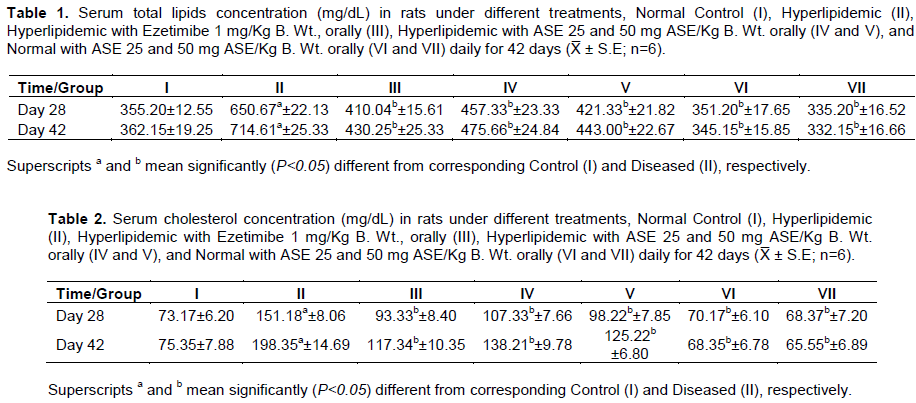
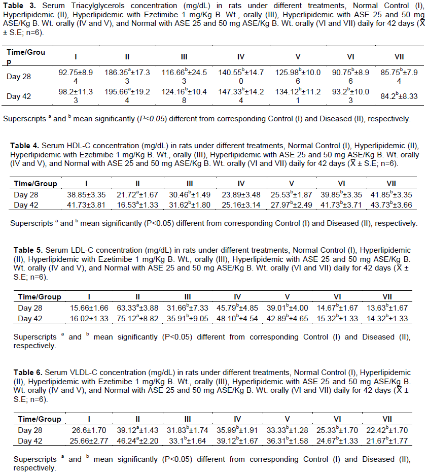
As shown in Tables 1, 2, 3, 5, 6, there were significant increases in serum total lipids, cholesterol, tri-acyl-glycerols, LDL-C, VLDL-C values in samples obtained from rats kept on high-fat diet, compared to those obtained from rats on balanced diet. While administration of ASE to control rats revealed insignificant alterations in these parameters throughout the experimental period, yet, its administration significantly (P<0.05) decreased their serum levels in samples of rats kept on high-fat diet in a dose-dependent manner. Nevertheless, as presented in Table 4, there was a significant (P<0.05) decrease in HDL-C values of samples of rats kept on high-fat diet. Such decrease was significantly (P<0.05) not only improved but also increased upon administration of ASE. Again, there was no significant changes in rats kept on basal diet.
Effect of ASE on Liver function profile
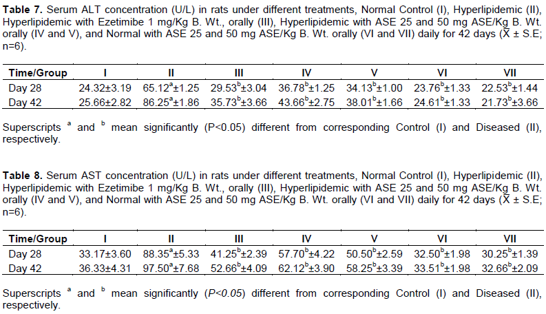
Data of the present study (Tables 7 and 8) demonstrate a significant elevation in serum ALT and AST activities in samples taken from rats kept on high-fat diet throughout the experiment, compared to those in samples taken from the normal control rats which were kept on balanced diet. Although administration of ASE to normal rats revealed insignificant alterations in liver enzyme activities throughout the experimental period; yet, it significantly (P<0.05) decreased their elevated serum activities in animals kept on high-fat diet compared to the diseased ones, upon its administration.
Effect of ASE on Liver structure
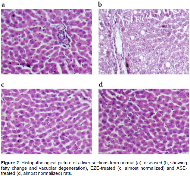
Data of the present study showed that there were no changes in liver samples picked out from rats of normal control group. Yet, subjecting experimental rats to high-fat diet (diseased-control group) resulted in some hepatic degenerative changes as hydropic and vacuolar degenerations and peri-portal fatty change of hepatocytes, as well as congestion of hepatic blood vessels. The severity and development of such changes were inhibited largely, in liver specimens picked out from ASE-treated groups and EZE-treated rats, as well (Figure 2a, b, c and d).
DISCUSSION
It is well established that alteration in lipid metabolic profile, especially long standing hyperlipidemia is a direct cause to various disease conditions including atherosclerosis, ischemic heart disease (Ross and Harker, 1976). This fact is later supported by Ross (1986) and Crowther (2005). Findings of the current experiment show that hyperlipidemia, induced by 42-day supplementation of high-fat (cholesterol and coconut oil 1 and 2% wt/wt, respectively) diet caused, as expected, marked alterations in the measured lipid parameters (Tables 1 to 6) of rat groups kept on such diet. In addition, the established dyslipidemia was associated with abnormally elevated liver function markers (AST and ALT; Tables 7 and 8), together with considerable hepatic histopathological changes (Figure 2). These rats were applied, in the present study, as a model for hyperlipidemia to evaluate the possible anti-hyperlipidemic potential of a mucilage extract prepared from Avena sativa green seeds and leaves.
Data presented in this study (Table 1) show a significant (P<0.05) increase in serum total lipid value of rats received high-fat diet throughout the 42 days of the experiment, compared to the normal control rats fed on a balanced diet. Similar findings were reported previously in rats (Csont et al., 2002; El-Mahmoudy et al., 2013; El-Mahmoudy et al., 2014) and rabbits (Diaz et al., 2000).
Administration of ASE significantly decreased serum total lipid concentration in animals received high-fat diet compared to the diseased untreated ones. The decrease in serum total lipid concentration in ASE-treated animals is logic after recording the improved serum values of TAGs, TC, LDL-C and VLDL-C that were observed simultaneously in this study. Lowering serum lipid profile recorded in the present study may be parallel with that reported by (Czerwiński et al., 2004) who found that oat and amaranth meals positively affect plasma lipid profile in rats fed cholesterol-containing diets. The authors attributed such effect to the contents of the bioactive components and the antioxidant activities of the studied plant samples.
Findings presented in the current study (Table 2) show a significant (P<0.05) elevation in serum TC concentration in samples from rats kept on high-fat diet throughout the 42-day experimental period, compared to that of the normal control animals. This finding is in accordance with that achieved by (Diaz et al., 2000) who found that rabbits kept on atherogenic diet exhibited marked elevation in TC in plasma.
Elevated serum cholesterol level is expected after high-fat diet supplementation and the inhibited clearing rate of LDL-C from the blood due to some defect in LDL receptors associated with elevated plasma total cholesterol values above normal levels (Zulet et al., 1999). Although administration of ASE to control rats revealed insignificant alteration in serum total cholesterol value throughout the experiment; yet, it significantly (P<0.05) decreased serum cholesterol concentration in animals received high-fat diet compared to the diseased untreated ones. The hypo-cholesterolemic effect of ASE may be explained based on decreasing cholesterol intestinal absorption with less dietary cholesterol is delivered to the pool of cholesterol to the liver; and/or greater clearance of LDL-cholesterol particles by the liver.
Findings presented in the current study (Table 3) show a significant (P<0.05) increase in TAGs in samples from animals kept on high-fat diet if compared with those of the normal control. Such significant elevation in serum TAGs may be explained on the basis of the diminished activity of lipase -insulin-dependent enzyme- contributing in TAGs clearance from blood by enhancing their hydrolysis to glycerol and free fatty acids (Yost et al., 1995). Daily oral administration of ASE significantly decreased serum TAGs concentration in animals received high-fat diet compared to that of diseased untreated ones. This finding may be parallel to those of Maier et al. (2000) and Czerwiński et al. (2004) in women and rats, respectively. The significant decrease in plasma TAGs has been explained previously by Bennani-Kabchi et al. (2000) who related that decrease to the higher rate of lipolysis mediated by enhanced plasma lipase activity. Nevertheless, more earlier, Griffin et al. (1982) stated that the lower level plasma TAGs might also reflect a lower rate of lipogenesis in the liver.
Tables 4, 5 and 6 present significant increases in serum LDL-C and VLDL-C and a significant decrease in HDL-C in rats kept on high-fat diet, compared to the corresponding normal control ones. The elevated serum LDL-C and VLDL-C seemed to occur upon over-production of LDL beyond the capacity of LDL-receptors expressed on hepatocyte cell membranes. In addition, the dietary fat and cholesterol may alter the serum lipoprotein pattern and increases the cholesterol content in VLDL (Mahley and Holcombe, 1977). ASE administration revealed significant decreases in serum LDL-C and VLDL-C concentrations with a significant increase in HDL-C when compared to those of the rats received high-fat diet. Such improving effect of ASE may be attributed to the decreased absorption of fats supplemented to rats and/or increased preipheral and hepatic breakdown of cholesterol esters from VLDL-C and LDL-C. The compositional change of HDL-C might be speculated due to a probable activation of Lecithin- cholesterol acyltransferase (LCAT) that is stimulated firstly by exogenous cholesterol.
Tables 7 and 8 present significant (P<0.05) elevations in ALT and AST activities in samples taken from rats kept on high-fat diet, if compared with those of the control. ASE administration significantly protected against elevations of these hepatic function biomarkers compared to the diseased untreated rats. This protecting effect may be explained on the bases of improved cholesterol hepatic metabolism as well as inhibiting its intestinal cholesterol absorption. Histopathological findings come supportive to the biochemical analysis, where the fatty degenerative changes observed in liver specimens from rats kept on high-fat diet were almost not observed upon concurrent administration of ASE (Figure 2). The recorded beneficial effects of ASE may be related to the active pharmacological constituents present in the extract, including β-glucan, avenanthramides, flavonoids, flavonolignans, triterpenoid saponins, sterols, and tocols. In addition, the mucilaginous nature of the extract may impede lipid absorption from the intestines upon oral administration (Singh et al., 2013; Miraj and Kiani, 2016).
CONCLUSION
The present findings suggest that A. sativa mucilage extract may protect the liver and the body against development of hyperlipidemia, indicated by clinicopathological analysis. The extract, therefore, may have a good health impact in dyslipidemia and concurrent illnesses.
CONFLICT OF INTERESTS
The authors have not declared any conflict of interests.
REFERENCES
|
Anderson JW, Baird P, Davis RH, Ferreri S, Knudtson M, Koraym A, Waters V, Williams CL (2009). Health benefits of dietary fiber. Nutrition Reviews 67(4):188-205. |
|
|
Aries M, Vaissiere C, Ceruti I, Charveron M, Gall Y (1999). Avena rhealba activity in cutaneous wound healing process. In: Proceedings of the Journal of Investigative Dermatology pp. 132-132. |
|
|
Bancroft JD, Gamble M (2008). Theory and practice of histological techniques Elsevier Health Sciences P 725 China. |
|
|
Bauer JD (1982). Clinical laboratory methods Cv Mosby, Portland, ME, USA. P 1235. |
|
|
Bennani-Kabchi N, Kehel L, El Bouayadi F, Fdhil H, Amarti A, Saidi A, Marquie G (2000). New model of atherosclerosis in insulin resistant sand rats: hypercholesterolemia combined with D2 vitamin. Atherosclerosis 150(1):55-61. |
|
|
Boyer F, Darne S C, Borrel M, Dupuy P, Gall Y (1998). Anti inflammatory properties of oatmeal extracts using a lauryl sulfate irritation model. Journal of Dermatological Science 16:S217. |
|
|
Bratt K, Sunnerheim K, Bryngelsson S, Fagerlund A, Engman L, Andersson RE, Dimberg LH (2003). Avenanthramides in oats (Avena sativa L.) and structure− antioxidant activity relationships. Journal of Agricultural and Food Chemistry 51(3):594-600. |
|
|
Capasso F (2003). Phytotherapy: A quick reference to herbal medicine. Springer Science and Business Media pp. 424 Heidelberg, Germany. |
|
|
Chabrol E, Castellano A (1961). SPV method for estimation of total serum lipid. Journal of Laboratory and Clinical Medicine 57:300. |
|
|
Crowther MA (2005). Pathogenesis of atherosclerosis. ASH Education Program Book (1):436-441. |
|
|
Csont T, Balogh G, Csonka C, Boros I, Horváth I, Vigh L, Ferdinandy P (2002). Hyperlipidemia induced by high cholesterol diet inhibits heat shock response in rat hearts. Biochemical and Biophysical Research Communications 290(5):1535-1538. |
|
|
Czerwiński J, Bartnikowska E, Leontowicz H, Lange E, Leontowicz M, Katrich E, Trakhtenberg S, Gorinstein S (2004). Oat (Avena sativa L.) and amaranth (Amaranthus hypochondriacus) meals positively affect plasma lipid profile in rats fed cholesterol-containing diets. The Journal of Nutritional Biochemistry 15(10):622-629. |
|
|
Das S, Mitra K, Mandal M (2016). Sample size calculation: Basic principles. Indian Journal of Anaesthesia 60(9):652. |
|
|
Dhillon P, Bhatia A (2008). Hypercholesterolemic and Immunomodulatory Effects of Oat Extracts containing ß-glucan. Research Journal of Immunology 1(1):29-35. |
|
|
Diaz M, Lopez F, Hernandez F, Urbina JA (2000). L-Carnitine effects on chemical composition of plasma lipoproteins of rabbits fed with normal and high cholesterol diets. Lipids 35(6):627-632. |
|
|
El-Mahmoudy AM, Abdel-Fattah FA, Abd El-Mageid A, Gheith IM (2014). Effect of the growth promotant mannan-oligosaccharide on the lipogram and organ function profile in hyperlipidemic albino rats. American Journal of Phytomedicine and Clinical Therapeutics 2:334-347. |
|
|
El-Mahmoudy AM, Shousha SM, Abdel-Maksoud H, AbouZaid O (2013). Effect of long-term administration of sildenafil on lipid profile and organ functions in hyperlipidemic rats. Acta Bio Medica Atenei Parmensis 84(1):12-22. |
|
|
Friedewald WT, Levy RI, Fredrickson DS (1972). Estimation of the concentration of low-density lipoprotein cholesterol in plasma, without use of the preparative ultracentrifuge. Clinical Chemistry 18(6):499-502. |
|
|
Griffin H, Whitehead C, Broadbent L (1982). The relationship between plasma triglyceride concentrations and body fat content in male and female broilers-a basis for selection? British Poultry Science 23(1):15-23. |
|
|
Harborne J (1973). Phytochemical methods, a guide to modern techniques of plant analysis, JB Harborne. Chapman & Hall. London. GB. p 4-13 |
|
|
Jenkins A, Jenkins D, Zdravkovic U, Wursch P, Vuksan V (2002). Depression of the glycemic index by high levels of beta-glucan fiber in two functional foods tested in type 2 diabetes. European Journal of Clinical Nutrition 56 (7):622. |
|
|
Karmally W, Montez MG, Palmas W, Martinez W, Branstetter A, Ramakrishnan R, Holleran SF, Haffner SM, Ginsberg HN (2005). Cholesterol-lowering benefits of oat-containing cereal in Hispanic Americans. Journal of the American Dietetic Association 105 (6):967-970. |
|
|
Kerckhoffs DA, Hornstra G, Mensink RP (2003). Cholesterol-lowering effect of β-glucan from oat bran in mildly hypercholesterolemic subjects may decrease when β-glucan is incorporated into bread and cookies. American Journal of Clinical Nutrition 78(2):221-227. |
|
|
Kumar A, Mazumder A, Saravanan V (2008). Antihyperlipidemic activity of Camellia Sinensis leaves in triton wr-1339 induced Albino rats. Pharmacognosy Magazine 4(13):60. |
|
|
Lipka LJ (2003). Ezetimibe: A Firstâ€inâ€Class, novel cholesterol absorption inhibitor. Cardiovascular drug reviews 21(4):293-312. |
|
|
Liu L, Zubik L, Collins FW, Marko M, Meydani M (2004). The antiatherogenic potential of oat phenolic compounds. Atherosclerosis 175 (1):39-49. |
|
|
Mahley RW, Holcombe K (1977). Alterations of the plasma lipoproteins and apoproteins following cholesterol feeding in the rat. Journal of Lipid Research 18(3):314-324. |
|
|
Maier SM, Turner ND, Lupton JR (2000). Serum lipids in hypercholesterolemic men and women consuming oat bran and amaranth products. Cereal Chemistry 77(3):297-302. |
|
|
Maizel J, Burkhardt H, Mitchell H (1964). Avenacin, an antimicrobial substance isolated from Avena sativa. I. Isolation and antimicrobial activity. Biochemistry 3(3):424-426. |
|
|
Meiattini F, Prencipe L, Bardelli F, Giannini G, Tarli P (1978). The 4-hydroxybenzoate/4-aminophenazone chromogenic system used in the enzymic determination of serum cholesterol. Clinical Chemistry 24(12):2161-2165. |
|
|
Miraj S, Kiani S (2016). Study of pharmacological effect of Avena sativa: A review. Der Pharmacia Lettre 8(9):137-140. |
|
|
Murray R (1984). Aspartate aminotransferase. Clinical Chemistry. Theory, analysis and correlation. Kaplan LA, Pesce AJ (Ed), CV Mosby Company:, St. Louis, Toronto, Princeton, pp: 1112-1116. |
|
|
Nelson RH (2013). Hyperlipidemia as a risk factor for cardiovascular disease. Primary Care: Clinics in Office Practice 40(1):195-211. |
|
|
Nie L, Wise M L, Peterson D M, Meydani M (2006). Avenanthramide, a polyphenol from oats, inhibits vascular smooth muscle cell proliferation and enhances nitric oxide production. Atherosclerosis 186(2):260-266. |
|
|
Patel SB (2004). Ezetimibe: a novel cholesterol-lowering agent that highlights novel physiologic pathways. Current Cardiology Reports 6(6):439-442. |
|
|
Ramakers JD, Volman JJ, Biörklund M, Önning G, Mensink RP, Plat J (2007). Fecal water from ileostomic patients consuming oat βâ€glucan enhances immune responses in enterocytes. Molecular Nutrition and Food Research 51(2):211-220. |
|
|
Robitaille J, Fontaine-Bisson B, Couture P, Tchernof A, Vohl M-C (2005). Effect of an oat bran-rich supplement on the metabolic profile of overweight premenopausal women. Annals of Nutrition and Metabolism 49 (3):141-148. |
|
|
Ross R (1986). The pathogenesis of atherosclerosis—an update. New England Journal of Medicine 314(8):488-500. |
|
|
Ross R, Harker L (1976). Hyperlipidemia and atherosclerosis. Science 193(4258):1094-1100. |
|
|
Singh R, De S, Belkheir A (2013). Avena sativa (Oat), a potential neutraceutical and therapeutic agent: an overview. Critical Reviews in Food Science and Nutrition 53 (2):126-144. |
|
|
Tabak C, Wijga A H, de Meer G, Janssen NA, Brunekreef B, Smit HA (2006). Diet and asthma in Dutch school children (ISAAC-2). Thorax 61(12):1048-1053. |
|
|
Ververidis F, Trantas E, Douglas C, Vollmer G, Kretzschmar G, Panopoulos N (2007). Biotechnology of flavonoids and other phenylpropanoid derived natural products. Part I: Chemical diversity, impacts on plant biology and human health. Biotechnology Journal: Healthcare Nutrition Technology 2(10):1214-1234. |
|
|
Xu JG, Tian CR, Hu QP, Luo JY, Wang XD, Tian XD (2009). Dynamic changes in phenolic compounds and antioxidant activity in oats (Avena nuda L.) during steeping and germination. Journal of Agricultural and Food Chemistry 57(21):10392-10398. |
|
|
Yost TJ, Froyd KK, Jensen DR, Eckel RH (1995). Change in skeletal muscle lipoprotein lipase activity in response to insulin/glucose in non-insulin-dependent diabetes mellitus. Metabolism-Clinical and Experimental 44(6):786-790. |
|
|
Young DS, Pestaner L, Gibberman V (1975). Effects of drugs on clinical laboratory tests (Vol. 4, No. 8). Washington, DC: AACC Press. |
|
|
Zhou M, Panchuk-Voloshina N (1997). A one-step fluorometric method for the continuous measurement of monoamine oxidase activity. Analytical Biochemistry 253 (2):169-174. |
|
|
Zulet MA, Barber A, Garcin H, Higueret P, Martinez JA (1999). Alterations in carbohydrate and lipid metabolism induced by a diet rich in coconut oil and cholesterol in a rat model. Journal of the American College of Nutrition 18(1):36-42. |
|
Copyright © 2024 Author(s) retain the copyright of this article.
This article is published under the terms of the Creative Commons Attribution License 4.0


