Full Length Research Paper
ABSTRACT
The aim of this study was to evaluate the effect of Berberis vulgaris water extract (BWE) against CCl4-induced hepatotoxicity and LPS/PCM induced- hepatic inflammation in the experimental animal models. CCl4 (100 ug/kg, oral) administration for 28 days as well as the co-administration of LPS (250 ug/kg bw) and PCM (2 g/kg bw) for 28 days resulted in massive elevation in serum and hepatic prooxidants and inflammatory parameters (thiobarbituric acid reactive substances (TBARS), tissue tumor necrosis factor–alpha (TNF-α), interleukin 6 (IL-6) and IL-12 and nitric oxide (NO) with depletion in antioxidants system (superoxide dismutase (SOD) and glutathione peroxidase (GPx). These alterations were combined with elevation of serum liver function tests (alanine transaminase (ALT), aspartate transaminase (AST) and alkaline phosphatase level and (ALP). Oral administration of the BWE (100 mg/kg) for 15 days showed hepatoprotective and anti-inflammatory effects against both CCl4 and LPS/PCM as it increased the activities of antioxidants enzymes, decreased the prooxidants and inflammatory markers levels and improved the serum liver function tests levels. The obtained findings thus suggest that B. vulgaris aqueous extract has protective roles against hepatic toxicity and hepatic inflammation due to its antioxidant and anti-inflammatory properties.
Key words: Carbon tetrachloride, paracetamol, lipopolysaccharide, glutathione peroxidase, alanine transferase.
INTRODUCTION
Berberis vulgaris L. (B. vulgaris) is a well-known medicinal plant which belongs to Berberidaceae family that is cultivated in Asia and Europe. The phytochemical investigations of various species of Berberis have led to the isolation of alkaloids, tannins, phenolic compounds, sterols and triterpenes (Mohammadzadeh et al., 2017). Berberine, the main isoquinoline alkaloid of Berberis, has several biological effects (Singh et al., 2011) with very low toxic and adverse reactions in human. Until now, berberine was not used as therapeutic compounds due to its poor oral bioavailability which is lower than 1% (Liu et al., 2016). Berberine poor oral bioavailability returns to its low water solubility, low intestinal permeation, binding with P-glycoprotein (P-gp) pumps and high extraction and distribution in the liver (Imenshahidi and Hosseinzadeh, 2016), therefore, the uses of permeation enhancers, P-gp inhibitors and lipid micro-particle delivery system improved the berberine bioavailability (Liu et al., 2016). All reported researches measured the biological effects of B. vulgaris ethanolic extract which showed potent antioxidative capacity in vitro (Abd El-Wahab et al., 2013) and in vivo (Xu and Zhou, 2010). B. vulgaris ethanolic extract in vitro and in vivo anti-inflammatory activity was noted by the reduction of proinflammatory cytokines as well as acute phase proteins (Xie et al., 2013) where it decreased TNF-α, IL-6, IL-1β, matrix metalloprotease 9 (MMP9), cyclooxygenase-2 (COX2), inducible NOS (iNOS), monocyte chemoattractant protein 1 (MCP-1) and C-reactive protein (CRP) and haptoglobin (HP) (Choi et al., 2006).
There are many comparative studies between the efficiency of the aqueous and organic extraction of the B. vulgaris as antiinflammation induced either in vitro or in vivo. Aliakbarlu et al. (2018) performed an investigation to evaluate the effect of different solvent extraction of B. vulgaris against inflammation. B. vulgaris is extracted by water, acetone and ethanol. The antioxidant of B. vulgaris is evaluated in terms of DPPH which is one of simplest methods to evaluate of antioxidant activity of the plant extract (Katalinic et al., 2006). Their investigation has showed that the scavenging activity of B. vulgaris water extract was higher than that of acetone extract or ethanolic extract (Aliakbarlu et al., 2018).
Vinay et al. (2014) found that both water and ethanolic extracts of Berberis aristata had in vitro anti-inflammatory activities as they prevented blood hemolysis and albumen denaturation; moreover, these extracts showed anti-inflammatory potential against carrageenan induced albino rats. These results proved that ethanol extract causes 80% inhibition of paw edema in comparison to that of aqueous extract which showed 72% inhibition at dose of 50 mg/kg.
Our experimental was designed to evaluate the protective effect of B. vulgaris water extract against acute liver injury, caused by chemical or immunologically agents. As well known, liver appears to be, due to its unique metabolic function, the most common target organ of toxicity. A wide variety of viruses, drugs and toxic chemicals can cause liver injury by means of their direct toxicity and/or endogenous toxic metabolic products. Carbon tetrachloride (CCl4) is a chemical hepatotoxin known for inducing in animal model the features similar to those of acute hepatitis in human. On the other hand, immunological liver injury model which was induced by LPS/PCM was found to reflect the clinical situation of hepatitis more accurately (Barman et al., 2016); hence both in vivo models were used in this study.
Liver toxicity can be induced by CCl4 because it converts into potent trichloroperoxyl radicals in hepatocytes which alters plasma membrane, endoplasmic reticulum, mitochondria, and Golgi apparatus structure which leads to fatty changes centrilobular steatosis, inflammation, apoptosis, and cell necrosis (Lutz et al., 2003; Sadeghi et al., 2008). PCM is a nonsteroidal anti-inflammatory compound used as analgesic and antipyretic drug (Aghababian, 2010). PCM high dose processes acute liver damage due to the formation of N-acetyl-p-benzoquinone imine reactive metabolite which causes oxidative stress, glutathione depletion (Shan et al., 2011) and NO production that cumulatively leads to hepatocytes necrosis (Abdullah et al., 2014). LPS, and glycoprotein on the gram-negative bacteria outer membrane, develops acute liver failure due to inflammatory response that occurred when it binds to Toll like receptor 4 (TLR4) that in turn activates transcription factor-nuclear factor-kappa B (NF-κB), which involves in the activation of different pro-inflammatory cytokines (Sainan et al., 2014).
This study aimed to estimate the activity of BWE as hepatoprotective against CCl4 induced hepatotoxicity and measure its anti-inflammatory properties against LPS/ Paracetamol induced hepatic inflammation in experimental animal models.
MATERIALS AND METHODS
Berberine standard, Cummen H2O2, reduced GSH, Salmonella typhimurium Lipopolysaccharide, Super oxide Dismutase standard and Thiobarbituric acid (TBA) were purchased from Sigma-Aldrich (St.Louis, MO). Carbon tetrachloride (CCl4) was purchased from Chemlab (Belgium), and Paracetamol was manufactured by Alexandria Pharmaceutical Company (Alexandria, Egypt). Pyrogallol, Sodium Nitrite, Sodium Nitroprusside and Sulphinamide were purchased from Fluka (Switzerland). The kits for determining serum aspartate aminotransferase (AST), alanine aminotransferase (ALT) and alkaline phosphatase activities were purchased from Chema Diagnostic (Italy). The tumor necrosis factor-(TNF-alpha) was purchased from Serva (Austria) .Interleukin-6 (IL-6) and Interleukin 12 assay kits were purchased from Ray Biotec (Italy).
Preparation and characterization of B. vulgaris water extract
Barberry root were bought and authenticated by Prof. Salma El-Dareir, Botany Department, Faculty of Science, Alexandria University, Egypt. The root was grinded and 500 g powder was soaked in 2 L of hot distilled water at 65°C, then incubated in shaker incubator at 50°C and 500 rpm for 24 h. The crude aqueous extract was collected by filtration, concentrated to 50 ml by using rotary evaporator (Büchi, Switzerland) and finally lyophilized to form powder water extract form (BWE, 60 gm, 12% yield) (DISHI, DS-DSH10, Xi’an Heb Biotechnology Co, China) (Ghareeb et al., 2017).
The water extract ingredients was analyzed using HPLC (Series 500 Bio-Tek Instrument, Milano, Italy, where the used column was Zorbax Eclipse XDB-C18 (250 × 4.6 mm i.d.,5 μm particle size) column (Agilent, Santa Clara, CA, USA). The BWE (1 mg/ml milli-Q water) was filtered through a 0.22-μm syringe filter prior to HPLC analysis. The operating temperature was maintained at 30°C and the detector was operated at a wavelength of 254 nm. The mobile phase was a mixture of two solvent compositions, solvent A (deionized water) and solvent B (methanol). Isocratic program was achieved with 40% solvent A and 60% solvent B at a solvent flow of 0.8 ml/min and injection volume of 20 μl (Qadir et al., 2009).
Experimental design
Sixty, two-months aged, albino male rats weighted 130 ± 8 g bought from Institute of Graduate Studies' Animal Facility, Alexandria University. Rats were randomly divided into two main groups where each group contained 30 rats for each animal model, where the first group was used to assess the BWE hepatoprotective effect on LPS/PCM induced hepatitis (Parmar et al., 2012) while the second group was used to measure the BWE effect on CCl4-induced hepatotoxicity (Feng et al., 2010). These rats were randomly housed as a group of 6 rats in metal cages (6 rats/cage) under normal conditions (room temperature was 23 to 25°C and a 12 h light/dark cycle) and freely accessed to normal basal diet and tap water. The experimental design was conducted according to Animal Ethics Committees (AEC) ethical instructions, The National Health and Medical Research Council (NHMRC), Egyptian Ministry of Health and Population (http://www.mohp.gov.eg). The study approval number was 15-1O-1018 which issued from the Pharmaceutical and Fermentation Industries Development Center, City for Scientific Research and Technology Applications, Alexandria, Egypt.
Hepatitis model
After one-week acclimation period, 30 rats were sub-divided into five groups (n=6); Negative control group which received intraperitoneal (IP) injection of 0.5 ml of saline; then after 2 h, they were orally administrated 1 ml of saline while the second one was BWE control (BWE-C) group which orally administrated 100 mg/kg BWE daily for two weeks. The third one was hepatitis group (LPS/PMC) which IP injected with 250 ug/kg LPS, then after 2 h, they were orally administrated 2 gm/kg PCM day by day for two weeks. The fourth one was Protection Group (BWE and LPS/ PMC) which orally administrated BWE for two weeks, then injected for another 2 weeks with LPS/PMC while the last group was Treatment group (LPS/PMC+BWE) which received firstly LPS/PMC for two weeks and then orally administered BWE for another 2 weeks.
Acute hepatotoxicity model
After one-week acclimation period, the other 30 rats were also divided into five groups. Negative Control group (C) in which rats were intraperitoneally injected with 0.5 ml of olive oil. BWE control (BWE-C) in which rats were orally administrated 100 mg /kg BWE daily for two weeks. Hepatotoxicity Group (CCl4) in which rats were intraperitoneally injected with 100 ul/kg CCl4 dissolved in olive oil for two weeks. Protection (BWE and CCl4) Group was orally administrated BWE for two weeks then IP injected with CCl4 for another two weeks. Finally, the treatment (CCl4+BWE) group was IP injected with CCl4 for two weeks which orally received BWE for another two weeks.
After one-month experimental period, all rats in both models were sacrificed. Blood was collected in EDTA tubes, allowed to stand in room temperature for 15 min and then centrifuged at 3000 rpm for 10 min to obtain plasma. Liver tissue was removed immediately, washed in iced saline solution, thereafter one gram of liver was homogenized with 9 ml of phosphate buffer, 0.1 M, pH 7.4, centrifuged at 3000 r.p.m. for 15 min at 4°C then the supernatant was collected and stored to be used as a liver homogenate (Ghareeb et al., 2017).
Biochemical parameters
Routine biochemical parameters
Liver function tests which were alanine aminotransferase (ALT), aspartate aminotransferase (AST) and alkaline phosphatase (ALP) were determined in plasma according to the instructions of commercial kits purchased from Chem Diagnostic (Italy).
Prooxidant/antioxidant parameters
TBARS as a marker for lipid peroxidation was determined in the form of malonaldehydehyde according to Tapel et al. (1959), and the concentration of plasma malonaldehyde was calculated by the following equation; (nmol/ml) =At × 0.156. While for tissue homogenate, the concentration was calculated by the following equation (nmol/ g wet tissue) = At × 0.156 × 10. SOD specific activity was assayed according to (Marklund and Marklund, 1974) in which the auto-oxidation of pyrogallol leads to formation of superoxide that will be decreased in the presence of SOD. One unit of SOD activity is defined as amount of the enzyme that inhibits the rate of auto oxidation of pyrogallol by 50%. GPx specific activity was assessed according to Paglia and Valentine (1967), in which the amount consumed GSH during cummen H2O2 reduction was calculated from subtraction of remaining GSH (At) from total GSH (Ac).
Inflammatory parameters
NO was determined in serum and liver homogenate according to the method of Montgomery and Dymock (1961). The hepatocytes cytokines (IL-1b and IL-12 and TNF-alpha) were measured by (ELISA) according to the manufacturer instructions.
Statistical analysis
Data were analyzed by one-way analysis of variance (ANOVA) using primer of Biostatistics (Version 5) software program. Significance of mean±SD was detected groups by the multiple comparisons Student- Newman- Keuls test as p ≤ 0.05.
RESULTS
Preliminary phytochemical screening of aqueous extract of B. vulgaris
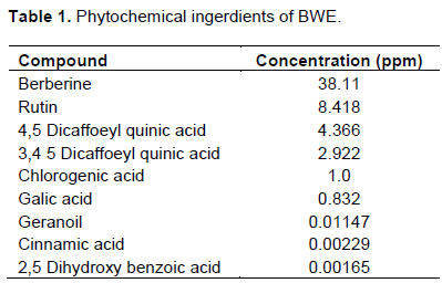
Preliminary phytochemical screening of the aqueous extract of B. vulgaris reveals the presence of Berberine, Rutin, 4,5 Dicaffoeyl quinic acid, 3,4 Dicaffoeyl quinic acid, Chlorogenic acid, Galic acid, Geranoil, Cinnamic acid and 2,5 Dihydroxy benzoic acid as shown in Table 1. Berberine is the highest concentration (38.11 ppm) while 2,5 Dihydroxy benzoic acid is the lowest one (1.65×10⁻³ ppm).
Protective and therapeutic effects of BWE on LPS/PMC induced hepatitis
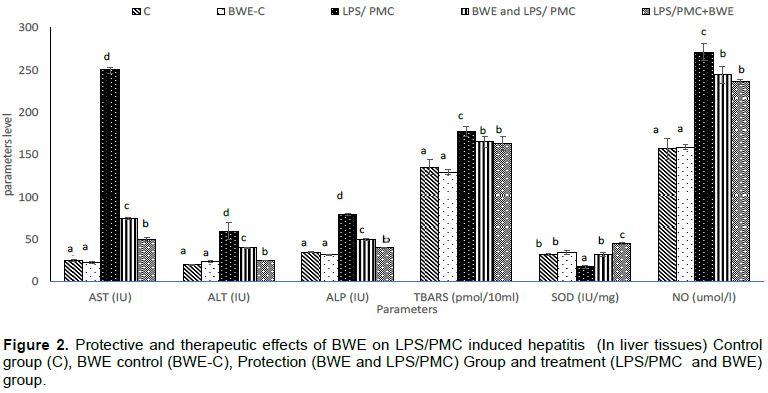
LPS and PCM administration for two weeks increased liver function parameters (AST, ALT and ALP activities) by 900, 200 and 128% respectively compared to the control group. The protection group showed that the liver function tests (AST, ALT, ALP) were lower than those of induction group level by 70, 33.3 and 37.5% respectively. Therapeutic group showed that liver function tests were lower than that of induction group by 80% for AST, 58.3% for ALT and 50.0% for ALP as shown in Figure 1.
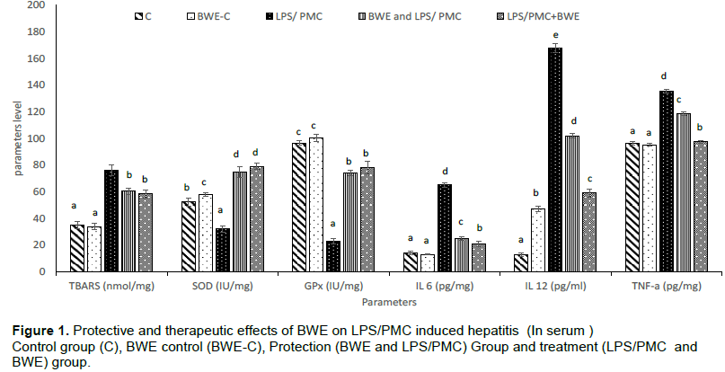
Upon administration of LPS/PCM to the experimental animals for two weeks, this led to induction of oxidative stress in both plasma and hepatic tissues. There was significant increment in the level of hepatic and plasma level of TBARS compared to the control group by 117 and 31% respectively, which was agreed with significant increment in the level of plasma nitric oxide in the induction group compared to the control group by 72.6%. Contrary to the effect of LPS/PCM as inducing of oxidative stress, there was decrement in the level of antioxidant enzyme activities (SOD and GPx). The level of hepatic and plasma enzymatic activity of SOD decreased by 44.6 and 38.8% respectively compared to the control group. The level of hepatic glutathione peroxidase activity decreased by 76% compared to the control group. There was a significant increment in the level of plasma cytokines (IL-6, IL-12 and TNF-alpha) when experimental animals induced by LPS/PCM as shown in Figure 2. Upon administration of the experimental animals by B. vulgaris water extract in the protection and therapeutic groups, there was retention in the activities of TBARS and NO in plasma and hepatic tissues. In the protection group, the level of hepatic and plasma TBARS were decreased by 20 and 7% respectively while for nitric oxide its plasma level decreased by 10%. In the therapeutic group, the level of hepatic and plasma TBARS were decreased by 22 and 8% respectively and for plasma nitric oxide there was decrement by 13% as shown in Figures 1 and 2.
Water extract administration to the experimental animals antagonizes the effect of LPC/PCM. In the protection group, there was increment in the activities of antioxidant enzyme as SOD and GPx in which the level of hepatic and plasma level of SOD was increased by 130 and 70% respectively compared to the induction group, and this was agreed by the increment in the level of plasma GPx by twice its value compared to the induction group. Inflammatory cytokines were decreased by the effect of the water extract of B. vulgaris in which the level of IL-6, IL-12 and TNF-alpha were decreased by 160, 65 and 14% respectively compared to the induction group. This effect was confirmed in the therapeutic group in which the level of IL-6, IL-12 and TNF-alpha were decreased by 200, 184 and 39% respectively compared to the induction group as shown in Figure 2.
Protective and therapeutic effects of BWE on CCl4 induced hepatotoxicity
Upon administration of the experimental animals by carbon tetrachloride (CCl4) for two weeks, this lead to significant increment in the level of liver function enzymatic (AST, ALT and ALP) activities than those of control level as shown in Table 2. In the protection group, the effect of water extract was significant in which there was improvement in the level of liver function test enzymes (AST, ALT, ALP) by 25, 18 and 25% respectively compared to the induction group. This was confirmed by its therapeutic effect in which the level of (AST, ALT and ALP) were decreased by 20, 24 and 43% respectively compared to the induction group. Administration of carbon tetrachloride for two weeks leads to induction of oxidative stress in which prooxidant markers (TBARS and NO). Hepatic and plasma TBARS were increased by 176 and 55% respectively compared to the control group. For plasma NO there was significant increment by 62% as shown in Table 2. Not only carbon tetrachloride leads to induction of prooxidant activity but also decrement in the antioxidant activity and this was significant in the levels of hepatic and plasma level of SOD and plasma GPx. The level of tissue and plasma SOD were decreased by 63% and 35% compared to control group. The level of hepatic GPx was decreased by 56% compared to control group as shown in Tables (1 and 2). CCl4 administration lead to significant increment in inflammatory cytokines (IL-6, IL-12 and TNF-alpha) compared to control group as shown in Table 3.
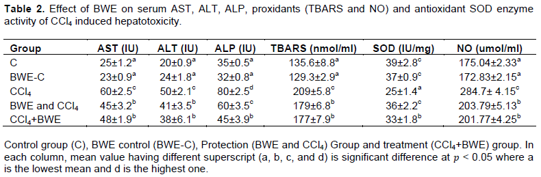
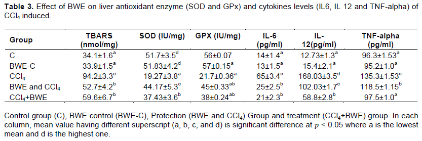
Administration of the B. vulgaris water extract to the experimental animals lead to significant improvement in the body vital enzymatic activities. In the protection and curing groups, there was improvement in the level of prooxidant enzymatic activities (TBARS and NO). In the protection group, tissue and plasma TBARS were decreased by 14 and 44% respectively; while for nitric oxide, the plasma level decreased by 28.5%. This was confirmed in the therapeutic group in which there was significant decrement in the level of tissue and plasma level of TBARS in which their level decreased by 37 and 14% respectively. For nitric oxide, its plasma level significantly decreased by 29% as shown in Tables 2 and 3. In addition to the positive effect of B. vulgaris extract on the prooxidant enzyme activities, there was improvement in the efficiency of the antioxidant enzyme activities (SOD and GPx) in both protection and therapeutic groups. In the protection group, the level of tissue and plasma SOD were increased by 44 and 131% respectively for GPx with its level increasing by twice compared to the induction group. This effect was agreed in the therapeutic group in which there was significant increment in SOD enzyme activities in tissue and plasma level as shown in Tables 2 and 3 whereas for plasma level of GPX, its level increased by 81% as shown in Table 2. The effect of B. vulgaris water extract as anti-inflammatory was reflected in the level of inflammatory cytokines in which the level of IL-6,IL-12 and TNF-alpha were significantly decreased in both protection and therapeutic group as shown in Table 3.
DISCUSSION
In the present study, we evaluated protective effect of the aqueous extract of B. vulgaris against acute liver injury, caused by chemical or immunologically agents. As it is well known, liver appears to be, due to its unique metabolic function, the most common target organ of toxicity. A wide variety of viruses, drugs and toxic chemicals can cause liver injury by means of their direct toxicity and/or endogenous toxic metabolic products. Carbon tetrachloride is a chemical hepatotoxin known for inducing in animal model the features similar to those of acute hepatitis in human. On the other hand, immunological liver injury model which was induced by LPS/Paracetamol was found to reflect the clinical situation of hepatitis more accurately therefore, both of these in vivo models were used in this study. The induction of the experimental animals by carbon tetrachloride lead to significant increment in the level of hepatic specific enzymatic biomarkers as AST, ALT and ALP which gave an evidence for considerable hepatocellular damage and resulting leakage of cytosolic contents into the systemic circulation. This gave an indication that carbon tetrachloride as a potent hepatotoxic agent, has been widely used to establish animal model to study liver injury which was characterized by typical centrilobular necrosis and was similar to the hepatotoxicity in human (Xiankui et al., 2017).
Liver damage induced by carbon tetrachloride (CCl4) involves biotransformation of free radical derivatives, increased lipid peroxidation and excessive cell death in liver tissue (Feng et al., 2010), which was confirmed by induction of the experimental animals with hepatotoxic agent as carbon tetrachloride and examining the activities of antioxidant defense enzymes, superoxide dismutase (SOD) and glutathione peroxidase (GPx), as well as the level of malondialdehyde (MDA) as an index of the extent of lipid peroxidation in liver tissue. The level of SOD was decreased in both serum and hepatic tissues while in case of glutathione peroxidase, its level decreased in the induction group. On the contrary, the level of serum and hepatic MDA as indicator to TBARS increased in the induction group. Numerous studies report that oxidative stress induced by CCl4 activate Kupffer cells which produce pro-inflammatory cytokines including TNF-α and IL-1ß and IL-6 (Kiso et al., 2012). TNF-α is considered as the main endogenous deleterious player in experimental liver injury model for its direct cytotoxicity and capacity to initiate inflammation cascades (Xiankui et al., 2017; Treadwell et al., 2012). Upon inducing the experimental animals by carbon tetrachloride in which pro-inflammatory cytokines as TNF-α and IL-6 were significantly increased. The cytokines released during the early phases of antigenic stimulation are well defined stimuli driving the development of naive CD4 cells into Th-1 or Th-2 effectors. In this respect, IL-12 is recognized to be an important differentiation cytokine, strongly promoting the development of Th-1-like responses and acting as an important growth factor for Th-1 cells (Giusepena et al., 2000). Its major function is the induction of a T- helper-1 cells and inhibition of the Th-2 (Hammerich and Tacke, 2014). Carbon tetrachloride activates the proinflammatory cytokines and this agreed with this study that the level of IL-12 increased the release of TNF-α from activated Kupffer cells up-regulates iNOS (induced Nitric oxide synthase) and stimulates production of reactive nitrogen species (RNS) such as NO (Debnath et al., 2013). This postulate was assured in our current study where the rats induced by carbon tetrachloride showed that the level of serum nitric oxide increased. This gave an indication that carbon tetrachloride lead to inducing expression of inflammatory agents as iNOS to produce nitric oxide which is an indicator for inflammatory incidence. The experimental animals subjected to the LPS/Paracetamol challenge developed a significant liver injury; this was assured by the significant elevation of specific plasma hepatic enzymes as ALT, AST and ALP. The co- administration of LPS and Paracetamol leads to significant changes in the oxidative stress state in the rats and was obvious in the levels of TBARS, SOD and Gpx activities. In the induction group, there was significant increment in the level of plasma and tissue level of TBARS. The increment in the plasma and tissue levels of TBARS accompanied with decrement in the SOD activity that give an insight for the oxidative stress that is performed by the LPS/Paracetamol administration. The level of SOD in plasma and liver tissues was significantly decreased. This proved by decline in the level of glutathione peroxidase in the induction group due to the effect of NAPQI as a product of paracetamol metabolism by CYP450 which lead to decrement in the expression of Glutathione peroxidase and increment in lipid peroxidation (Tajua et al., 2010). LPS/Paracetamol act as inflammatory stimuli of Reactive oxygen species (ROS) which lead to releasing NF-κB that promotes the transcription of a wide variety of genes such as inducible nitric oxide (iNOS) and cyclooxygenase-2 (COX-2) (Allen and Tresini, 2000; Alderton et al., 2001). This concept agreed with our current study where the level of nitric oxide was increased in the experimental animals induced by LPS/Paracetamol. LPS/Paracetamol not only lead to release of NF-kb but also leads to activation of inflammatory cells (mostly macrophages, including Kupffer cells) produce pro-inflammatory mediators and cytokines. TNF-alpha is a multifunctional cytokine that activate the production of IL-6, IL-12 and other inflammatory cytokines, Paracemol as an inducer of oxidative stress leading to the generation of ROS, which themselves induce cytokine production via the activation of nuclear factor-κB (NF- κB), which is a major transcription factor for tumor necrosis factor alpha (TNF-α), interleukin-6, and interleukin-8 that lead to oxidative damage of all the biological molecules present in our body, such as nucleic acids, proteins, lipids, and the initiation or aggravation of diverse pathological states (Mohamed et al., 2015).
Upon oral administration of B. vulgaris aqueous extract to the experimental animals in both experimental designs, the vital processes that were negatively affected by the aid of the hepatotoxic agents as carbon tetrachloride or hepatic inflammatory agents as LPS/Paracetamol co-injection were retained back due to the effect of the Berberine which was considered to be the major constituents in the B. vulgaris. The oral administration of aqueous extract of B. vulgaris showed a significant decrement in the hepatotoxicity induced by CCl4, In the protection and treatment groups, the aqueous extract decreased plasma concentrations of AST, ALT and ALP in both protection and treatment groups. These current results suggest that berberine can prevent acute hepatotoxicity induced by CCl4 through activation of Adenisone Monophosphate kinase (AMPK) that leads and blockage of the NADPH oxidase by attenuating of its expression level that lead to hepatic lesion (Li et al., 2014) and also preserves the structural integrity of hepatocellular membrane, and this agreed with previous studies that berberine had hepatoprotective effect (Ye et al., 2009).
The hepatoprotection of B. vulgaris aqueous extract against hepatic inflammation induced by LPS/paracetamol co-administration was proved by the significant improvement in the level of hepatic specific enzymes, which was obvious in the protection and treatment groups showed significant decrement in the plasma levels of AST, ALT and ALP in both protection group and treatment. Our results coincide with an experimental design conducted by Mehrazadi et al. (2018) where upon induction of the rats by methotrexate as inducer of hepatotoxicity and hepatic inflammation and injection of the rats by berberine standard of 100 mg/kg as treatment dose, the level of specific hepatic enzymes as AST, ALT and ALP by 30, 35 and 10% respectively compared to the rats that are induced by methotrexate. It is speculated that antioxidant and hepatoprotective effects of B. vulgaris extracts fundamentally linked to their phenol and flavonoid components (Maryam et al., 2016). In our study, the administration of B. vulgaris aqueous extract to the experimental animals improved the oxidative stress parameters performed by LPS/ Paracetamol. The plasma and tissue levels of TBARS were decreased in protection and treatment groups. The improvement in the level of TBARS was accompanied with increase in the plasma and hepatic tissues level of SOD in both protection and treatment groups. Glutathione peroxidase activity was increased upon administration of B. vulgaris which gave an indication for the activity of the extract to decrease oxidative stress performed by LPS/paracetamol. The level of GPx was significantly increased in both protection and treatment groups, in agreement with the postulates that polyphenolic compounds are important antioxidants. These compounds, especially flavonoids, also had a protective effect on the liver against damage caused by free radicals and toxins in the liver. The inhibitory effects of berberine extract on oxidative stress have been recently reported (Taheri et al., 2012). The anti-inflammatory activity of B. vulgaris was detected by the reduction of pro- inflammatory cytokines such as TNF-α, IL-12, IL-6, and IFN-γ. It has been reported that B. vulgaris is able to completely antagonize the TNF-α- mediated barrier defects in cell models, which is related to tyrosine kinase, pAkt and NF-κB pathways (Zuo et al., 2017), in agreement with this study where upon administration of B. vulgaris, led to significant decrement in the inflammatory cytokines in both protection and treatment groups.
CONCLUSION
From the current study, B. vulgaris is considered as safe therapeutic medical plant with low side effects. The comparison between the aqueous and organic solvent extraction of the B. vulgaris with both of them acting as anti-inflammatory and anti-oxidants and save for hepatic enzymes but the usage of the aqueous extract give it an opportunity to be more safe and cheaper and easily handled than that of organic solvent usage.
The aqueous extract of B. vulgaris has a promising effect against hepatic inflammation and hepatic toxicity either as therapeutic or curative route of administration.
CONFLICT OF INTERESTS
The authors have not declared any conflict of interests.
REFERENCES
|
Abdullah K, Bunyami O, Mehmet IT, Ismayil Y, Ismail D, Sabri SA, Ebubekir B, Halis S (2014). Damage induced by paracetamol compared with N-acetylcysteine, Journal of the Chinese Medical Association 77(9):463-468. |
|
|
Abd El-Wahab AE, Ghareeb DA, Sarhan EE, Abu-Serie M.M, El Demellawy MA (2013). In vitro biological assessment of Berberis vulgaris and its active constituent, berberine: antioxidants, anti- acetylcholinesterase, anti-diabetic and anticancer effects. BMC Complementary and Alternative Medicine 13:1. |
|
|
Aghababian RV (2010). Essentials of emergency medicine. Burlington: Jones & Bartlett Publishers P 814. |
|
|
Alderton WK, Cooper CE, Knowles RG (2001). Nitric oxide synthases: structure, function and inhibition. Biochemical Journal 357(3):593-615. |
|
|
Aliakbarlu J, Ghiasi S, Bazargani-Gilani B (2018). Effect of extraction conditions on antioxidant activity of barberry (Berberis vulgaris L.) fruit extracts. Veterinary Research Forum 9(4):361-365 |
|
|
Allen RG, Tresini M (2000). Oxidative stress and gene regulation. Free Radical Biology and Medicine 28(3):463-499. |
|
|
Barman PK, Mukherjee R, Prusty BK, Suklabaidya S, Senapati S, Ravindran B (2016). Chitohexaose protects against acetaminophen-induced hepatotoxicity in mice. Cell Death and Disease 7(5):e2224-e2224. |
|
|
Choi BH, Ahn IS, Kim YH, Park JW, Lee SY, Hyun CK, Do MS (2006). Berberine reduces the expression of adipogenic enzymes and inflammatory molecules of 3T3-L1 adipocyte. Experimental and Molecular Medicine 38(6):599-605. |
|
|
Debnath S, Ghosh S, Hazra B (2013). Inhibitory effect of Nymphaea pubescens Willd. flower extract on carrageenan-induced inflammation and CCl4-induced hepatotoxicity in rats. Food and Chemical Toxicology 59:485-491. |
|
|
Feng Y, Siu KY, Ye X, Wang N, Yuen MF, Leung CH, Kobayashi S (2010). Hepatoprotective effects of berberine on carbon tetrachloride-induced acute hepatotoxicity in rats. Chinese Medicine 5(1):1-6. |
|
|
Ghareeb DA, Shaban NZ, Habashy NH, El-Demella MA, El-Rashidy FH (2017). Constructions of hepatitis C Virus prophylactic vaccine candidate using Berberis vulgaris stimulated and nonstructural protein 3 loaded dendritic cells. Vaccine Research 4(3):55-63. |
|
|
Giusepena P, Tortorella C, Schiraldi O, Antonaci S (2000). Relationship between interferon-γ, interleukin-10, and interleukin-12 production in chronic hepatitis C and in vitro effects of interferon-α. Journal of Clinical Immunology 20(1):54-61. |
|
|
Hammerich L, Tacke F (2014). Interleukins in chronic liver disease: lessons learned from experimental mouse models. Clinical and Experimental Gastroenterology 7:297-306. |
|
|
Imenshahidi M, Hosseinzadeh H (2016). Berberis vulgaris and berberine: an update review. Phytotherapy Research 30(11):1745-1764. |
|
|
Katalinic V, Milos M, Kulisic T, Jukic M (2006). Screening of 70 medicinal plant extracts for antioxidant capacity and total phenols. Food Chemistry 94(4):550-557. |
|
|
Kiso K, Ueno S, Fukuda M, Ichi I, Kobayashi K, Sakai T, Kojo S (2012). The role of Kupffer cells in carbon tetrachloride intoxication in mice. Biological and Pharmaceutical Bulletin 35(6):980-983. |
|
|
Li CH, Wu DF, Ding H, Zhao Y, Zhou KY, Xu DF (2014). Berberine hydrochloride impact on physiological processes and modulation of twist levels in nasopharyngeal carcinoma CNE-1 cells. Asian Pacific Journal of Cancer Prevention 15(4):1851-1857. |
|
|
Liu CS, Zheng YR, Zhang YF, Long XY (2016). Research progress on berberine with a special focus on its oral bioavailability. Fitoterapia 109:274-282. |
|
|
Lutz WD, Meinrad B, Andrease S (2003). Hepatotoxicity and mechanism of action of haloalkanes: carbon tetrachloride as a toxicological model. Critical Reviews in Toxicology 33:105-136. |
|
|
Marklund S, Marklund G (1974). Involvement of the superoxide anion radical in the autoxidation of pyrogallol and a convenient assay for superoxide dismutase. European Journal of Biochemistry 47(3):469-474. |
|
|
Mehrazadi S, Fatemib I, Esmaeilizadehd M, Ghaznavie H, Kalantar H, Goudarzif M (2018) Hepatoprotective effect of berberine against methotrexate induced liver toxicity in rats. Biomedicine and Pharmacotherapy 97:233-239. |
|
|
Mohamed K, Gaëlle S, Raphael L, Laurent M, Abdelfattah E, Nicolas L (2015). Antioxidant and anti-inflammatory effects of Ruta chalepensis L. extracts on LPS-stimulated RAW 264.7 cells. In Vitro Cellular and Developmental Biology - Animal 51(2):128-141 |
|
|
Mohammadzadeh N, Mehri S, Hosseinzadeh H (2017). Berberis vulgaris and its constituent berberine as antidotes and protective agents against natural or chemical toxicities. Iranian Journal of Basic Medical Sciences 20(5):538-551. |
|
|
Montgomery HAC, Dymock JF (1961). Determination of nitrite in water. Analyst 86(102):414. |
|
|
Paglia DE, Valentine WN (1967). Studies on the quantitative and qualitative characterization of erythrocyte glutathione peroxidase. The Journal of Laboratory and Clinical Medicine 70(1):158-169. |
|
|
Parmar HB, Das SK, Gohil KJ (2012). Hepatoprotective activity of Macrotyloma uniflorum seed extract on paracetamol and D-galactosamine induced liver toxicity in albino rats. International Journal of Pharmacological Research 2(2):86-91. |
|
|
Qadir SA, Kwon MC, Han JG, Ha JH, Chung HS, Ahn J, Lee HY (2009). Effect of different extraction protocols on anticancer and antioxidant activities of Berberis koreana bark extracts. Journal of Bioscience and Bioengineering 107(3):331-338. |
|
|
Sadeghi H, Nikbakht M, Ghaitasi I, Sabzali S (2008). Hepatoprotective effect of Cichorium intybus on CCl4-induced liver damage in rats. African Journal of Biochemistry Research 2:141-144. |
|
|
Sainan Z, Naibin Y, Shunlan N, Wenyuan L, Lanman X, Peihong D, Mingqin L (2014). Pretreatment of lipopolysaccharide (LPS) ameliorates D-GalN/LPS induced acute liver failure through TLR4 signaling pathway. International Journal of Clinical and Experimental Pathology 7(10):6626-6634. |
|
|
Singh A, Bhat TK, Sharma OP (2011). Clinical Biochemistry of Hepatotoxicity. Journal of Clinical Toxicology S4:001. |
|
|
Shan WJ, Huang L, Zhou Q, Meng FC, Li XS (2011). Synthesis, biological evaluation of 9-N-substituted berberine derivatives as multi-functional agents of antioxidant, inhibitors of acetylcholinesterase, butyrylcholinesterase and amyloid-β aggregation. European Journal of Medicinal Chemistry 46(12):5885-5893. |
|
|
Taheri S, Zarei A, Ashtiyani SC, Rezaei A, Zaheiri S (2012). Evaluation of the effects of hydroalcoholic extract of Berberis vulgaris root on the activity of liver enzymes in male hypercholesterolemic rats. Avicenna Journal of Phytomedicine 2(3):153. |
|
|
Taju G, Jayanthi M, Basha AN, Nambi KSN, Sivaraj A (2010). Hepatoprotective effect of Indian medicinal plant Psidium guajava Linn leaf extract on paracetamol induced liver toxicity in Albino rats. Journal of Pharmacy Research 3(8):1759-1763. |
|
|
Treadwell T, Kleinman HK, Crockford D, Hardy MA, Guarnera GT, Goldstein AL (2012). The regenerative peptide thymosin β4 accelerates the rate of dermal healing in preclinical animal models and in patients. Annals of the New York Academy of Sciences 1270(1):37-44. |
|
|
Vinay G, Archana P, Abhishek M (2014). Screening of solvent extracts of Berberis aristata for isolation of anti-inflammatory compound. Journal of Chemical and Pharmaceutical Research 6(6):1196-1206. |
|
|
Xiankui Li, Lei W, Cai C (2017). Effects of exogenous thymosin β4 on carbon tetrachloride-induced liver injury and fibrosis. Scientific Report 7(1):1-13. |
|
|
Xie X, Chang X, Chen L, Huang K, Huang J, Wang S, Huang H (2013). Berberine ameliorates experimental diabetes-induced renal inflammation and fibronectin by inhibiting the activation of RhoA/ROCK signaling. Molecular and Cellular Endocrinology 381(1-2):56-65. |
|
|
Xu DH, Zhou CH. (2010). Antioxidative effects of berberine pre-treatment on hydrogen peroxide-induced PC12 cell toxicity. Neural Regeneration Research 5(18):1391-1395. |
|
|
Ye X, Feng Y, Tong Y, Ng KM, Tsao S, Lau GK, Sze C, Zhang Y, Tang J, Shen J, Kobayashi S (2009). Hepatoprotective effects of Coptidis rhizoma aqueous extract on carbon tetrachloride-induced acute liver hepatotoxicity in rats. Journal of Ethnopharmacology 124(1):130-136. |
|
|
Zuo K, Zhao L, Zhang Y, Zhang H, Li B, Zhu W, SHI J, Qi J, Li Y (2017). Advances in the study of berberine and its derivatives: a focus on anti-inflammatory and anti-tumor effects in the digestive system. Acta Pharmacologica Sinica 38(2):157-167. |
|
Copyright © 2024 Author(s) retain the copyright of this article.
This article is published under the terms of the Creative Commons Attribution License 4.0


