Full Length Research Paper
ABSTRACT
Adequate studies have been done using proton pump inhibitors and H2-receptor antagonist and only few studies for cyto-protective and gastric acid secretions have been done in Nigeria. Therefore this work studied the cyto-protective and gastric acid secretory effects of rabeprazole, ranitidine, omeprazole and cimetidine in wistar rats. 28 male wistar rats of weights 300 to 400 g were recruited and randomly divided into seven experimental groups of 4 rats each. Ulcers were induced via oral administration of a mixture acid alcohol (Ethanol and HCl). Group A: Ulcer alone; Group B: 20 mg/kg Rabeprazole + Ulcer; Group C: 20 mg/kg Rabeprazole + 20 mg/kg Ranitidine + Ulcer. Group D: Normal control group received clean drinking water ad libitium. Group E: 20 mg/kg Omeprazole + Ulcer. Group F: 20 mg/kg ranitidine + ulcer. Group G: 100 mg/kg cimetidine + ulcer. At the end of the treatment and induction, volume of gastric acid secreted, pH values, Ulcer index, stomach and body weights were analyzed statistically. There were significant decrease (P<0.05) in the volume of gastric acid secreted for the groups that received the ranitidine and rabeprazole compared to group A (ulcer alone). The pH values of the groups that received the proton pump inhibitors were neutralized at the end of the experiment which shows a better cyto-protective effects of the drugs and there were significant differences (P<0.05) among those groups E, F and G compared to group A. The animals with lesser stomach weights have more ulcers index compared to those with higher stomach weights. This research showed that groups treated with a combination of rabeprazole and ranitidine has a better potency for the management of gastric ulcer patients.
Key words: Ulcer, acid-alcohol, Rabeprazole, Ranitidine, Omeprazole, Ranitidine, Wistar rats.
INTRODUCTION
Gastric ulcer is a deep defect in the gastric (stomach) wall penetrating the entire mucosal thickness and muscaris mucosa (Adeniyi et al., 2016). It is the most common prevalent gastrointestinal disorder ever known accounting 15 mortalities from 15,000 complications yearly in the world. An ulcer in the gastrointestinal tract is a deep necrotic region penetrating the entire mucosal thickness and muscularis mucosae.
Ulcer healing is an active process of filling the mucosal defect with proliferating and migrating epithelial and tissue cells. At the margin, epithelial cells proliferate and migrate unto the granulation tissue to cover (repitheliliaze) the ulcer and also invade granulation tissue to reconstruct the glandular structures within the ulcer scar. The epithelilization and reconstruction of glandular structure is controlled by growth factor. Gastric protection means protection against mucosal injury by mechanisms order than inhibition or neutralization of gastric acid (Souza and Dhume, 1991). Protection against mucosal injury includes tight intercellular junctions, mucus secretion and mucosal blood flow, cellular restitution, prostaglandin E2. epithelial renewal. Drugs such as sucralfate, colloidal bismuth and aluminum containing antacids (Yuan et al., 2015).Gastric ulcers have long been rated as one of the most common diseases affecting humans and young people in particular (Saad et al., 2016).
Peptic Ulcer disease cause high rate of morbidity particularly in the population of non-industrialized countries like Nigeria, where Helicobacter pylori affects about 50% of the population (Sidahmed et al., 2013). About 81.4% of the ulcer patients diagnosed with peptic ulcer disease had H. pylori infection in south western Nigeria (Adeniyi et al., 2012). Nigeria is listed as an area of high peptic ulcer disease (PUD) prevalence with perforation being most frequent indication for surgery (Felix et al., 2013). Mortality risk for post perforation surgeries in a Nigeria study was found to be 5 to 15% (Sidahmed et al., 2013). PUD is high in sub-Saharan African due to the H. pylori and HIV infections that affect about 50% and 15% of the population, respectively (Hestvik et al., 2011). Patients with gastric ulcers are also at risk of developing gastric malignancy (Hansson et al., 1996). Peptic ulcer disease is a looming health challenge in sub-Saharan Africa with over 50% of the population exposed to aggressive factors; hence the need for this research (Saad et al., 2016). PUD is the most common gastrointestinal disease affecting humans with cases of complex surgeries following perforations involving 10% in every 30 hospitalized cases of PUD (Modirat et al., 2018).
Some aggressive factors that causes ulceration include chronic intake of Non- steroidal anti-inflammatory drugs such as aspirin etc, helicobacter pylori infection, excessive consumption of alcohol, bile salts, acid and pepsin, severe physiologic stress and some lifestyle factors, tobacco use, stress, depression, anemia, social deprivation, hypersecretory states and genetic factors (Drini, 2017; Parveen and Michael, 2012). This study is aimed at assessing the cytoprotective and gastric acid secretory effects of rabeprazole, ranitidine, omeprazole and cimetidine in acid-alcohol induced ulcer in wistar rats with the objectives to determining the possible gastric acid protective effect of proton pump inhibitors and H2-receptor blockers on acid alcohol induced gastric ulcers in wistar rats.
MATERIALS AND METHODS
Drugs and chemicals
Drugs used for this research work were purchased from Open heaven pharmacy limited. Parklane avenue, G.R.A, Enugu, while the chemicals of analytical grades were obtained from Sigma Aldrich USA.
Experimental animals
A total of 28 male adult Wistar rats of weights between 150 to 300 g were purchased from the animal house unit of the College of Medicine, Enugu State University of Science and Technology, Parklane Enugu. The research was conducted in the Research Laboratory Animal House Unit of the College of Medicine, Enugu State University of Science and Technology, Parklane Enugu. The rats were housed in standard rat’s cages and acclimatized for 7 days with a 12 h dark/light cycle at a temperature of 26.1±2°C and relative humidity of 56±0.2 during which they were provided with hybrid feed and clean tap drinking water ad libitium.
Experimental design
The 28 wistar rats were divided into seven experimental groups of four rats each and they include;
Group 1: Ulcer alone; Group 2: 20 mg/kg Rabeprazole + Ulcer; Group 3: 20 mg/kg Rabeprazole + 20 mg/kg Ranitidine + Ulcer; Group 4: Normal control group received clean drinking water ad libitium; Group 5: 20 mg/kg Omeprazole + Ulcer; Group 6. 20 mg/kg ranitidine + Ulcer and Group 7: 100 mg/kg Cimetidine +Ulcer.
Methods used in the induction of ulcers
The animals were anesthetized with intraperitoneal (IP) administration of 25% Urethane (Sigma Aldrich USA) at a dose of 0.6 ml/100 g one hour before induction of ulcer. The induction of ulcer was done according to the method of [7]. Ulcer was induced by oral administration of 1 ml/200 g of acid alcohol (50% solution of absolute ethanol and 50% solution of HCl) through oesophagogastric cannula.
Measurement of gastric acid secretion and gastric pH
After anesthesia, the trachea was cannulated using polyethene oral tubing after an incision was made at the trachea. The oral tube inserted into the trachea and then ligated using a thread to ensure proper aeration of the animal during the experimental period (Adeniyi et al., 2012). Abdomen was opened and the gastric content evacuated, followed by ligation of the pylorus.
The animals were given 1 hr to acclimatize before collection of basal acids secretion. The basal acid secretions were collected 10 ml/15 mins, from each animal and recorded accordingly. 4 hrs after acid-alcohol administration, stomach contents were collected and centrifuged at 1000 rpm for 15 mins. The volume of the supernatants were measured and expressed as ml/100 g and the pH of the supernatants was measured using a digital pH meter according to the method of (Modirat et al., 2018).
Determination of gastric acid output
The gastric acid output was determined in the supernatant (2 ml) by titration with 0.0025N NaOH using Toepfers reagent as indicator. The concentration of the acid were calculated using the formula below according to the method of (Saheed et al., 2015).
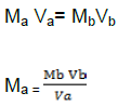
where, Ma= Normalty of acid in effluent sample, Va= Volume of acid in effluent sample, Nb= Normality of base (NaoH = 0.0025N) and Vb= Volume of base (NaOH).
Cyto-protective studies
Briefly cleaned stomachs were pinned on a corkboard and the ulcer were scored using dissecting microscope with square grid eyepiece based on grading on 0 to 5 scale (depicting severity of vascular congestion lesions/hemorrhage as presented. 0 means no lesion. 1 represents vascular congestion, 2, 3, 4 and 5 represents one or two lesions, severe lesions, very severe lesions and mucosa full of lesions of marked sizes, respectively (Saheed et al., 2015).
Mean ulcer scores/indices
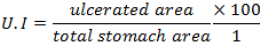
Mean ulcer scores for each animal were expressed as ulcer index (U.I) and the percentage of inhibition against ulceration was determined using the expression by (Szabo and Hollander, 1995).
Statistical analysis
Data were presented as mean ± standard error of mean (SEM). Data were analyzed using statistical computer software (SPSS version 21) one way analysis of variance (ANOVA) with Tukey post hoc test for further multiple comparisons. Value of p<0.05 was considered to be statistically significant except otherwise stated.
Ethical clearance
The experiment was approved by the Animal Research Ethics Committee of the College of Medicine, Enugu State University of Science and Technology, Enugu and handling of animals followed the internationally accepted procedures according to the Institute of Laboratory Animal Research guide for the care and use of laboratory Animals.
RESULTS
Table 1 shows the result of the basal volume of gastric acid secreted at the 10 mins intervals for 30 mins. At the 10th min of gastric acid secretion, it can be observed from the table that group A secreted the maximal volume of gastric acid while the least volume was secreted by group E. There were significant differences (P<0.05) among all the groups compared with group A (Ulcer alone), at the end of the 10th min of secretion.
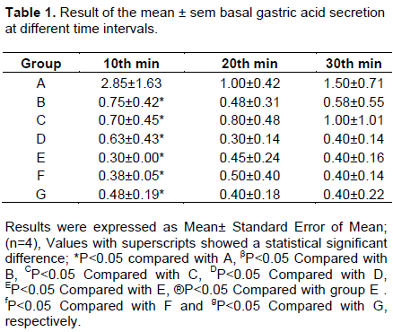
The effluent was in a decreasing order starting from group A followed by group B and C respectively but on the 20 mins, group A also recorded the highest volume (1.00±0.42) ml with group D as the least. Hence, at the 30th min, group A (1.550±0.71 ml) still recorded the highest volume of gastric acid secreted followed by group B as usual. There was no significant difference (P>0.05) at the 20th and 30th mins of the gastric acid secretion respectively. Table 2 shows the volume of the gastric acid secreted after one hour post treatment and induction of the gastric ulcer by the use of acid alcohol.
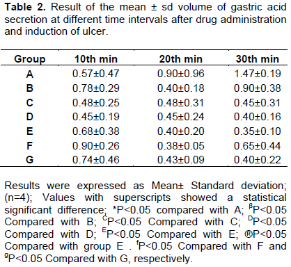
After the first 10 mins of the post ulcer induction, the volume of gastric acid secreted was more in group F (0.9±0.2 ml), followed in a decreasing order by group B (0.78±0.29 ml), G (0.74±0.46 ml), E (0.68±0.38) etc., as seen in the table 2 below. Hence, at the end of the 20th mins of post treatment and induction of ulcer, it was observed that group A (0.90±0.96 ml), recorded the highest volume with the least value in group B and E with values of 0.40±0.18 and 0.40±0.20 ml respectively. At the 30th mins, the volume secreted was more in group A (ulcer alone) group without anti-ulcer agents with the least recorded by group by group E (0.35 ±0.10 ml). There was no significant (P>0.05) difference among the groups.
Table 3 also shows the basal pH levels of gastric acid secreted by all the experimental groups at different time intervals. At the 10th min, the pH of the solution still falls within the normal physiological ranges for gastric acid. Groups A, B and C were more acidic than groups E, F and G at the 10th min. The same patterns were observed in the 20th and 30th mins, respectively. There were significant differences (P<0.05) among some of the groups compared with one another. At the 20th min of basal secretion, there were significant differences (P<0.05) among groups E and G compared with group C but on the 30th min, there was significant difference between group E and F compared with group C respectively. Moreover, Table 4 shows the result of the pH after post induction of ulcer and treatment of administration of the drugs. Group A was found to be more acidic followed by group D and B respectively. Groups E and F were slightly alkaline and group G showed a neutral pH of 7.0. There were significant differences (P<0.05) among all the groups compared with group A as shown in the table 4 below. Hence, at the end of the 30th mins, Group A still maintained its acidic nature with a PH of 4.83±0.29 while group C became totally alkalinized with a pH of 8.10 ±0.00 as seen in the table below. Group E, F and G were neutralized with PH stabilized at 7.00.
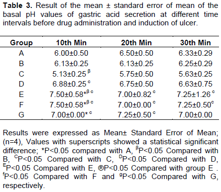
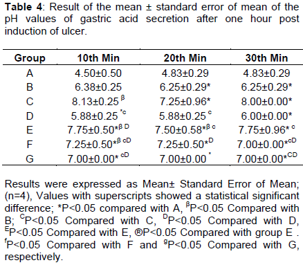
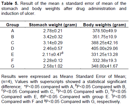
Table 5 shows the rats in group D have the highest body weights (405±29.06 g) and this was followed by rats in groups C, A, B,G and E with group F as the least weight. The body weights were compared statistically and there was no significant difference (P>0.05) among the groups. The stomach weights did not follow similar pattern as the body weight as the stomach weight recorded the highest weight in group B and C respectively. They were followed by groups A, G, D and E with group E as the least in their stomach weights values. There was also a significant difference (P<0.05) between group E when compared with group B. From the table, it has been reported that animals with smaller stomach weight has more ulcers than those with higher stomach weight and there is a correlation as depicted by the ulcer index table and that of the stomach weight.
From Table 6, it was observed that 3 rats in group A have multiple linear ulcer of mark size and one hemorrhagic erosion greater than 5 mm size while in group B, one rats has hemorrhagic erosion of less than 5 mm and no ulcer was observed in group C and D. In group E, two rats have hemorrhagic erosion of less than 5mm and one rat has hemorrhagic erosion greater than 5 mm and no ulcer was seen in rats 4 as presented in the table above. In group F, three rats have hemorrhagic erosion of less than 5 mm while one rat has hemorrhagic erosion greater than 5 mm. Finally, in group G, rat 2 has many linear ulcers of smaller sizes that are greater than 2 mm while two rats have hemorrhagic erosion of less than 5 mm and no ulcer was seen in rat 4 of the same group.
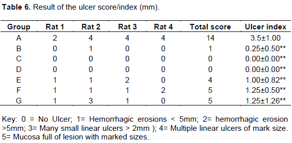
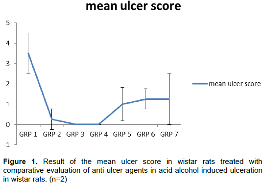
Figure 1 represents the mean±SD of the ulcer score/ index for all the experimental groups. From the graph, the ulcer score was more pronounced in group 1 with mean value of 3.5±1.00 and this was followed by group F and G with values of 1.25± 1.26 and 1.25±0.5, respectively. Groups C and D recorded the least ulcer score. These differences in ulcer indices were compared statistically and observed that there was significant difference (P<0.05) between groups B to G compared to group A (Ulcer alone).
DISCUSSION
This research work compared the cytoprotective effects of some antiulcerative agents (rabeprazole, ranitidine, omeprazole and cimetidine, etc.) on acid alcohol induced gastric ulceration in Wistar rats. The objectives involve determining the possible gastric acid protective effect of proton pump inhibitors and H2_Receptor blockers and to assess their possible synergistic effects on acid alcohol induced gastric ulcers in wistar rats. In this present study, the significant increase in ulcer index and gastric acid volume following oral administration of cimetidine and ranitidine may be attributed to either free radical formation or inhibition of prostaglandin synthesis (Parveen and Michael, 2012).
Decreased prostaglandin level has been attributed to impaired gastroprotection and increased gastric acid secretion which are important event in the etiology of mucosal ulceration (Parveen and Michael, 2012). This agrees with the reports of Saheed et al. (2015) and Szado and Hollander (1995), where indomethacin was reported to have caused alteration in gastric secretion of rats. Conversely, pretreatment with the antiulcer agents significantly reduced these parameters. In fact, the effect noticed with pH compared favorably with normal control and indeed suggestive of the possible gastroprotective attributes of the anti-ulcer agents. A combination of events including released preformed mucus, wound retraction and epithelilization is involved in ulcer protective process after toxicological injury (Modirat et al., 2018; Bech et al., 2000).
Besides, providing significant buffering capacity for neutralization of luminal acids, the mucus also offer protection against both endogenous aggressors and exogenous gastro toxic agents such as acid-alcohol, thereby enhancing the rate of local healing process. In this study, decreased cyto-protective activity in acid alcohol ulcerated rats indicated reduced protective ability of the mucosal membrane against hemorrhagic erosion, thus resulting in tissue damage. This implied the decreased ability of gastric mucosa to withstand the offensive onslaught of acid alcohol. Thus antiulcer drugs such as rabeprazole, ranitidine, used, inhibit the secretion and synthesis of gastric acid and thus protects against gastric ulcer damage and this could be attributed to its synergistic effects.
Pre-treatment with the antiulcer agents however gave cyto-protective effects which is associated with decreased pepsin activity and elevated mucus level in the gastric mucosa. Thus these drugs shielded the gastrointestinal membrane by abrogating the catastrophic influence of acid alcohol in ulcerative rats (Bech et al., 2000). This is indication of enhanced mucus secretory potential of the drugs and suggestive of their significant role in cyto-protection against gastric acid damage to the mucosal endothelium. Cyto-protection of mucosa epithelial cells was prominently displayed by a synergistic administration of 20 mg/kg rabeprazole and ranitidine thereby depicting a better cyto-protective capacity than cimetidine and ranitidine alone.
CONCLUSION
The attenuation of gastric affronts of acid alcohol by the administration of 20 mg/kg dose of a combination of Rabeprazole and ranitidine showed an excellent cyto-protective effect on the gastric mucosa of wistar rats. These cyto-protective effects of the combination of Rabeprazole and Ranitidine could be a possible synergistic efficacy.
ACKNOWLEDGEMENT
The authors sincerely acknowledge the efforts of Mr. Ani, Celestine Okafor (Principal Laboratory Technologist) and all the staff of Physiology Department, College of Medicine, Enugu State University of Science and Technology, Parklane, Enugu, Nigeria.
CONFLICT OF INTERESTS
The authors have not declared any conflict of interests.
REFERENCES
|
Adeniyi B, Jesse A, Lawal A (2012).Prevalence of helicobacter pylori infection among dyspepsia patients in Ibadan, South west Nigeria. African Journal of Microbiology Research 6(14):3399-3402. |
|
|
Adeniyi OS, Omale J, Egwuje RJ, Ajayi OS (2016). Effect of selenium treatment on healing of Acetic Acid induced gastric ulcer in albino wistar rats. American Journal of Biomedical Research 4(1):18:22. |
|
|
Bech PL, Xavier R, Lu N, Nanda NN, Dinaeur M, Podolsky DK (2000). Mechanism of NSAID induced gastrointestinal injury defined using mutant mice. Gastroenterology 119(3):699-705. |
|
|
Drini M (2017).Peptic ulcer and non-steroidal anti-inflammatory drugs. Australian Prescriber 40(3):91-93. |
|
|
Felix OO, Bamidele OA, Tunde A, David OA (2013). Perforated duodenal ulcer; management in resource poor, semi-urban Nigeria hospital. Nigerian Journal of Surgery 19:13-15. |
|
|
Hansson LE, Nyren O, Hsung AW, Bergstrom R, Josefsson S, Chow WH, Fraumeni JF Jr, Adami HO (1996). The risk of stomach cancer in patients with gastric or duodenal ulcer disease. New England Journal of Medicine 335(4):242-249. |
|
|
Hestvik E, Tylleskev T, Ndeezi G, Grahnquist L, Olafsdottir E, Tumwine JK and Deograttias HK (2011). Prevalence of Helicobacter pylori in HIV-infected, HAART-naïve Ugandan children; a hospital based survey. Journal of the International AIDS Society 14(1):34. |
|
|
Modirat AA, Akomolafe OS, Alabi OK, Ogundipe L, Omole JG, Olanisoye KP (2018). Protective effect of methanol extract of Vernonia amygdalina (del) leaf on aspirin induced Gastric ulceration and oxidative mucosal damage in rat's model of Gastric injury. Dose Response 16(3):1559325818785087. |
|
|
Parveen K, Michael C. (2012). Clinical Medicine 8th edtn. Pub.Saunders Elsevier Ltd. Edinburgh London, New York pp. 247-251. |
|
|
Saad BA. Nagala AE, Ayim TA, Umamu AB, Soad SA, Soad KA (2016). Antioxidant, Anti-inflammatory and Anti-ulcer potentials Manuuks Honey against Gastric ulcer in rats. Oxidative Medicine and Cellular Longevity. |
|
|
Saheed S, Taofeeq G, Taofik S, Emmanuel A, Abdulhakeem S, Ismaila N, Balogun A (2015). Indomethacin induced gastric ulceration in rats; Protective role of spondias mombira and fiscus exasperate. Toxicology Reports 2:261-267. |
|
|
Sidahmed HMA, Abdelwahab SI, Mohan S, Mahmood A, Manal ME, Najiha MH (2013). α-Mangostin from Cratoxylum arborescens (Vahl) Blume demonstrates anti-ulcerogenic property: a mechanistic study. Evidence-Based Complementary and Alternative Medicine. |
|
|
Souza D', Dhume VG (1991). Gastric cytoprotection. Indian Journal of Physiology and Pharmacology 35(2):88-98. |
|
|
Szabo S, Hollander D (1995). Pathways of gastrointestinal protection and repair; mechanism of action of sulcrafate. American. Journal of Medicine 86(6A):23-31. |
|
|
Yuan XG, Xie C, Chen J, Xie Y, Zhang kh, Lu NH (2015). Seasonal changes in gastric mucosal factors associated with peptic ulcer bleeding. Experimental and Therapeutic Medicine 9(1):125-130. |
|
Copyright © 2024 Author(s) retain the copyright of this article.
This article is published under the terms of the Creative Commons Attribution License 4.0


