Full Length Research Paper
ABSTRACT
Cochlospermum planchonii is traditionally used in Togo in treating several diseases, including burn wounds which achievement requires anti-infective and good cicatrisation processes. This study aims to evaluate the antimicrobial properties of hydroethanolic extracts from leaves and roots of C. planchonii and its phytochemical composition. Five (05) bacterial strains were suspended in liquid media with plant extracts at various concentrations and then spread, after 24 h incubation, on solid media. The minimum inhibitory (MIC) and minimal microbicidal (MMC) concentrations were assessed. Phytochemical characterization tests were performed and flavonoids, tannins and total phenol contents were also determined. Hydroethanolic extracts from the leaves and roots of C. planchonii were active on all the germs tested, with MICs ranging from 0.782 to 50 mg/mL and MMCs ranging from 1.563 to 100 mg/mL. The bests activities were recorded with root extract against Staphylococcus aureus: MIC=0.782 mg/mL and MMC =1.563 mg/mL. The MMC/MIC ratios are all equal to 2, indicating that both roots and leaves have bactericidal activities rather than inhibitory activities. Phytochemical tests have demonstrated the presence of metabolites such as flavonoids, tannins, carbohydrates, sterols, triterpenes, saponosides, and an absence of alkaloids. Total phenols, tannins and total flavonoids amounts found were respectively 85.466 ± 0.289; 33.566 ± 0.503; 218.333 ± 0.577 µgRE/mg in root extracts, and 75.833 ± 0.289; 26.533 ± 0,053; 183.666 ± 0.577 µgRE/mg in leaf extracts. Both root and leaf hydroethanolic extracts of C. planchonii could be potential natural antimicrobial remedies.
Key words: Phytochemical screening, antimicrobial activity, Cochlospermum planchonii, flavonoids, tannins.
INTRODUCTION
Cochlospermum planchonii is a medicinal plant used in West Africa to treat a number of diseases. It has been described as anti-plasmodial (Benoit-Vical et al., 2001), anti-inflammatory (Benoit-Vical et al., 2003; Anaga and Oparah, 2009), anti-bacterial (Isah et al., 2013), hepatoprotective (Aliyu et al., 1995), and anti-diabetic (Nafiu et al., 2011) agent. A previous work in our laboratory has shown that C. planchonii hydroethanolic root extract (2.5 and 5% in gel) administered topically has significant in vivo burn wound healing activity in mice (Nakpane et al., 2020). Since infection installation is the most common and inevitable impediment to wound healing (Church et al., 2006; Negut et al., 2018), wound dressings loaded with antimicrobial agents have emerged as viable options to reduce wound bacterial colonization and infection, in order to improve the healing process. Gram-positive cocci such as Staphylococcus aureus, and Gram-negative bacilli such as Escherichia coli, Pseudomonas aeruginosa, and Klebsiella pneumonia are among the most common pathogenic bacteria isolated from wounds (Pallavali et al., 2017; Atef et al., 2019). It became necessary to investigate the antimicrobial activities of C. planchonii root extract against these bacterial strains. In addition, the utilization of roots could affect the plant survival. According to the necessity to preserve the biodiversity of the species, it was imperative to include leaves in this study. Since infection is one of the causes of delayed healing and especially death due to burn wounds (Krishnan et al., 2013; Negut et al., 2018), the objective of this study is to evaluate the phytochemical composition of hydroethanolic extracts from leaves (EBF) and roots (EBR) of C. planchonii and their antimicrobial properties.
MATERIALS AND METHODS
Plant material
The roots and leaves of C. planchonii were harvested in Kabou (TOGO) in September 2018. A voucher specimen was identified and deposited in the herbarium of the Laboratory of Botany and Plant Ecology under the number Togo15501.
Preparation of the hydroethanolic extracts
The samples collected were dried under air conditioning in the Laboratory of Physiology/Pharmacology, Faculty of Sciences, University of Lomé (Togo). The dried roots and leaves of C. planchonii were ground to powder and 300 g of each was macerated in 4 L of ethanol-water mix (5:5.v/v) for 72 h. The filtrate was then evaporated under vacuum at 40°C using a rotavapor (Buchi R- 210) (Trease and Evans, 1989). The extraction yields were calculated following equation:
Yield (%) = (W1 × 100)/W2
Where, W1 is the weight of the extract residue obtained after evaporation and W2 is the weight of dried roots or leaves powder used.
Reagents
Mayer’s reagent, methanol, acetic anhydride, gallic acid and polyvinylpyrrolidone (PVP) were provided by VWR International (Haasrod, Belgium). Rutin hydrate, chloroform, and Folin- Ciocalteu reagent were obtained from Sigma Chemicals (St. Louis, USA). All other chemicals and reagents used were of the analytical grade.
Phytochemical analysis
Qualitative tests
An aliquot (1 mL) of each extract (1 mg/mL in ethanol) obtained from leaves and roots of C. planchonii was subjected to qualitative phytochemical analysis to ascertain the presence of secondary metabolites such as alkaloids, flavonoids, tannins, carbohydrates, sterols, triterpenes, and saponosides. The compound classes were characterized using adequate techniques and specific reagents according to the respective methods (Karumi, 2004; Edeoga et al., 2005) described below.
Test for alkaloids
The hydroethanolic extracts were evaporated to dryness and the residues were heated with 2% hydrochloric acid on a boiling water bath. The extracts were cooled, filtered and treated with the Mayer’s reagent. The samples were then observed for the presence of yellow precipitation or turbidity.
Test for flavonoids
1.5 ml of 50% methanol was added to 4 ml of crude hydroethanolic extracts. The solution was warmed and metal magnesium was added. Then 5 to 6 drops of concentrated hydrochloric acid were added to the solutions and observed for red coloration.
For tannins
To 0.5 ml of crude hydroethanolic solutions, 1 ml of distilled water and 1 to 2 drops of Ferric chloride solution were and it was observed for blue or green black coloration.
Test for Phenolic compounds: 2 ml of ethanol was added to the test solutions and few drops of ferric chloride solution were added and observed for coloration.
Test for Saponosides: 2 ml of distilled water was added to 2 ml of the test solutions and well shaken and observed for frothing.
Test for reducing compounds (carbohydrates): Two milliliter of crude hydroethanolic solutions was placed in test tube and 5 mL mixture of equal volumes of Fehling’s solution A and B were added and boiled in a water bath for 2 min. The tests tubes were observed for brick red precipitate.
Identification of sterols and triterpenes: 10 mL of crude hydroethanolic solutions was placed in small beakers and evaporated to dryness. The residues were dissolved in acetic anhydride (0.5 mL) and chloroform (0.5 mL). The solution was transferred into dry tests tubes and concentrated sulfuric acid (2 mL) was added. Brownish red or violet rings at the zone of the contact with the supernatant and green or violet coloration denoted the presence of sterols and triterpenes.
Determination of total phenolic content
Protocol of Pourmorad et al. (2006) was used with some modifications. The principle of the assay is based on the oxidation of phenolic compounds by the Folin- Ciocalteu reagent. The extracts were prepared in distilled water-methanol mixture (V: V). Samples (500 μL) of each extract at 1 mg/mL were mixed with 1 mL of diluted Folin-Ciocalteu reagent (1:10 in water). After 4 min, this mixture received 5 mL of saturated solution sodium carbonate (75 mg/mL), followed by 15 min of incubation at room temperature (protected from light). The absorbance was measured at 765 nm. Gallic acid (0–500 mg/L) was used as standard in a calibration curve. The results were expressed as mg of gallic acid equivalents (mgGAE)/g dry weight of plant extract.
Determination of tannins content
The protocol is the same as that described for the determination of total phenolic content. The tannins are first fixed by polyvinylpyrrolidone (PVP). Samples (500 μL) of each extract at 1 mg/mL were brought into contact with 10 mg of PVP and 500 µl of 95% methanol. The mixture is shaken with a vortex and incubated on ice for 30 min. After decanting, 500 µl of the supernatant is brought into contact with 5 ml of Folin-Ciocalteu reagent and 5 ml of sodium carbonate. The absorbance was read at 765 nm and a calibration curve was made using gallic acid solution range as preview. The results are expressed as mg of gallic acid equivalents (mgGAE)/g dry weight of plant extract.
Determination of flavonoids content
The quantification of total flavonoid content in each extract followed the methodology proposed by Woisky and Salatino (1998). In brief, to 0.5 mL of diluted samples (1 mg) was added 0.5 mL of 2% AlCl3 (w/v) solution prepared in methanol. After 30 min of incubation at room temperature, protected from light, the absorbance was measured at 420 nm. All measurements were done in triplicate. The results were expressed as mg of rutin equivalent mgRE/g dry weight of plant extract.
Antimicrobial assays
Microorganisms
Five microbial strains obtained from the strain bank of the bacteriology laboratory of the National Institute of Hygiene of Togo were used. These strains are referenced as follows: Escherichia coli, ATCC 25922; Pseudomonas aeruginosa, ATCC 27853; Klebsiella pneumonia, ATCC 700603; Staphylococcus aureus, ATCC 29213; and Candida albicans, ATCC 35659.
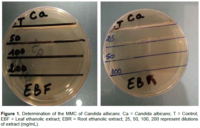
Determination of minimum inhibitory concentration (MIC) and minimal microbicidal Concentration (MMC)
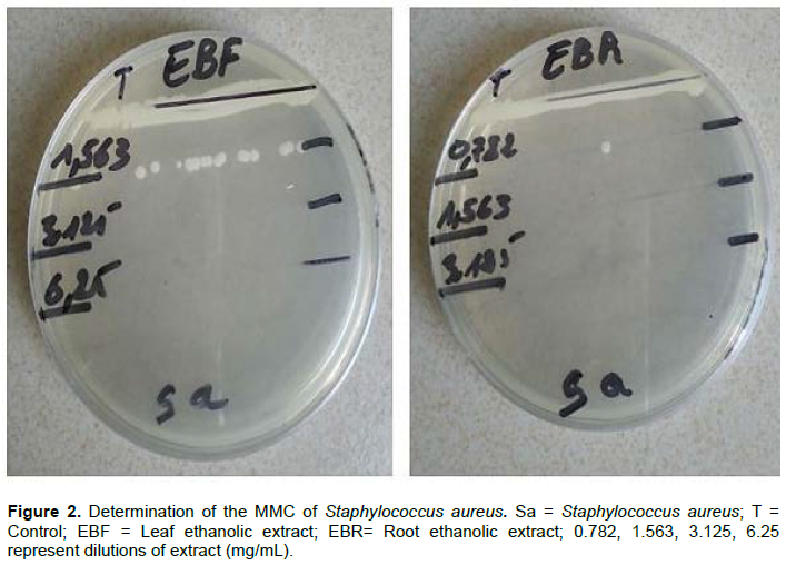
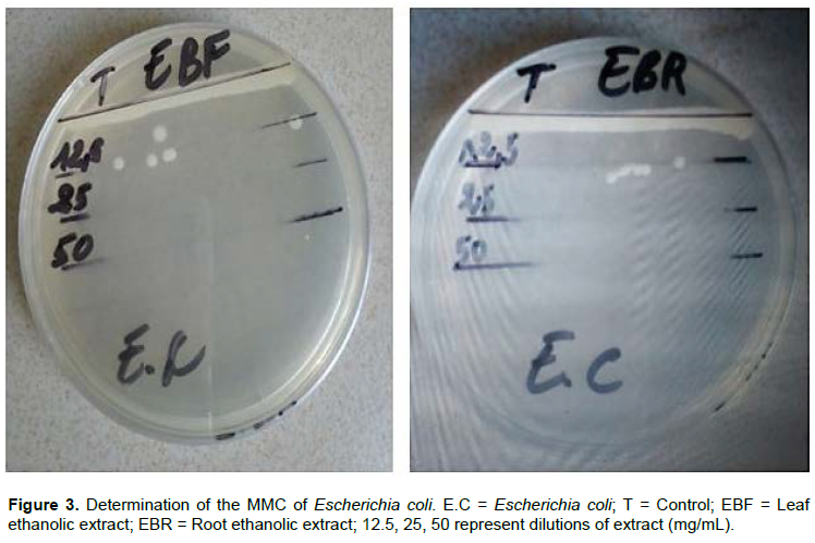
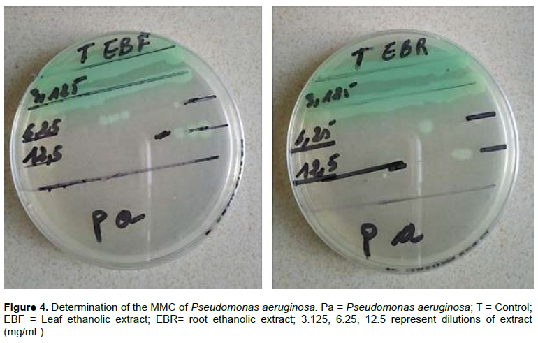
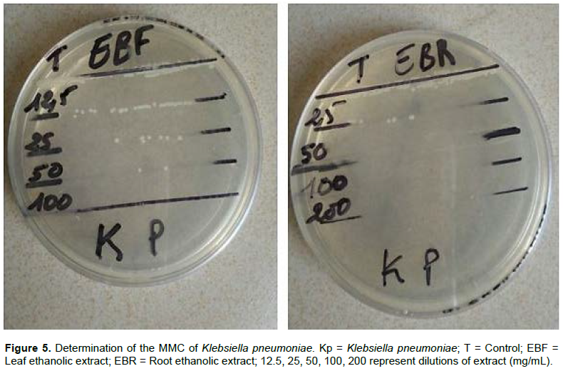
The minimum inhibitory (MIC) and minimal microbicidal (MMC) concentrations were determined by The de Souza dilution method (de Souza et al., 1993). Sabouraud Chloramphenicol agar (GSC) was used for Candida albicans and Muller-Hinton agar (GMH) for bacterial strains. Tested microorganisms were standardized by the McFarland turbidity scale equivalent to the tube 0.5, corresponding to a concentration of approximately 107 CFU/mL for yeasts and 108 CFU/mL for bacteria. Solutions of extracts ranging from 0.782 to 200 mg/mL were prepared and sterilized by fixing with 95° alcohol. The microbial suspensions were contacted with the extract in liquid Muller Hinton medium (MH) and incubated at 37° C for 24 h and then spread on solid medium (GMH or GSC). The protocol consists of introducing 0.1 ml of the microbial suspension in 0.5 ml of the extract prepared in Muller Hinton. For the controls, the Muller Hinton medium is without the extract. The tests and controls were then smeared on GMH for bacteria and on GSC for Candida albicans. All boxes were incubated at 37°C for 24 h for bacteria and 25°C for 48 h for Candida albicans. The absence of growth after incubation indicates the sensitivity of the extract on the microorganism. The MIC corresponds to the lowest concentration of extract for which no microbial growth has been observed with the naked eye. The MMC corresponds to the large dilution for which growth is zero or 0.1% of the growth control.
Statistical analysis
GenStat software (VSN International, UK) was used for calculations. Results are presented as percentages and means with standard error on the mean (M ± SEM).
RESULTS
Extraction yields
Yields obtained were 27.21% for root extracts (EBR) and 16.25% for leaf extracts (EBF).
Phytochemical analyses of the samples from leaves and roots of C. planchonii
Phytochemical screening
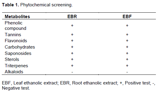
The qualitative phytochemical analysis detected both in root and leaves the presence of flavonoids, tannins, carbohydrates, sterols, triterpenes, and saponosides. Alkaloids were not detected. The results of the qualitative tests are presented in Table 1.
Quantitative analyses
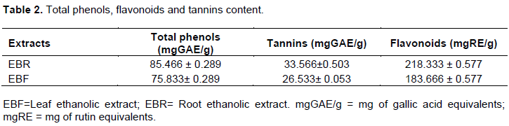
C. planchonii was found to possess flavonoids, tannins and total phenols. Analysis showed that the C. planchonii roots contain the highest quantities, in comparison with the leaves. The results of the quantitative tests are presented in Table 2.
DISCUSSION
The phytochemical study revealed the presence of phenolic compounds, tannins, flavonoids, saponosides, carbohydrates, and triterpenes, both in the leaves and roots of C. planchonii (Table 1). Alkaloids were characterized neither in the roots nor in the leaves. The quantitative analysis of these two extracts (Tables 2) confirms the richness of the leaves and roots of C. planchonii in phenolic compounds, particularly tannins and flavonoids. This observation is correlated with the conclusions of many studies that have highlighted the variations in concentration of secondary metabolites from plant to plant species as well as in the different parts of a plant, leaves and roots being the preferential sites of accumulation of these compounds (Hyder et al., 2002; Springer et al., 2002; Isah et al., 2013; Kantati et al., 2016). But compared to leaves, utilization of root parts highly affects the survival and ecological aspect of the plant so leaves should be preferred in order to protect this plant species. In addition, the presence of so important pharmacological groups would justify the traditional use of the plant in the treatment of several diseases; burn, snake bites, malaria (Evans and Gaiere, 2017), palpitations, typhoid fever, urinary tract infections, hypertension, jaundice, wounds, viral hepatitis (Togola et al., 2008), hepatitis, diarrhea dysentery, infertility, diabetes (Igoli et al., 2005).
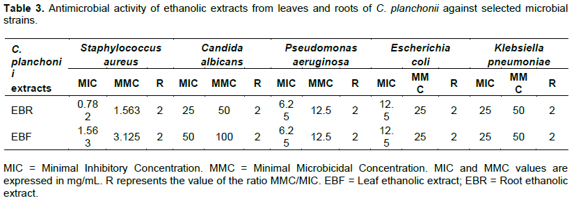
Elsewhere, this study also showed that the hydroethanolic extracts from the leaves and roots of C. planchonii were active on all the germs tested. Based on the bacterial parameters MIC and MMC (Figures 1 to 5; Table 3), it can be said that the antibacterial activity of leaf and root extracts of C. planchonii is stronger on Staphylococcus aureus strain with a MIC of 0.782 mg/mL and an MMC of 1.563 mg/mL for the root extract and a MIC of 1.563 mg/mL and an MMC of 3.125 mg/mL for the leaf extract. Candida albicans was the microbial strain least sensitive to hydroethanolic leaf and root extracts of C. planchonii with a MIC of 25 mg/mL and an MMC of 50 mg/mL for the root extract and a MIC of 50 mg/mL and an MMC of 100 mg/mL for the leaf extract. It should be also noted that the MMC/MIC ratios are all equal to 2 (Table 3). It can therefore be said that hydroethanolic extracts of roots and leaves of Cochlospermum planchonii have bactericidal activities on the strains studied (Okou et al., 2018). These results are confirmed by those of Isah et al. (2013) who demonstrated that the hydroethanolic extract of young leaves of C. planchonii, at the concentration of 80 mg/mL was sensitive on Pseudomonas aeruginosa, Staphylococcus aureus and Escherichia coli. This antibacterial activity of the plant is also reported by the work of Alain et al. (2014) who found that the plant extracts are active on several germs, including Pseudomonas aeruginosa and Staphylococcus aureus. The antibacterial activity observed would be related to the metabolites found in the plant. Indeed, flavonoids and tri-terpenes have a known antimicrobial activity (Ayeni, 2018), anti-ulcer properties (Madivoli et al., 2018), and anti-inflammatory properties (Özçelik et al., 2011). Tannins are known for their antiseptic, anti-microbial and anti-fungal activity (Nissiotis and Tasioula-Margari, 2002). Tannins are generally endowed with an astringent power which confers them vasculo- protective and healing properties (Hennebelle et al., 2004). These biological properties of the compounds found in the plant could justify the antimicrobial activities shown in this study. But as it have been noticed in upper lines, it should be preferable to promote the utilization of C. planchonii leaves which have proven to be as effective as the roots, in order to protect this plant. A controlled agriculture operation of C. planchonii could also contribute to the economic development of the regions where it has been harvested.
CONCLUSION
This study shows that the C. planchonii collected in Kabou (TOGO) contains a lot of compounds such as polyphenols, flavonoids, tannins, sterols, triterpenes, saponosides and anthocyanins. The quantification of polyphenols, flavonoids and tannins revealed that both roots and leaves of C. planchonii contain large amount of these metabolites, probably responsible of the antimicrobial activities observed. Further studies are needed for possible new antimicrobial drug discovery from this plant. It is therefore recommended, in accordance with ecological imperatives, to use the leaves of C. planchonii in order to preserve this plant species.
CONFLICT OF INTERESTS
The authors have not declared any conflict of interests.
ACKNOWLEDGMENTS
The authors are grateful to the staff members of the National Institute of Hygiene of Togo for their assistance in antimicrobial tests.
REFERENCES
|
Alain KY, Oronce DL, Mahudro Y, Pascal AD, Paul TF, Alain AG, Koko SD (2014). Caractérisation chimique, activités antiradicalaire et antibactérienne des extraits de l'écorce de racine de Cochlospermum planchonii du Bénin [Chemical characterization, antiradical and antibacterial activities of extracts of the root bark of Cochlospermum planchonii of Benin]. International Journal of Innovation and Applied Studies 7(4):1582. |
|
|
Aliyu R, Okoye ZSC, Shier WT (1995). The hepatoprotective cytochrome P-450 enzyme inhibitor isolated from the Nigerian medicinal plant Cochlospermum planchonii is a zinc salt. Journal of Ethnopharmacology 48(2):89-97. |
|
|
Anaga AO, Oparah N (2009). Investigation of the methanol root extract of Cochlospermum planchonii for pharmacological activities in vitro and in vivo. Pharmaceutical Biology 47(11):1027-1034. |
|
|
Atef NM, Shanab SM, Negm SI, Abbas AY (2019). Evaluation of antimicrobial activity of some plant extracts against antibiotic susceptible and resistant bacterial strains causing wound infection. Bulletin of the National Research Centre 43:144. |
|
|
Ayeni MJ, OyeyeMi SD, KAyODe J, Abanikanda AI (2018). Phytochemical, Proximate and Mineral Analyses of the Leaves of Bambusa vulgaris L. and Artocarpus Altilis L. Ghana Journal of Science 59:69-77. |
|
|
Benoit-Vical F, Valentin A, Da B, Dakuyo Z, Descamps L, Mallie M (2003). N'Dribala (Cochlospermum planchonii) versus chloroquine for treatment of uncomplicated Plasmodium falciparum malaria. Journal of Ethnopharmacology 89(1):111-114. |
|
|
Benoit-Vical F, Valentin A, Mallié M, Bessière JM (2001). Antiplasmodial activity of Cochlospermum planchonii and C. tinctorium tubercle essential oils. Journal of Essential Oil Research 13(1):65-67. |
|
|
Church D, Elsayed S, Reid O, Winston B, Lindsay R (2006). Burn wound infections. Clinical Microbiology Reviews 19(2): 403-434. |
|
|
De Souza C, Ameganvi KK, Koumaglo K, Gbeassor M (1993). Etude de l'activité antimicrobienne des extraits aqueux totaux de dix plantes médicinales. Revue de Médecines et Pharmacopées Africaines 2(7):107-115. |
|
|
Edeoga HO, Okwu DE, Mbaebie BO (2005). Phytochemical constituents of some Nigerian medicinal plants. African Journal of Biotechnology 4(7):685-688. |
|
|
Evans EC, Gaiere Y (2017). Effect of solvent extraction on phytochemical composition of selected Nigerian medicinal plants. Scientia Agriculturae 20(1):23-31. |
|
|
Hennebelle T, Sahpaz S, Bailleul F (2004). Polyphénols végétaux, sources, utilisations et potentiel dans la lutte contre le stress oxydatif. Phytothérapie 2(1):3-6. |
|
|
Hyder PW, Fredrickson EL, Estell RE, Tellez M, Gibbens RP (2002). Distribution and concentration of total phenolics, condensed tannins, and nordihydroguaiaretic acid (NDGA) in creosotebush (Larrea tridentata). Biochemical Systematics and Ecology 30(10):905-912. |
|
|
Igoli JO, Ogaji OG, Tor-Ayiin TA, Igoli NP (2005). Traditional medicine practice amongst the Igede people of Nigeria. Part II. African Journal of Traditional, Complementary and Alternative Medicines 2(2):134-152. |
|
|
Isah Y, Ndukwe IG, Ayo RG (2013). Phytochemical and antimicrobial analyses of stem-leaf of Cochlospermum planchonii. Journal of Medicinal Plant and Herbal Therapy Research 1:13-17. |
|
|
Kantati YT, Kodjo KM, Dogbeavou KS, Vaudry D, Leprince J, Gbeassor M (2016). Ethnopharmacological survey of plant species used in folk medicine against central nervous system disorders in Togo. Journal of Ethnopharmacology 181:214-220. |
|
|
Karumi Y (2004). Identification of Active Principles of M. balsamina (Balsam Apple) Leaf Extract Y. Karumi," PA. Onyeyili and "VO Ogugbuaja. Journal of Medical Sciences 4(3):179-182. |
|
|
Krishnan P, Frew Q, Green A, Martin R, Dziewulski P (2013). Cause of death and correlation with autopsy findings in burns patients. Burns 39(4):583-588. |
|
|
Madivoli ES, Maina EG, Kairigo PK, Murigi MK, Ogilo JK, Nyangau JO, Kipyegon C (2018). In vitro antioxidant and antimicrobial activity of Prunus africana (Hook. f.) Kalkman (bark extracts) and Harrisonia abyssinica Oliv. extracts (bark extracts): A comparative study. Journal of Medicinal Plants for Economic Development 2(1):1-9. |
|
|
Nafiu MO, Akanji MA, Yakubu MT (2011). Effect of aqueous extract of Cochlospermum planchonii rhizome on some kidney and liver functional indicies of albino rats. African Journal of Traditional, Complementary and Alternative Medicines 8(1): 22-26. |
|
|
Nakpane F, Metowogo K, Kantati YT, Kodjo A, Tchin D, Eklu-Gadegbeku K, Aklikokou AK (2020). Burn wound healing effects of the root hydroethanolic extract of Cochlospermum planchonii in mice. [Unpublished]. Laboratory of Physiology/Pharmacology, University of Lomé, Togo. |
|
|
Negut I, Grumezescu V, Grumezescu AM (2018). Treatment strategies for infected wounds. Molecules 23(9):2392. |
|
|
Nissiotis M, Tasioula-Margari M (2002). Changes in antioxidant concentration of virgin olive oil during thermal oxidation. Food Chemistry 77(3):371-376. |
|
|
Okou OC, Yapo SES, Kporou KE, Baibo GL, Monthaut S, Djaman AJ (2018). Évaluation de l'activité antibactérienne des extraits de feuilles de Solanum torvum Swartz (Solanaceae) sur la croissance in vitro de 3 souches d'entérobactéries. Journal of Applied Biosciences 122(1):12287-12295. |
|
|
Özçelik B, Kartal M, Orhan I (2011). Cytotoxicity, antiviral and antimicrobial activities of alkaloids, flavonoids, and phenolic acids. Pharmaceutical Biology 49(4):396-402. |
|
|
Pallavali RR, Degati VL, Lomada D, Reddy MC, Durbaka VRP (2017). Isolation and in vitro evaluation of bacteriophages against MDR-bacterial isolates from septic wound infections. PLoS ONE 12(7):e0179245. |
|
|
Pourmorad F, Hosseinimehr SJ, Shahabimajd N (2006). Antioxidant activity, phenol and flavonoid contents of some selected Iranian medicinal plants. African Journal of Biotechnology 5(11):1142-1145. |
|
|
Springer TL, McGraw RL, Aiken GE (2002). Variation of Condensed Tannins in Roundhead Lespedeza Germplasm. Crop Science 42(6):2157-2160. |
|
|
Togola A, Austarheim I, Theïs A, Diallo D, Paulsen BS (2008). Ethnopharmacological uses of Erythrina senegalensis: a comparison of three areas in Mali, and a link between traditional knowledge and modern biological science. Journal of Ethnobiology and Ethnomedicine 4(1):6. |
|
|
Trease GE, Evans WC (1989). Pharmacognosy (13th edn). Bailliere Tindall, London pp.176-180. |
|
|
Woisky RG, Salatino A (1998). Analysis of propolis: some parameters and procedures for chemical quality control. Journal of Apicultural Research 37(2):99-105. |
|
Copyright © 2024 Author(s) retain the copyright of this article.
This article is published under the terms of the Creative Commons Attribution License 4.0


