Full Length Research Paper
ABSTRACT
Coffea arabica (Arabica) and Coffea canephora (Robusta) are the economic plants in Thailand that are widely cultivated in Northern and Southern Thailand. This study aims to evaluate the antioxidant, anti-tyrosinase activities, toxicity, stability and identify chemical components of the coffee bean extracts. The best extract that showed good biological activities will be further used to develop cosmeceutical products. Green and roasted coffee beans from two species were extracted with hexane following ethanol by maceration. Their antioxidant activities were detected by 2,2-diphenyl-1-picryl hydrazyl (DPPH), 2,2’-Azino-bis(3-ethylbenzthiazoline-6-sulphonic acid) (ABTS) and lipid peroxidation inhibition assays. In addition, anti-tyrosinase activity was also evaluated. The results revealed that the ethanolic coffee bean extracts showed a higher level of antioxidant activity than in the hexane extracts. All extracts also possessed a considerable anti-tyrosinase activity, but less potent than kojic acid and arbutin. Chemical compounds of these extracts were determined using caffeine and chlorogenic acid as standards of reference by the thin layer chromatography and the high performance liquid chromatography. The green coffee bean extracts consisted of caffeine and chlorogenic acid while the roasted coffee bean extracts presented only caffeine due to a few chlorogenic acid content after the roasting process. The ethanolic coffee bean extracts that showed good activities were selected to be evaluated on toxicity and stability. The selected extracts were kept at various storage conditions to evaluate their stability using DPPH assay and anti-tyrosinase activity assay. The result showed that the extracts were not toxic to cells. Therefore, the extracts were safe to be components in skin care products. After the stability test, the extracts indicated a good stability and activities. These results led to the conclusions that the coffee bean extracts possess a good biological activities and are assumed to be promising natural active ingredients with a good stability profile for further development of cosmeceutical or anti-aging products.
Key words: Coffea arabica, Coffea canephora, green coffee bean, roasted coffee bean, antioxidant activity, anti-tyrosinase activity.
INTRODUCTION
Many factors such as environmental conditions, UV radiation, foods, stress as well as pollutants are all causes of free radicals formation in the body. Free radicals can induce many diseases such as different types of cancer, coronary artery disease, nervous system diseases, lung diseases and also rheumatoid arthritis (Devasagayam et al., 2004; Pham-Huy et al., 2008). Moreover, they play an important role in tissue aging, including skin aging (Farage et al., 2008; Poljsak et al., 2012). It is a never-ending endeavor for researchers in attempt to find the new active ingredients to counteract the aging process, especially the focus on antioxidant or anti-free radical capability and also anti-tyrosinase activity; which are involved in the prevention of skin aging and help to generate skin brightening. Numerous Thai plants have been used as health care and cosmetic products for many decades.
Coffee is one of the economic plants which is widely grown in Thailand. It is a native plant of Africa in Rubiaceae family and it is very popular around the world, especially Southeast Asia (Charrier and Berthaud, 2012). Coffea arabica (Arabica) is popularly cropped in the Northern part of Thailand while Coffea canephora (Robusta) is mostly cultivated in Southern Thailand. They are different in the seed shape, smell and taste (Chuakul et al., 1997). Robusta coffee is a major production in Thailand, with about 80,000-85,500 tons per year, whereas Arabica coffee production is only approximately 800-850 tons per year. Sixty percent of the Robusta coffee is exported and mostly used for instant coffee production. Most of Arabica coffee is used in roasted and ground coffee for the domestic market.
Previous studies showed that drinking coffee could reduce risk of Parkinson, Alzheimer, hypertension, diabetes type 2 and cancers, and also promote the liver function (Chu et al., 2011; Cano-Marquina et al., 2013; O’Keefe et al., 2013).
In addition, coffee beans serves as antioxidant, anti-inflammatory, for inhibition of albumin denature, UV radiation protection, and in anti-bacterial activities (Antonio et al., 2011; Wagemaker et al., 2011; Almeida et al., 2012; Chandra et al., 2012; Moreira et al., 2013; Liang et al., 2016). Therefore, coffee beans are an interesting option to select for the development of cosmeceutical products in the future. Previous phytochemical studies of coffee indicated that green coffee beans consisted of caffeine, caffeic acid, chlorogenic acid and trigonelline, whereas roasted coffee beans are composed of caffeine, trigonelline, chlorogenic acid, and melanoidin (Liu et al., 2011; Vignoli et al., 2011; Moreira et al., 2013). The chemical components that are mentioned above indicate that coffee beans are a great source for antioxidant.
The data from this research will be used to develop further cosmeceutical products. Therefore, the aims of to select the best extract from antioxidant, lipid peroxidation inhibition and anti-tyrosinase activities. This study are to choose the good solvent extraction and research also shows toxicity of selected coffee bean extracts and stability at various storage conditions. Moreover, the research attempts to identify the chemical constituents of coffee bean extracts by thin layer chromatography and high performance liquid chromatography to confirm active compounds in the extracts.
MATERIALS AND METHODS
Plant materials, chemicals and enzymes
Green and roasted coffee beans (Arabica and Robusta) were obtained from a coffee farm in Chiang Mai province in the northern part of Thailand. The best geography and environment for cultivating coffee include clay soil with high potassium, pH range between 4.5 and 6.5, and rainfall 1,500 and 2,300 ml per year. Arabica coffee is grown with the open-system without shade, the temperature of 15 and 26°C, 80% humidity at 1,000 to 1,700 m above sea level in Chiang Mai, Thailand. Arabica coffee cherries were harvested in October, they were prepared by the pulping process, the wet fermentation process, and the sun drying process. Green coffee beans were then transferred from a high efficiency hulling machine where the final layer of parchment was completely removed. Robusta coffee is grown with the open-system with shade, the temperature of 23 – 32°C, 90% humidity at 700 to 1,000 m above sea level in Chumphon province, Thailand. Robusta coffee cherries were harvested in November. Green coffee beans were prepared the same way as Arabica green coffee beans. Roasted coffee beans were prepared in a high quality, fully automated roaster and sealed in 4-layer-foil bags embedded with one way air valves at 210 - 240°C for 10 to 20 min (medium roast). The green and roasted coffee beans were stored away from light at the room temperature.
Turmeric extract and mangosteen extract were obtained from a cosmetic laboratory at Chiang Mai University, Chiang Mai, Thailand. Caffeine, chlorogenic acid, mushroom tyrosinase and L-tyrosine were purchased from Sigma-Aldrich, USA. L-dopa was purchased from Isotec. Trolox, gallic acid, quercetin, 2,2-diphenyl-1-picryl hydrazyl (DPPH), Folin-Ciocalteu reagent and linoleic acid were purchased from Sigma Chemical Co., (USA). 2,2’-Azino-bis(3-ethylbenzthiazoline-6-sulphonic acid) (ABTS) and 2, 2' azobis 2-amidinopropane dihydrochloride (AAPH) were purchased from Wako Pure Chemical Industries, Japan. RAW 264.7 cells were purchased from American Type Culture Collection (USA). MTT dye was purchased from Bio Basic (Markham, Canada). Dulbecco's Modified Eagle Medium (DMEM) was purchased from Gibco. Acetonitrile and acetone were purchased from RCI Labscan Ltd., Thailand.
Extractions
Green and roasted coffee beans were grounded into powder before being extracted with hexane by maceration for three days. Then filtered with Whatman No. 1 filter paper and the filtrates were evaporated to concentrated extracts by rotary evaporator. The obtained extracts were named as hexane green Arabica bean extract (HGA), hexane roasted Arabica bean extract (HRA), hexane green Robusta bean extract (HGR) and hexane roasted Robusta bean extract (HRR).
After that, each residue after hexane extraction was dried and extracted with 95% ethanol by maceration for three days, filtered and evaporated by rotary evaporator. The obtained extracts in this part were named as ethanolic green Arabica bean extract (EGA), ethanolic roasted Arabica bean extract (ERA), ethanolic green Robusta bean extract (EGR), and ethanolic roasted Robusta bean extract (ERR). All the extracts were kept in light resistant well-closed container in a freezer of a refrigerator for further investigations.
Determination of total phenolic content
The coffee bean extracts were determined for total phenolic content by Folin-Ciocalteu assay (Johnson et al., 2008; Garzón et al., 2009). Each sample was dissolved in ethanol (1 mg/ml) and then the 500 µl was transferred into a test tube, mixed with Folin-Ciocalteu reagent then Na2CO3 7.5% w/v was added. The mixtures were mixed with a vortex mixer and incubated for 30 min in the dark. The absorbance was measured at 765 nm using a spectrophotometer (Shimadzu UV-Vis 2450, Japan). The concentration of total phenolic content in all extracts was calculated as gallic acid equivalent (GAE), in milligram gallic acid/gram of a dry sample.
Determination of antioxidant activities
DPPH radical scavenging assay
The stable free radical DPPH (DPPH·) reacted with antioxidants and produced colorless 2,2-diphenyl-l-picryl hydrazine. The more colorless sample indicated the high antioxidant activity. Different concentrations of extracts were dissolved in ethanol and tested with freshly prepared 180 µl of DPPH• in ethanol. The mixtures were then mixed with a vortex mixer and incubated in the dark at room temperature for 30 min. The absorbance was measured spectrophotometrically at 520 nm with a microplate reader (DTX 880 multimode detector) (Brem et al., 2004). The percentage of inhibition was calculated by the equation:
Inhibition (%) = [(Acontrol – Asample) / Acontrol] x 100
Where, Acontrol is the absorbance of the control reaction and Asample is the absorbance of the test sample. The half maximal inhibitory concentration (IC50) was calculated from the curve between the percentage of inhibition and the concentration of extract. Gallic acid, trolox and quercetin were used as standard antioxidants.
ABTS cation radical scavenging assay
ABTS stock solution was prepared by mixing 7 mM ABTS with 140 mM K2S2O8 and kept in the dark at room temperature for 16 h before use (Tang et al., 2004). The ABTS stock solution was diluted with deionized water to obtain the absorbance of 0.9±0.1 at 734 nm. The extracts were dissolved in ethanol and then 10 µl of each sample was mixed with 1 ml of ABTS solution. The mixture was kept for 6 min and was then measured for the absorbance at 734 nm using the spectrophotometer.
The absorbance was used to calculate percentage inhibition of antioxidant and IC50 value when compared with gallic acid, trolox and quercetin.
Lipid peroxidation inhibition (linoleic acid) assay
The extracts were diluted with ethanol before used. Each sample (200 µl) was mixed with 800 µl of phosphate buffer (pH 7.0), 200 µl of ethanol, 400 µl of deionized water, 400 μl of 2.5% linoleic acid and 80 µl of AAPH in a test tube. The mixture was incubated in the dark at 37°C for 24 h to generate the lipid peroxidation. After that, the mixture was tested by the ferric thiocyanate method. The mixture reacted with FeCl2 and ammonium thiocyanate for 5 min. The absorbance was measured at 500 nm using a spectrophotometer.
The absorbance was used to calculate the percentage in the inhibition of lipid peroxidation activity and IC50 value when compared with gallic acid, trolox and quercetin.
Determination of mushroom tyrosinase inhibition activity
Each extract was dissolved in ethanol at the concentration of 2.5, 100 µl of each sample was added to the 96-well plate and then 40 µl of 2.5 mM L-dopa or 2.5 mM L-tyrosine solution were added to the well plate, then incubated at 37°C for 5 min before adding 60 µl of mushroom tyrosinase enzyme (Pomerantz et al., 1963). The mixture was incubated again at 37°C for 15 min before determining the absorbance at 450 nm with the microplate reader. Kojic acid, ellagic acid, α-arbutin and β-arbutin were used as reference tyrosinase inhibitors. The percentage inhibition of tyrosinase activity was calculated as followed:
inhibition (%) = [(Aa-Ab)/ Aa] × 100
Where Aa = absorbance without a test sample and Ab = absorbance with a test sample.
Cell culture and MTT assay
The cell culture was adapted from the previous study of Mueller et al. (2010). Briefly, RAW 264.7 cells were seeded at a density of 2 × 10⁶ cells per well in 24 well plates, and incubated for 24 h at 37°C. On the following day, the extracts in ethanolic solution were added, and cells were incubated for a further 24 h at 37°C. Then, the media was removed and MTT was added to the cells, and the cells were incubated for 2 h at 37°C. The supernatant was then removed, and the cells were lysed with lysis buffer (10% SDS in 0.01 N HCl). The optical density at 570 nm, corrected by the reference wavelength 690 nm, was measured using a microplate reader.
Determination of TLC chromatogram
The extracts with good antioxidant and anti-tyrosinase activities were selected for TLC analysis. Caffeine and chlorogenic acid were used as standards. The extracts were performed for TLC fingerprints on Merck Silica gel 60 F254 plates. The solvent system was toluene : ethyl acetate : water : formic acid (15:90:5:5) (Adham, 2015). Then, the chromatogram was detected under short wavelength UV (246 nm) and Rf values were calculated when compared with caffeine and chlorogenic acid. The Rf values were calculated from the equation:
Rf = distance traveled by substances/distance traveled by solvent
Identification of chemical components of extracts using HPLC
Chlorogenic acid and caffeine were determined using HPLC model
1100 (Agilent®, USA). All samples were filtered with 0.45 µm filter paper. Ten microliters of samples was injected into a C18 column (Mightysil®, Japan). The mobile phase consisted of acetonitrile and 1% acetic acid (pH 3) with ratio of 15:85 at a flow rate of 1.0 ml/min (Ayelign et al., 2013). Chromatograms were recorded at 280 nm. Identification of chlorogenic acid and caffeine in extracts was performed by comparing the retention time and chromatogram with their reference standard compounds.
The stability of coffee bean extracts
The extracts with good biological activities were selected for a stability study in which the extracts were kept at various storage conditions: room temperature (RT), room temperature in the dark (DRT), 4 and 45°C for 3 months. In addition, they were kept in accelerated conditions: heating-cooling cycling: 45°C for 48 h and then moved to 4°C for 48 h (1 cycle) for 6 cycles. After each condition, the extracts were analyzed on their antioxidant activity by DPPH assay and anti-tyrosinase activity.
Statistical analysis
All the experiments were done in triplicate and data were showed as mean ± standard deviation (sd). One-way analysis of variance (ANOVA) was carried out to determine the significant difference of the data between the green and roasted coffee bean extracts and standards at the level of p-value < 0.05 using software SPSS (Version 19.0, IBM).
RESULTS AND DISCUSSION
The yield of extracts
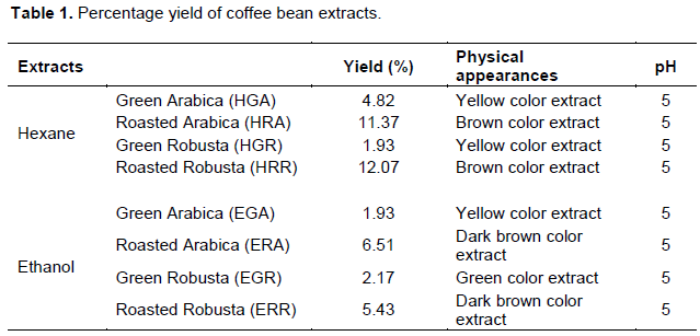
The coffee bean extracts obtained from hexane and ethanol maceration were calculated with percentage yield which ranged between 1.93 and 12.07% as shown in Table 1. The results showed that HGA, HGR and EGA were semisolid with a yellow color and unique odor. HRA, HRR, ERA and ERR were semisolid with brown or dark brown color and coffee odor whereas the EGR was green semisolid with a unique odor. All the extracts had pH of 5 which is suitable for skin care application. HRR possessed the highest percentage yield (12.07%) while HGR and EGA showed the lowest (1.93%). The hexane extracts from both green and roasted coffee beans showed higher percentage yield than ethanolic extracts. This might be due to the non-polar property of hexane that could extract most of the lipid contents from the coffee bean. Additionally, the roasted coffee bean showed higher lipid contents than the green coffee bean in both species corresponding to their percentage yield (Farah, 2012).
Determination of total phenolic content
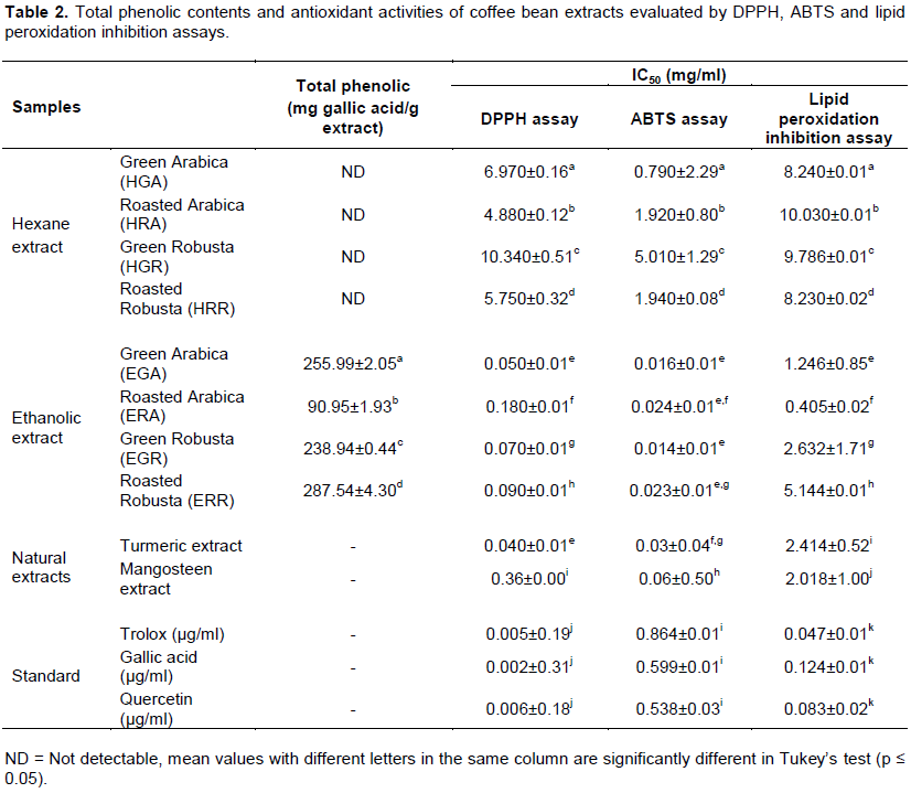
Total phenolic contents of all the extracts were determined by Folin-Ciocalteu assay. The total phenolic contents of both ethanolic green and roasted coffee extracts were statistically different. From the results, ERR presented the highest phenolic content (287.54 mg gallic acid/g extract) followed by EGA, EGR and ERA, respectively (255.99, 238.94 and 90.95 mg gallic acid/g extract) as shown in Table 2. In contrast, for hexane extracts, their total phenolic contents were not detectable. The results showed that total phenolic content of the green coffee bean extract was significantly higher than roasted coffee beans, except ERR. This might be due to auto-oxidation or degradation during the roasting process, leading to the decreased of polyphenol level in roasted coffee beans (Cheong et al., 2013). Generally, many research papers presented that the phenolic compounds were the good free radical scavenger. In addition, previous studies showed that coffee bean contained many polyphenolic compounds such as chlorogenic acid, mangiferin and hydroxycinnamic acid esters (Vignoli et al., 2011; Campa et al., 2012; Moreira et al., 2013). The major phenolic acid in all coffee samples was chlorogenic acid (Cheong et al., 2013). Therefore, the extracts that revealed a high total phenolic content tends to present a high level of antioxidant activity.
The determination of antioxidant activities
The coffee bean extracts’ antioxidant activity was evaluated by DPPH, ABTS and lipid peroxidation inhibition (linoleic acid) assays when compared with natural extracts (turmeric extract and mangosteen extract) and standards: trolox, gallic acid and quercetin. DPPH assay is widely used for testing the ability of compounds that act as free radical scavengers or hydrogen donors. Turmeric extract and mangosteen extract are widely used as active ingredients in anti-aging products due to their antioxidant activity. Therefore, researchers selected these extracts to compare biological activities with coffee bean extracts. The results are shown in Table 2. A lower IC50 value revealed a good antioxidant activity. Ethanolic extracts showed the higher antioxidant activity was significantly different from hexane extracts due to the presence of phenolic compounds that could be extracted by a more polar solvent (Prieto and Vázquez, 2014). Therefore, the research focus on the results of ethanolic extracts. Ethanolic green coffee bean extracts showed higher activity than ethanolic roasted coffee bean extracts in the same species that may be related to the higher polyphenol contents, especially chlorogenic acid (Yashin et al. 2013). Chlorogenic acid is a major component in green coffee beans and is reduced by the roasting process. There are many antioxidant experiments which prove that the phenolic compounds were the good free radical scavenger as mentioned above (Sendra, 2009). These results also strongly indicated that phenolic compounds in coffee bean are major contributors to their antioxidant capacity. The results also showed no significant differences in the antioxidant capacity of EGA and turmeric extract. Additionally, the ethanolic extracts of both species revealed a better antioxidant activity than in the mangosteen extract. The results from ABTS assay exhibited the same trend as DPPH assay. The hexane extracts revealed IC50 value much significantly higher than the ethanolic extracts. Additionally, the EGA and EGR presented better activity than turmeric extract while all ethanolic extracts presented a significantly higher level of activity than in the mangosteen extract. The results from lipid peroxidation inhibition assay also showed that ethanolic extracts significantly inhibited lipid peroxidation better than hexane extracts. The ethanolic roasted arabica bean extract showed a better activity than green arabica bean extract. This result might be due to the roasted coffee bean containing higher caffeine (lipophilic agent) than the green coffee bean that could better react with linoleic acid and inhibit lipid peroxidation. On the other hand, the ethanolic green Robusta bean extract exhibited high activity than the roasted Robusta bean extract due to synergism effect of phenolic compounds. Moreover, EGA and ERA showed a good anti-lipid peroxidation activity as compared to turmeric and mangosteen extracts. However, all the extracts showed a lower antioxidant activity than the standards. The ethanolic coffee bean extracts revealed good antioxidant activity with different assays as mentioned earlier. They could also inhibit lipid peroxidation which is a major cause of skin aging. Therefore, the ethanolic extracts were selected for further study.
Determination of mushroom tyrosinase inhibition activity
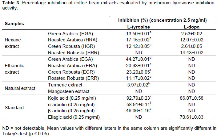
Tyrosinase enzyme plays an important role in melanin synthesis. It can change tyrosine to L-dopa, then convert to dopaquinone and with several polymerization reactions, eumelanin and pheomelanin are formed (Chang, 2009). Compounds that can inhibit tyrosinase enzyme are used as skin brightening agent. The results are shown in Table 3. When tyrosine was used as substrate, EGA revealed the highest activity (%inhibition = 44.27%). HRR, mangosteen extract and ellagic acid showed no activity. The coffee bean extracts presented a higher activity than turmeric extract. However, all the extracts presented a lower activity than kojic acid and arbutin. These indicated that antioxidant compounds might promote the tyrosinase inhibition activity due to their antioxidative synergistic (Chang, 2009). Therefore, the extracts which consist of high amounts of total phenolic compounds possessed a good inhibition to tyrosinase enzyme. In the part of L-dopa substrate, HRR showed the highest percentage of inhibition, whereas the ethanolic extracts showed no activity. Interestingly, the hexane extracts could inhibit tyrosinase enzyme in the step of converting L-dopa to dopachrome while α-arbutin and β-arbutin could not. It may be due to the components of triglycerides in the hexane extracts that are binding with some sites of the tyrosinase enzyme (Chang, 2009). It could be concluded that the ethanolic coffee bean extracts were the alternative ingredients in whitening products or mixing with other brightening natural ingredients.
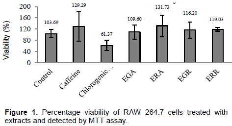
The effect of coffee bean extracts on cell viability
The cytotoxicity of coffee bean extracts was measured in RAW 264.7 cells using MTT assay. Percentage of cell viability between samples and the control at the same concentration (100 mg/ml) is shown in Figure 1. Caffeine and chlorogenic acid were used as controls. The results revealed that all extracts showed no toxicity on cells including caffeine, whereas chlorogenic acid presented only 61.37% of cell viability due to its acidity. Additionally, the extracts showed a higher percentage of cell viability than 100 which is in accordance with the effect of caffeine on cell viability. This result improves the assertion that the selected extracts are safe and can be developed as skin care products.
Determination of TLC chromatogram and the identification of chemical components of extracts using HPLC
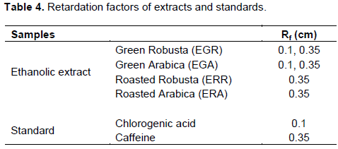
The ethanolic extracts showed good biological activities, therefore they were selected to further analyze major constituent by TLC. Coffee bean extracts, caffeine and chlorogenic acid were spotted on Merck Silica gel 60 F254 plate and developed with the mobile system of toluene: ethyl acetate : water : formic acid (15:90:5:5). Then, the chromatograms were detected under a short UV wavelength (246 nm). The TLC plates emitted green light where the compounds absorbed the light, and indicated as the dark areas. All the coffee bean extracts showed a deep dark spot with the same retardation factors with caffeine (Rf = 0.35) as shown in Figure 2 and Table 4. In addition, the chlorogenic acid, EGR and EGA showed the dark spot at the same distance (Rf = 0.1).
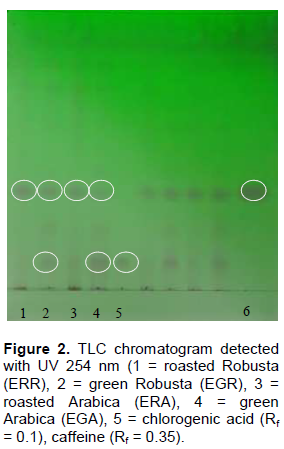
According to the results from TLC, caffeine was found in all extracts, whereas chlorogenic acid could be found only in the green coffee bean extracts due to the low amount in roasted coffee bean extracts. These results are related to the previous study which stated that caffeine was found in both green and roasted coffee beans. The previous study also indicated that chlorogenic acid was found in a higher amount in green coffee bean than roasted coffee bean. This may be due to its degradation by heat (Farah, 2012). It could be assumed that caffeine and chlorogenic acid are key compounds in coffee bean that serve as antioxidant and anti-tyrosinase ingredient.
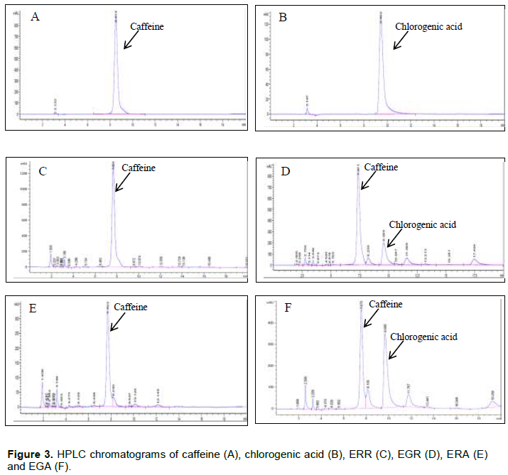
The ethanolic extracts were evaluated by HPLC using caffeine and chlorogenic acid as reference standards. The retention time of caffeine reference was 8.514 min, while retention time of chlorogenic acid was 9.450 min. The HPLC chromatogram of ERR and ERA showed a peak of caffeine, whereas EGR and EGA presented
both peaks of caffeine and chlorogenic acid as shown in Figure 3. The HPLC chromatograms are related to the results from TLC chromatogram. The roasted coffee bean extract loss of chlorogenic acid may be due to high temperature during the roasting process.
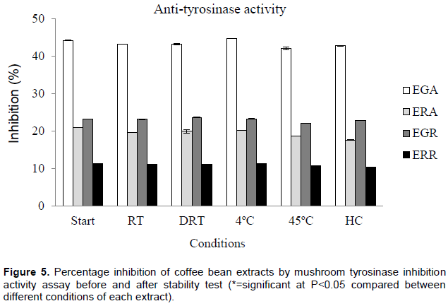
The stability of coffee bean extracts
The ethanolic extracts were kept in various storage conditions: room temperature (RT), the room temperature in the dark condition (DRT), 4 and 45°C for 3 months and heating-cooling (HC) for 6 cycles. After stability test, the extracts were analyzed by DPPH assays and mushroom tyrosinase inhibition activity assay. The results are shown in Figures 4 and 5. The percentage of inhibition of green Arabica (EGA), green Robusta (EGR) and roasted Robusta (ERR) extracts did not change after being stored in all conditions. Whereas, roasted Arabica (ERA) extract showed a significant decrease in the percentage of inhibition (P<0.05) after being stored at all conditions except 4°C. In contrast, the results from the mushroom tyrosinase inhibition activity assay showed that the percentage of inhibition did not change after being stored at various conditions. The results are related to their chemical compositions. Previous report indicated that Arabica coffee beans consist of coffee oil (cafestol and kahweol), triglycerides, fatty acids and tocopherol that are sensitive to heat, light and oxygen (Farah, 2012). Therefore, these compounds degrade after a stability test leading to a decrease in the antioxidant activity.
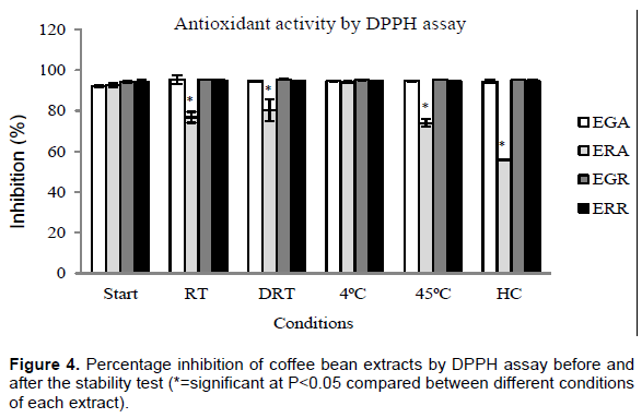
Therefore, the extracts should be kept to avoid light and heat to protect the degradation of active compounds.
CONCLUSION
In this study, the green and roasted coffee bean extracts from Arabica and Robusta beans were extracted with hexane and then followed by ethanol with maceration. The hexane extracts showed higher percentage of yields than in the ethanolic extracts; this may be due to high lipid contents. However, the ethanolic extracts possessed higher total phenolic contents and an enhanced level of antioxidant activity than in the hexane extracts. All the extracts except HRR could inhibit tyrosinase activity when using L-tyrosine as a substrate, whereas the hexane extracts showed anti-tyrosinase activity when L-dopa was used as a substrate. Antioxidant and anti-tyrosinase activities of extracts are related to the amount of caffeine and polyphenol contents. The higher caffeine and polyphenol contents generated higher biological activities. The ethanolic extracts that indicated good biological activities and non-toxicity were chosen for a further study. From TLC and HPLC chromatograms, the selected ethanolic extracts consisted of caffeine, while chlorogenic acid was found only in the green coffee bean extracts. The extracts also possessed good activities after being stored at various conditions for 3 months. Therefore, the ethanolic coffee beans are a promising source of natural antioxidant and anti-tyrosinase agent, and should be further developed into cosmeceutical products such as anti-aging or brightening products.
CONFLICT OF INTEREST
The authors have not declared any conflict of interest
ACKNOWLEDGEMENTS
The authors are grateful to Chiang Mai University, Chiang Mai, Thailand for financial support. They also thank the Faculty of Pharmacy, Chiang Mai University for all the facilities.
REFERENCES
|
Adham AN (2015). Simultaneous estimation of caffeic and chlorogenic acid content in Ammi Majus seed by TLC and HPLC. Int. J. Pharm. Pharm. Sci. 7(6):263-267. |
|
|
Almeida AAP, Naghetini CC, Santos VR, Antonio AG, Farah A, Gloria MBA (2012). Influence of natural coffee compounds, coffee extracts and increased levels of caffeine on the inhibition of Streptococcus mutans. Food Res. Int. 49: 459-461. |
|
|
Antonio AG, Iorio NLP, Pierro VSS, Candreva MS, Farah A, Santos KRN, Maia LC (2011). Inhibitory properties of Coffea canephora extract against oral bacteria and its effect on demineralisation of deciduous teeth. Arch. Oral Biol. 56:556-564. |
|
|
Ayelign A, Sabally K (2013). Determination of Chlorogenic Acids (CGA) in Coffee Beans using HPLC. Am. J. Res. Commun. 1(2):78-91. |
|
|
Brem B, Seger C, Pacher T, Hart M, Hadacek F, Hofer O, Vajrodaya S, Greger H (2004). Antioxidant dehydrotocopherols as a new chemical character of Stemona species. Phytochemistry 65:2719-2729. |
|
|
Campa C, Mondolot L, Rakotondravao A, Bidel LPR, Gargadennec A, Couturon E, Fisca PL, Rakotomalala J, Jay-Allemand C, Davis AP (2012). A survey of mangiferin and hydroxycinnamic acid ester accumulation in coffee (Coffea) leaves: biological implications and uses. Ann. Bot. 1-19. |
|
|
Cano-Marquina A, Tarin JJ, Cano A (2013). The impact of coffee on health. Maturitas 75:7-21. |
|
|
Chandra S, Chatterjee P, Dey P, Bhattachary S (2012). Evaluation of in vitro anti-inflammatory activity of coffee against the denaturation of protein. Asian Pac. J. Trop. Biomed. pp. 178-180. |
|
|
Chang TS (2009). An Updated Review of Tyrosinase Inhibitors. Int. J. Mol. Sci. 10:440-2475. |
|
|
Charrier A, Berthaud J (2012). Botanical classification of coffee; in Clifford MN, ed., Coffee botany, biochemistry and production of beans and beverage. The AVI Publishing Company Inc., Westport, Connecticut. pp. 13-47. |
|
|
Cheong MW, Tong KH, Ong JJM, Liu SQ, Curran P, Yu B (2013). Volatile composition and antioxidant capacity of Arabica coffee. Food Res. Int. 51:388-396. |
|
|
Chu YF, Chen Y, Black RM, Brown PH, Lyle BJ, Liu RH, Ou B (2011). Type 2 diabetes-related bioactivities of coffee: Assessment of antioxidant activity, NF-KB inhibition, and stimulation of glucose uptake. Food Chem. 124: 914-920. |
|
|
Chuakul W, Saralamp P, Paonil W, Temsiririrkkul R, Clayton T (1997). Medical plants in Thailand, Siambook and Publications Co., Ltd., Bangkok. |
|
|
Devasagayam TPA, Tilak JC, Boloor KK, Sane KS, Ghaskadbi SS, Lele RD (2004). Free radicals and antioxidants in human health: current status and future prospects. J. Assoc. Physicians India. 52:794-804. |
|
|
Farage MA, Miller KW, Elsner P, Maibach HI (2008). Intrinsic and extrinsic factors in skin ageing: a review. Int. J. Cosmet. Sci. 30:87-95. |
|
|
Farah A (2012). Coffee constituents; in Chu YF. ed., Coffee: Emerging Health Effects and Disease Prevention. John Wiley & Sons, Inc. pp. 21-58. |
|
|
Garzón GA, Riedl KM, Schwartz SJ (2009). Determination of anthocyanins, total phenolic content, and antioxidant activity in Andes Berry (Rubus glaucus Benth). J. Food Sci. 74(3):227-232. |
|
|
Johnson CE, Oladeinde FO, Kinyua AM, Michelin R, Makinde JM, Jaiyesimi AA, Mbiti WN, Kamau GN, Kofi-Tsekpo WM, Pramanik S, Williams A, Kennedy A, Bronner Y, Clarke K, Fofonoff P, Nemerson D (2008). Comparative assessment of total phenolic content in selected medicinal plants. Niger. J. Nat. Prod. Med. 12:40-42. |
|
|
Liang N, Lu X, Hu Y, Kitts DD (2016). Application of Attenuated Total Reflectance−Fourier Transformed Infrared (ATR-FTIR) Spectroscopy To Determine the Chlorogenic Acid Isomer Profile and Antioxidant Capacity of Coffee Beans. J. Agric. Food Chem. 64:681-689. |
|
|
Liu Y, Kitts DD (2011). Confirmation that the maillard reaction is the principle contributor to the antioxidant capacity of coffee brews. Food Res. Int. 44: 2418-2424. |
|
|
Moreira MEC, Pereira RGFA, Dias DF, Gontijo VS, Vilela FC, Moraes GOI, Giusti-Paiva A, Santos MH (2013). Anti-inflammatory effect of aqueous extracts of roasted and green Coffea arabica L. J. Funct. Foods 5:466-474. |
|
|
Mueller M, Hobiger S, Jungbauer A (2010). Anti-inflammatory activity of extracts from fruits, herbs and spices. Food Chem. 122(4):987-996. |
|
|
O'Keefe JH, Bhatti SK, Patil HR, DiNicolantonio JJ, Lucan SC, Lavie CJ (2013). Effects of habitual coffee consumption on cardiometabolic disease, cardiovascular health, and all-cause mortality. J. Am. Coll. Cardiol. 62(12):1043-1051. |
|
|
Pham-Huy LA, He H, Pham-Huy C (2008). Free radicals, antioxidants in disease and health. Int. J. Biomed. Sci. 4(2):89-96. |
|
|
Poljsak B, Dahmane RG, Godic A (2012). Intrinsic skin aging: The role of oxidative stress. ACTA dermatovenerol APA. 21:33-36. |
|
|
Pomerantz SH (1963). Separation, purification and properties of two tyrosinases from hamster melanoma. J. Biol. Chem. 238:2351-2357. |
|
|
Prieto MA, Vázquez JA (2014). In vitro determination of the lipophilic and hydrophilic antioxidant capacity of unroasted coffee bean extracts and their synergistic and antagonistic effects. Food Res. Int. 62:1183-1196. |
|
|
Sendra ME (2009). Total phenolic content and antioxidant activity of myrtle (Myrtus communis) extracts. Nat. Prod. Commun. 4:819-824. |
|
|
Tang SY, Whiteman M, Peng ZF, Jenner A, Yong EL, Halliwell B (2004). Characterization of antioxidant and antiglycation properties and isolation of active ingredients from traditional chinese medicines. Free Radic. Biol. Med. 36(12):1575-1587. |
|
|
Vignoli JA, Bassoli DG, Benassi MT (2011). Antioxidant activity, polyphenols, caffeine and melanoidins in soluble coffee: The influence of processing conditions and raw material. Food Chem. 124:863-868. |
|
|
Wagemaker TAL, Carvalho CRL, Maia NB, Baggio SR, Filho OG (2011). Sun protection factor, content and composition of lipid fraction of green coffee beans. Ind. Crops Prod. 33:469-473. |
|
|
Yashin A, Yashin Y, Wang JY, Nemzer B (2013). Antioxidant and Antiradical Activity of Coffee. Antioxidants 2:230-245. |
|
Copyright © 2024 Author(s) retain the copyright of this article.
This article is published under the terms of the Creative Commons Attribution License 4.0


