Full Length Research Paper
ABSTRACT
The ethanolic extract of the stems of Massularia acuminata is widely used in several regions of Côte d’Ivoire for the improvement of the male sexual performance. The study was carried out with a view to investigate the effect of long term use of the plant on rat. For this purpose, four groups of 6 rats each, aged 8 weeks and weighing on average 125 ± 23.16 g. Groups 2, 3 and 4 were treated repeatedly with the ethanolic extract of the stem of M. acuminata at doses of 40, 80 and 160 mg/kg of body weight for 28 days respectively. Group 1, the control group, received only distilled water. In addition to the testicular histological study, body mass and relative testicular mass, sperm motility, concentration and viability were evaluated. This study showed that 40 and 80 mg/kg of ethanolic extract of stems of M. acuminata were better tolerated by the testes. However, the 160 mg/kg body weight dose caused a significant reduction in body mass (-4.68 vs +1.30%) and relative testicular mass (1.27 vs 0.45) on day 28 of the experiments. In addition, the result of the study revealed a significant decrease in motility (16.29 vs 41.71%), sperm concentration (10.33 vs 138.83 sperm/µl) and viability compared to the control group. This study showed that at high doses, the ethanolic extract of the stems of M. acuminata can disrupt spermatogenesis in rats.
Key words: Massularia acuminata, toxicity, rat, testis, spermatozoa.
INTRODUCTION
In all developing countries such as Côte d'Ivoire, the use of medicinal plants is the most common way of solving public health problems, especially in rural areas. According to the World Health Organisation (WHO), more than 80% of the African population uses traditional medicine for their primary health care because of their proximity and accessibility (WHO, 2002).
Medicinal plants for aphrodisiac use are increasingly coveted in our society, especially among men, to develop, preserve their sexual capacity or stimulate sexual desire. Among the many aphrodisiac preparations in Côte d'Ivoire, is the ethanolic extract of the stems of Massularia acuminate. This preparation is sold in cabarets under the name '4 o'clock in the morning'.
The species M. acuminata (G. Don) Bullock ex Hoyl. belonging to the Rubiaceae family, is a plant used in traditional African medicine against various indications, in particular against hernia, sterility, ovarian disorders, fevers, difficult deliveries, rheumatic pains, oral infections, haematuria, poison, dysentery, coughs and especially sexual weaknesses (Aladesanmi et al., 2007; Bouquet, 1969; Kayode and Omotoyinbo, 2008; Neuwinger, 2000; Singh et al., 2011; Yakubu et al., 2008). The phytochemical screening revealed that the ethanolic stem extract of M. acuminata contains coumarins, flavonoids flavonoids, tannins, triterpenes, triterpeneaponins, Glycoside, Saponin, Tannin, Flavonoid, Anthocyanin and Anthraquinone sterols (Bankole et al., 2012; Maloueki et al., 2015; Gbogbo et al., 2021).
Pharmacological studies have shown good antibacterial and antifungal activity of ethanolic extracts of M. acuminata on Staphylococcus, Streptococcus, Proteus, Aspergillus favus, Candida tropicalis, Candida albican (Enwa et al., 2016; Olusola et al., 2020).
Other studies have demonstrated the aphrodisiac activity of the roots and stems of M. acuminata. These authors observed, in a dose-dependent mode, a significant increase in the frequency of sexual mounts, erections, ejaculations and a decrease in the latency time between two consecutive sexual mounts (Yakubu and Akanji, 2011; Gbogbo et al., 2021). Despite these promising results, there are few data on the effect of its prolonged use on the testes. It is in this context that the present study aims to evaluate the subacute testicular toxicity of the ethanolic extract of M. acuminata stems in rats.
MATERIALS AND METHODS
Plant material
The plant material collected in Bonoua, in the department of Grand-Bassam in the South-Comoé region (Côte d'Ivoire), in November 2020. The plant was identified at the National Floristic Centre of the Félix Houphouët Boigny University where a specimen was deposited and identification number (UCJ15291) was collected.
Animals
For this study, male rats Rattus norgevicus, aged eight weeks, weighing an average 125 ± 23.16 g were used for the experiment. The rats were bred in the animal house of the Physiology, Pharmacology and Pharmacopoeia Laboratory of the Research Unit of Nangui Abrogoua University. A total of 24 rats were divided into four groups animals were subjected to a temperature of 25 ± 2°C and to an alternation of 12 h of light and 12 h of darkness. The diet consisted of IVOGRAIN® pellets and the rats were provided with tap water. The experimental protocol and animal handling procedures were conducted according to good laboratory practice (OECD, 1998).
Preparation of ethanolic extract
For the preparation of the extract, two hundred grams (200 g) of powder obtained from the stem of M. acuminata were macerated in 2.5 L of 96% (v/v) ethanol for 24 h under continuous stirring. The resulting macerate was filtered and then concentrated under reduced pressure at 40°C using a rotary evaporator. The concentrated filtrate was dried in an oven at 40°C. The dry extract obtained constituted the ethanolic extract and was kept for the experiments (Gbogbo et al., 2021).
Toxicity assessment
The subacute toxicity study was determined using OECD guideline 407 (OECD, 2008) which involved the daily oral administration of extracts in increasing doses to four groups of animals, one dose per group for 28 days. The doses of 40, 80 and 160 mg/kg were given to Groups 2, 3 and 4 respectively. Group 1 (control) received only distilled water. Prior to administration of the extracts, the animals in each group were individually marked and weighed. They received a volume of solution of 2 mL/100 g body weight once a day for 28 days by oral gavage using a cannula. The animals were observed individually every morning throughout the study. The influence of the different doses administered was assessed on the basis of the rats' body mass, cytological and histological examinations.
Body mass and relative testicular mass
The body mass of each rat was measured on days D0, D14 and D28. After 28 days of treatment, all rats were anaesthetised and sacrificed by cervical dislocation and the testes were removed and weighed (AVMA, 2020).
Effect of the extract on spermatozoa
Sperm evaluation (sperm motility, concentration and viability)
After the rats were sacrificed, the caudal epididymis of the right testis of each rat was removed, cut into small pieces and transferred to 1 ml of 0.9% sodium chloride solution (0.9%). The mixture was gently shaken to allow the sperm to disperse in the sodium chloride solution, and the sperm suspension was transferred to an Eppendorf tube as described by Talebi et al. (2011). Each sample was then temporarily incubated at 37°C, for 30 min for subsequent sperm analysis.
Sperm motility
Sperm motility was analysed by adding 6 µl of the sperm suspension to the KOVA®-Slide chamber hemocytometer to determine the proportion of motile sperm. The slide was examined under a light microscope at magnification × 400 and motility was recorded in 10 small squares of different grid cells. Sperm showing any degree of movement were considered motile and were counted in groups (motile and immobile). Sperm motility was calculated as follows:
Motility (%) = [Number of motile sperm/total number of sperm] × 100 and expressed as percentage motility (WHO, 1999).
Sperm concentration
Sperm concentration was determined by counting sperm in 10 fields at 400x magnification. For this purpose, 6 µl of the sperm suspension was introduced into the KOVA®-Slide chamber hemocytometer. The sperm concentration was the total average of the sperm in 10 small squares of different grid cells by the following formula:
Sperm count/µl= (Total number of sperm in 10 squares / 10) × 90 (KI, 2021).
Assessment of sperm viability
In a haemolysis tube, 50 µl of sperm suspension was mixed with 50 μl of 0.5% eosin dye. The mixture was shaken for 30 s and then 100 µl of nigrosin is added to the first mixture. A drop of the stained sperm suspension was placed on a slide and covered with a coverslip. Observation is carried out under a light microscope at the x400 objective. Dead spermatozoa appeared purple while live spermatozoa had a blue outline (Dieusaert, 1994). A total of 200 spermatozoa were counted and the proportions of live and dead spermatozoa were calculated.
Histological examinations
At the end of the 28-day experiment, the testes removed and preserved in 10% formalin prior to histological examination on the male reproductive system. The method used was the paraffin embedding technique (Hould, 1984).
Statistical analysis
The statistical study was carried out using the XLSTAT-PRO 7.1 statistical analysis software. The results were analysed using Tukey's and Dunnett's post hoc tests combined with a one-factor Anova. Values are given as the mean followed by the standard error of the mean. Some of the results of the cytological studies were presented as proportions and their analysis was performed using the parametric k-proportion test (G-test). For the study of the relative mass of the organs, we used the following ratio (Yakubu et al., 2008):
Relative mass = (absolute mass of the organ (g)) / (body mass of the animal on the day of sacrifice (g)) × 100.
These tests give us the degree of significance for p < 0.05.
RESULTS
Evolution of body mass
The results indicated a loss of body mass in rats treated with ethanolic extract of M. acuminata stem at 40 and 160 mg/kg body weight compared to the control group on day 14 of the experiment. A gain in body weight was observed in the group treated with the ethanolic extract at the dose of 80 mg/kg. However, this gain remained below that obtained by the control group (Table 1). On day 28, a gain in body mass was observed in all groups without significant difference except for the rats treated with the 160 mg/kg extract where a loss of mass was observed compared to the control batch.
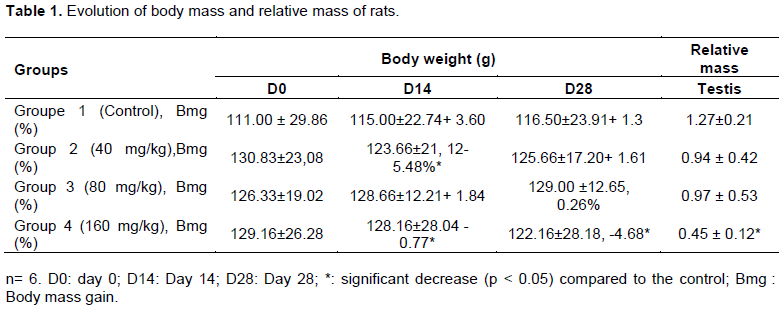
Relative testicular mass
The results showed a general decrease in the relative mass of the testes in all rats treated with the ethanolic extract of M. acuminata stem compared to the mass value observed in the controls. This decrease was particularly significant in rats treated with the 160 mg/kg dose compared to the control (Table 1).
Effect of the extract on spermatozoa
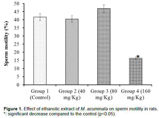
Sperm motility
Assessment of sperm motility did not reveal a significant change in the proportion of motile sperm in rats from the control group compared to those treated with the extract at 40 and 80 mg/kg. In contrast, a significant decrease in the proportion of motile spermatozoa was observed in rats treated with the extract at 160 mg/kg compared to the control group (Figure 1).
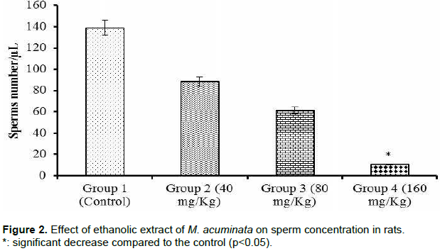
Sperm concentration
The results showed (Figure 2) a dose-dependent decrease in sperm concentration in treated rats compared to the control group. Group 4 showed a significantly (p < 0.05) lower sperm concentration compared to the control group (10.33 vs. 138.83 sperms/µl).
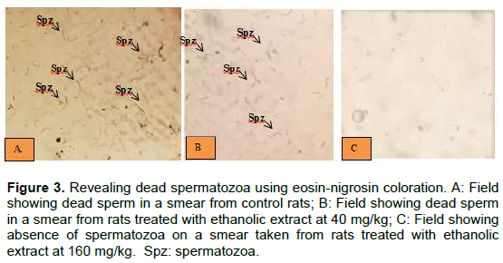
Sperm viability
The eosin-nigrosin stain was used to observe mainly dead sperm from the rats on the smears. Figure 3A shows dead sperm in a smear from control rats. A decrease in sperm count is observed in micrograph 3B compared to the control. In contrast, a near absence of observable spermatozoa was observed in the smears of rats treated with ethanolic extract at a dose of 160 mg/kg (Figure 3C).
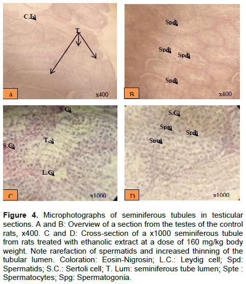
Histological study
Histological sections of the testes showed different cells from the different stages of spermatogenesis in the control group, resulting in the presence of spermatids, spermatocytes and spermatogonia (Figure 4A and B). In addition, the microphotographs show Sertoli cells between the germ cells. The lumen of the seminiferous tube is very small. In contrast, histological sections from the group of rats treated with ethanolic extract at 160 mg/kg body weight show disturbances. The disturbances included a rarefaction of spermatids, a greater thinning of the tubular lumen and a disorganisation of the germ cells in reduced numbers compared to the histological sections in the control rats.
DISCUSSION
This study was conducted to test the effect of prolonged consumption of an ethanolic extract of M. acuminata stems in the rats. The results of this study showed that the ethanolic extract of M. acuminata stems generally caused a loss of body weight gain in the treated groups compared to the control batch. These results are similar to those obtained by Oloyede et al. (2020) who observed a significant decrease in body mass of mice after oral administration of ethanolic extract of Lecaniodiscus cupanioides (Sapindaceae), a plant with aphrodisiac virtue (Olaide and Ajiboye, 2012), at the dose of 100 mg/kg body weight over a period of 30 days.
This decrease in body weight of rats observed in the study could be explained by the presence of tannin and flavonoid in ethanolic extracts of M. acuminata (Gbogbo et al., 2021). These authors demonstrated the presence of these secondary metabolites in the ethanolic extract of M. acuminata. Indeed, according to some authors, the consumption of plants containing tannins in moderate amounts influences growth (Rochfort et al., 2008; Gutiérrez-Salmeán and Pérez, 2015) and it has been demonstrated that flavonoids can reduce body mass via fatty acid synthesis and thus contribute to the improvement of energy expenditure.
Concerning the testis, this study generally revealed a decrease in their relative mass in all rats treated with ethanolic extract of M. acuminata stem compared to the value of the mass observed in controls. These results are similar to those obtained by Dayang and Mahanem (2015) who reported a significant decrease in the relative mass of the testis in rats treated with the 800 mg/kg body weight dose of the methanolic leaves extract of Andrographis paniculata (Burm. f.) Wall. Ex (Acanthaceae) in rats. The reduction in relative testicular mass found in this study may be due to a decrease in androgen production. This steroid hormone is responsible for the induction of differentiation and maturation of the male reproductive organs (O'Donnell et al., 2017). Repeated administration of the ethanolic extract of M. acuminata stem could therefore inhibit testosterone production by the Leydig cells of the testis, located around and between the seminiferous tubules (Zhou et al., 2019). This study also showed a decrease in sperm motility, concentration and viability, particularly at a dose of 160 mg/kg body weight. These results corroborate those observed in the reduction of testicular mass. A decrease in testosterone production would lead to a disruption of spermatogenesis.
According to studies by Amaral et al. (2014), the decrease in sperm motility is probably due to ATP activity in sperm by uncoupling oxidative phosphorylation from the respiratory chain and preventing the phosphorylation of ADP to ATP, thus rendering sperm immobile. However, motility is considered a key function of good sperm quality as the sperm has to move through the female reproductive tract to fertilise oocyte II. The low sperm concentration and viability of spermatozoa could be due to free radical attacks or oxidative stress. More than any other cell, the spermatozoon is susceptible to free radical damage. The sensitivity of the male gamete to free radical attack is indeed increased by the particular lipid composition of its plasma membrane, rich in polyunsaturated fatty acids, prime targets of reactive oxygen species (Migdal and Serres, 2011). An excessive accumulation of reactive oxygen species can then lead to cell death (Ford, 2004). The administration of ethanolic extract of M. acuminata stems would therefore lead to a decline in the number of spermatogonia and their proliferative activity.
With regard to the histology of the rat testicles, the results revealed a disorganised testicular structure with a rarefaction of spermatids, a greater thinning of the tubular lumen and a disorganisation of the germ cells in reduced numbers. These observations confirm the previous results obtained in this study. The administration of 160 mg/kg body weight to rats induces changes in spermatogenesis; the dysfunction of the endocrine system, both pituitary and testicular, is competent enough to influence spermatogenesis negatively (Verhoeven et al., 2010). The toxicity caused by the administration of the ethanolic extract of M. acuminata stems would therefore be dose-dependent, as at low doses the effects are less significant than at high doses.
CONCLUSION
The present study showed that at the high dose of 160 mg/kg body weight, ethanolic extract from the stems of M. acuminata could cause a significant decrease in body mass and relative testicular mass of rats compared to the control group. In addition, a decrease in motility, sperm concentration and viability was observed at the same dose. The histological study revealed a disorganised testicular structure with rarefaction of spermatids, increased thinning of the tubular lumen and cellular disorganisation in the seminiferous tubule. Lower doses should be chosen during repeated use of the ethanolic extract of M. acuminata stem.
CONFLICT OF INTERESTS
The authors have not declared any conflict of interests.
REFERENCES
|
Aladesanmi AJ, Iwalewa EO, Adebajo AC, Akinkunmi EO, Taiwo BJ, Olorunmola FO, Lamikanra A (2007). Antimicrobial and antioxidant activities of some Nigerian medicinal plants. African Journal of Traditional, Complementary and Alternative Medicines 4(2):173-184. |
|
|
Amaral A, Paiva C, Attardo Parrinello C, Estanyol JM, Ballescà JL, Ramalho-Santos J (2014). Identification of proteins involved in human sperm motility using high-throughput differential proteomics. Journal of Proteome Research 13(12):5670-5684. |
|
|
American Veterinary Medical Association Guidelines for euthanasia of animals (AVMA) (2020). Guidelines for the euthanasia of animals: Edition 121 p. |
|
|
Bankole PO, Adekunle AA, Oyede RT, Faparusi F, Adewole A (2012). Antimicrobial Activities and Phytochemical Screening of two Tropical Nigerian Chewing Sticks. International Journal of Applied Science and Technology 6(2). |
|
|
Bouquet A (1969). Fetishists and traditional medicine in Congo (Brazzaville). Memory ORSTOM 36, Paris P 49. |
|
|
Dayang NF, Mahanem MN (2015). Fertility Suppression in Male Sprague-Dawley Rats by Administration of Methanolic Extract of Hempedu bumi (Andrographis paniculata). Sains Malaysiana 44(9):1249-1255. |
|
|
Enwa FO, Iyamu MI, Micheal O, Success MC (2016). Antibacterial screening of Massularia acuminata aqueous and ethanolic stem extracts against selected oral isolates. Journal of Basic and Applied Research International 16(4):270-277. |
|
|
Ford WC (2004). Regulation of sperm function by reactive oxygen species. Hum Reprod Update10(5):387-399. |
|
|
Gbogbo M, N'Guessan OHA, Koné M, Ahoulou AS, Aboli TF, Yapo AP (2021). Phytochemical screening and aphrodisiac effect of ethanolic extract of Massularia acuminata (G. Don) bullock ex hoyl. (Rubiaceae) in male wistar rats. International Journal of Advanced Research. 9(9):653-659. |
|
|
Gutiérrez JT, Pérez E (2015). Significant quality factors in the chocolate processing: Cocoa Post Harvest, and in its Manufacture. In: Chocolate: Cocoa Byproducts Technology, Rheology, Styling, and Nutrition. New York: Nova Science Publisher, Inc; pp. 1-47. |
|
|
Hould R (1984). Histopathology and cytopathology techniques. Maloine edit. Paris 399 p. |
|
|
Kayode J, Omotoyinbo AM (2008). Cultural erosion and biodiversity: conserving chewing stick knowledge in Ekiti State, Nigeria. African Scientist 9(1):41-51. |
|
|
KI (2021). Calculation method for diluted body fluids. |
|
|
Maloueki U, Kunyima KP, Mbomba ID, Dani NA, Lukuka KA, Lami NJ, Mpiana PT, Ngbolua KN, Ndimbo KSP, Mbomba NB, Muganza CD (2015). Antioxidant and antiplasmodial activities of extracts of Massularia acuminata (Rubiaceae). Phytotherapie 13(6):389-395. |
|
|
Migdal C, Serres M (2011). Reactive oxygen species and oxidative stress. Medicine Sciences 27(4):405-412. |
|
|
Neuwinger HD (2000). African traditional medecine: a dictionaryof plant use and applications. Search system for diseases. Med-pharm Scientific Publishers, Stuttgart 589 p. |
|
|
O'Donnell L, Stanton P, de Kretser DM (2017). Endocrinology of the Male Reproductive System and Spermatogenesis. In: Feingold KR, Anawalt B, Boyce A, et al., editors. Endotext [Internet]. South Dartmouth (MA): MDText.com, Inc.; 2000. |
|
|
Olaide NO, Ajiboye TO (2012). Aqueous root extract of Lecaniodiscus cupanioides restores the alterations in testicular parameters of sexually impaired male rats. Asian Pacific Journal of Reproduction 1(2):120-124. |
|
|
Oloyede AM, Anyika CC, Ottu B (2020). Reproductive Toxicity of Ethanolic Extract of Lecaniodiscus cupanioides on Male Albino Mice. Herb. Herbal Medicines Journal 5(1):31-36. |
|
|
Olusola A, Bakare O, Ekun O, Olusola A (2020). A Comparative Study on the Effect of Massularia acuminata Fractions against Bacterial and Fungal Isolates from the Oral Cavity. Health 12(7):904. |
|
|
Organisation for Economic Co-operation and Development (OECD) (1998). Series on the principles of good laboratory practice and verification of compliance with these principles. ENV/MC/CHEM 17(98):22-23. |
|
|
Organisation for Economic Co-operation and Development (OECD) (2008). Guidelines for chemical testing - 28-day repeated dose oral toxicity study in rodents 24 p. |
|
|
Rochfort S, Parker AJ, Dunshea FR (2008). Plant bioactives for ruminant health and productivity. Phytochemistry 69(2):299-322. |
|
|
Singh R, Pallavi KJ, Singh S (2011). Aphrodisiac agentsfrom medicinal plants: a review. Journal of Chemical and Pharmaceutical Research 3(2):911-2130. |
|
|
Talebi AR, Sarcheshmeh AA, KhaliliMA, Tabibnejad N (2011). Effect of ethanol consumption on chromatin condensation and DNA integrity of epididymal sperm in rat. Alcohol 45(4):403-409. |
|
|
Verhoeven G, Willems A, Denolet E, Swinnen JV, Gendt K (2010). Androgens and spermatogenesis: lessons from transgenic mouse models. Philosophical Transactions of the Royal Society B: Biological Sciences; 365(1546):1537-1556. |
|
|
World Health Organization (WHO) (1999). WHO laboratory manual for the examination of human semen and semen-cervical mucus interaction. Cambridge University Press, Cambridge. |
|
|
World Health Organization (WHO) (2002). WHO Strategy for Traditional Medicine 2002-2005. Geneva, WHO/EDM/TRM/2002.1:65. |
|
|
Yakubu MT, Akanji MA (2011). Effect of Aqueous Extract of Massularia acuminata Stem on Sexual Behaviour of Male Wistar Rats. Evidence-Based Complementary and Alternative Medicine, pp. 1-10. |
|
|
Yakubu MT, Akanji MA, Oladiji AT (2008). Androgenicpotentials of aqueous extract of Massularia acuminata (G. Don) Bullock ex Hoyl. Stem in male Wistar rats. Journal of Ethnopharmacology 118(3):508-513. |
|
|
Zhou R, Wu J, Liu B, Jiang Y, Chen W, Li J, He Q, He Z (2019). The roles and mechanisms of Leydig cells and myoid cells in regulating spermatogenesis. Cellular and Molecular Life Sciences 76(14):2681-2695. |
|
Copyright © 2024 Author(s) retain the copyright of this article.
This article is published under the terms of the Creative Commons Attribution License 4.0


