Full Length Research Paper
ABSTRACT
This study assessed the microbiological quality of meat from free rage-produced chicken processed in an informal slaughter facility. The total viable counts (TVC), total coliform counts, coagulase positive Staphylococcus aureus, Streptococcus species, Salmonella species and Campylobacter species were used as indicators. A cross-sectional sampling of chicken carcasses at informal slaughter facility was carried out. Whole carcass rinse fluid was prepared from 40 randomly obtained freshly dressed carcasses. Fluid samples were cultured in selective media to isolate and enumerate the specific bacteria. S. aureus was further identified by coagulase test, Streptococci by serotyping into Lancefield groups, Campylobacter by DNA analysis and Salmonella by biochemical tests and serology. Bacterial concentrations in the carcasses were calculated as colony forming units (CFU) per ml and CFU/cm2. The mean carcass CFU/ml concentration was 1.59 × 10⁷, 1.44 ×10⁵, 3.2 × 10⁴ and 1.06 × 10⁴ for TVC, Coliforms, S. aureus and Streptococci, respectively. All the mean concentration values were higher than the limits recommended by the Codex Alimentarius Commission (CAC). Coagulase-positive Staphylococcus was isolated from 12 (30%) carcasses and Streptococci from 35 (87%). Majority Streptococci were Lancefield Group D (48.57%) followed by Group G (17.14%), and Group F (14.28%). Campylobacter genus was identified in 11 carcasses (27.5%) and Campylobacter jejuni in three (7.5%). On the other hand, Salmonella was not isolated from any carcass. The results of the study indicated that the low hygienic standard in non-regulated slaughter houses exposed the chicken meat to microbial contaminants which may pose a risk to the consumers. Improvement of slaughter infrastructure and capacity-building of slaughter personnel is therefore critically required to ensure food safety and enable access to high value markets.
Key words: Slaughter, free-range chicken, bacterial quality.
INTRODUCTION
Food spoilage and foodborne diseases are important economic and health concerns and raw poultry products are frequently contaminated by spoilage organisms and human pathogens. Contamination may occur exogenously or endogenously and the health of birds at farm level, transportation, slaughter facilities and processing are critical (Shahdan et al., 2017; Rasschaert et al., 2020). Contaminated products undergo rapid spoilage especially when post-slaughter and processing preservation is inadequate, as a result of growth and metabolic activities of contaminant bacteria. Spoilage may lead to large economic losses, impacting on the economy of the poultry production sector (Rouger et al., 2017).
Bacterial contaminants may include pathogenic species and human infections results from handling raw products, undercooking or cross-contamination of other foodstuffs at retail or household level (Kennedy et al., 2011; Sirsat et al., 2014; Mkhungo et al., 2018). Campylobacter and Salmonella are the two most important human zoonotic gastrointestinal bacterial pathogens. Poultry meat is believed to be the main source of human Campylobacter infection worldwide (EU, 2017; Kuria et al., 2018; Carron et al., 2018) and an important source of non-typhoid salmonellosis (Antunes et al., 2016; WHO, 2018). Other important pathogens that may be associated with poultry foodborne disease include Staphylococcus aureus, verotoxigenic Escherichia coli and fecal Streptococci (Rouger et al., 2017; Svobodová et al., 2012; Vaidya et al., 2005).
Free range chicken production in Kenya constitutes the largest proportion of the national poultry population, and serves as a source nutritional needs and income to resource-poor farmers in developing countries. However, production, transportation, slaughter and processing are carried out under poor sanitary conditions (King’ori et al., 2010; Ipara et al., 2019). The poor hygienic processing conditions in informal facilities hinder the chicken products access to high value markets and expose consumers to health risks. Data on microbiological quality of chicken carcasses have been limited mainly to intensive production and conventional processing systems in Europe and North America and data on free range indigenous chicken processed under informal facilities has been scanty. Ensuring microbial safety in food products is important in the context of increasing production and consumption as well as safeguarding human health. Consequently, the Codex Alimentarius Commission (CAC) has set the limits for microbial organisms in foods (CAC, 1997). This study aims to assess the bacterial quality of meat carcasses from free range-produced chicken slaughtered in non-conventional facility in Nairobi, Kenya.
MATERIALS AND METHODS
Study area
The study comprised collection and laboratory analysis of chicken carcasses. Burma Maziwa market (Elevation: 1,795 M, Coordinates: S-01.2921°, E-036.8219°) in Nairobi County is one of several informal markets for indigenous free-range produced chickens. It receives chicken from several parts of the country and serves as a significant outlet of chicken destined to other smaller live bird-markets in the county. There are no chicken housing facilities and the chickens are held in wooden transport cages for short durations. Dressed carcasses are supplied to lower-class supermarkets and butcheries, and also homes. Slaughter facility consists of a single stone-walled room with concrete floor, a concrete work-bench and no running water. The slaughter method involves manual strangulation, dry-plucking and evisceration. Laboratory analysis was carried out at the faculty of Veterinary Medicine, University of Nairobi.
Sample collection
Five (5) freshly dressed chicken carcasses were randomly obtained from the slaughter house on each of 8 sampling days. A total of 40 carcasses were collected. The carcasses were put in sterile double polythene bags, labeled, put in a cool box and transported to the laboratory within 4 to 5 h. In the laboratory, the carcasses were weighed and then rinse-washed with 400 ml of buffered peptone water (pH 7.2) following the method described (NACMCF, 2007). The rinse fluid was then analyzed for the microorganisms of interest.
Enumeration of total viable counts (TVC) and coliforms
Tenfold serial dilutions of rinse fluid from each carcass were prepared in peptone water. One milliliter of each of the highest 4 consecutive serial dilutions (10-3 to 10-6) was inoculated into Plate Count Agar (PCA) (HIMEDIA M091, India) in triplicates, using the pour plate overlay method. The plates were then incubated at 37°C for 48 h and the number of colonies formed per dilution recorded after 24 and 48 h of incubation. The mean CFU/ml and CFU/cm2 were then calculated as described (Brichta-Harhay et al., 2008) and transformed into log CFU/ml and log CFU/cm2. Total coliforms were similarly enumerated, using Violet Red Bile Lactose Agar (VRBA) media (OXOID CM 0968, UK).
Enumeration and confirmation of coagulase positive Staphylococcus
Using spread plate method, 0.5 ml of each 4 consecutive 10 fold serial dilutions (10-1 to 10-4) of the rinse fluid were inoculated in triplicates into mannitol salt agar (Oxoid CM 085, UK). The plates were incubated at 37°C for 24 h and the number of yellow colonies recorded per dilution. CFU/ml, CFU/cm2 and the respective logs were then calculated. For confirmation of coagulase positive S. aureus, four suspect colonies per dilution were sub-cultured onto blood agar (OXOID CM0271, UK) and incubated at 37°C for 24 h. Golden yellow and cream white colonies were tested for catalase and coagulase activity.
Enumeration and serotyping of Streptococci
Sodium azide blood agar (OXOID CM 259, UK) was inoculated with 0.5 ml of each of the 4 consecutive 10 fold serial dilutions of the rinse fluid (10-1 to 10-4), in triplicates, using the spread plate method. The plates were incubated at 37°C for 48 h and the number of pin point βhemolytic colonies recorded per dilution. CFU/ml, CFU/cm2 and the respective logs were calculated. The colonies were purified by sub-culturing four β hemolytic colonies per plate onto blood agar plates and incubating for 24 h at 37°C. Characteristic βhemolytic colonies were serotyped into Lancefield groups using streptex* kit (Oxoid, TSMX7829, UK), following the manufacturer’s instructions.
Isolation of Campylobacter species
Campylobacter blood-free medium containing antibiotics and supplement (mCCDA, Oxoid CM739, UK) was used for isolation of thermophilic Campylobacter spp. All samples were cultured directly on the media (OIE, 2008) within 6 h of collection. Tenfold serial dilutions of the rinse-wash fluid were prepared in peptone water and 0.5 ml of 4 consecutive serial dilutions, 100 to 10-3, inoculated into the media using the spread plate method. Inoculated plates were incubated at 42°C, for 48 h in candle extinction jar (Kuria et al., 2018). Suspect Campylobacter colonies were then selected for further analysis by Gram stain, catalase and oxidase biochemical tests. The Gram stain was performed using reagents prepared according to WHO (2009) method. Suspect isolates were confirmed by DNA analysis.
Campylobacter DNA analysis
DNA extraction
A loopful of suspect Campylobacter colonies was harvested and suspended in 200 µl of sterile distilled water in labeled 0.5 ml Eppendorf tubes. The tubes were then heated in boiling a water bath at 100°C for 10 min, cooled immediately on ice for 5 to 10 min and then centrifuged (Eppendorf, Gerãtebau, West Germany) at 11,000×g for 5 min. The supernatant was stored at -20°C and used as DNA templates. Confirmation of Campylobacter genus was done using PCR analysis for 16S rRNA gene (Linton et al., 1997) and identification of Campylobacter jejuni and Campylobacter coli by multiplex PCR using species specific primers. The primers, C412F and C1228R; C412F and C1228R; ENg03F and ENg04R for Campylobacter genus, C. jejuni and C. coli, respectively were based on nucleotides sequences of monospecific probes from DNA fragments library (WHO, 2009).
DNA amplification
Amplification of Campylobacter genus DNA was performed in a 25 µl reaction volume per sample. Briefly, aliquots of 12.5 µl of Taq Master Mix (Qiagen GmbH, Limburg, Netherlands), 10 pmol of each primer (Bioneer, Inc. USA), 5 µl of DNA template and 7.3 µl of molecular grade water (Qiagen GmbH, Limburg, Netherlands) were put in labeled sterile PCR tubes, and placed in a thermocycler (MJ Research, Watertown, MA, USA). The samples were subjected to initial denaturation temperature of 95°C for 10 min followed by 35 cycles of denaturation at 95°C for 30 s, annealing at 59°C for 90 s, extension at 72°C for 60 s and a final extension of 72°C for 10 min. Amplification of C. jejuni and C. coli species DNA was performed in a 50 µl multiplex reaction volume per sample as follows: briefly, 25 µl Taq PCR Master Mix (Qiagen GmbH, Limburg, Netherlands), 5 µl of DNA template, 60 pmol of C. coli primers (Bioneer, Inc. USA), 25 pmol of C. jejuni primers (Bioneer, Inc. USA) and 18.3 µl of molecular grade water (Qiagen GmbH, Limburg, Netherlands) were put into labelled PCR tubes. The PCR protocol included initial denaturation temperature of 94°C for 5 min; 2 cycles of 1 min at 94°C, 1 min at 64°C, and 1 min at 72°C; 2 cycles of 1 min at 94°C, 1 min at 62°C, and 1 min at 72°C; 2 cycles of 1 min at 94°C, 1 min at 60°C, and 1 min at 72°C; 2 cycles of 1 min at 94°C, 1 min at 58°C, and 1 min at 72°C; 2 cycles of 1 min at 94°C, 1 min at 56°C, and 1 min at 72°C; 30 cycles of 1 min at 94°C, 1 min at 54°C, and 1 min at 72°C; and a final extension step of 10 min at 72°C.
Agar gel electrophoresis
Amplicons were analysed by gel electrophoresis in agarose (Ultra PURETM, BRL, and Gaithersburg, MD) containing ethidium bromide (77 µl/100 ml) and submerged in 1x Tris-acetate buffer solution. Electrophoresis of Campylobacter genus and species amplicons was performed in 1.3 and 1% agarose gel, respectively. The PCR products were mixed with the loading dye (4:1) and loaded into the gel wells. A 100 bp DNA molecular ladder was used as size reference. Genomic DNA from C. jejuni (Kenya Medical Research Institute (KEMRI) 4529 and 4478 and C. coli (KEMRI 4443 and 4543) were used as positive control in all the PCR assays. Electrophoresis was conducted at 100 V for 1.5 h after which the amplicons were viewed and photographed under UV-transilluminator (VilberLourmat, Germany).
Isolation of Salmonella species
Salmonella was isolated by incubating 200 ml of the rinse wash fluid for 18 to 20 h at 37°C for pre-enrichment followed by inoculation of 1 ml of the broth into 10 ml of tetrathionate broth (HIMEDIA M032, INDIA) for selective enrichment (ISO 6579: 2002). The broth was incubated at 37°C for 18 to 24 h. A loopful of the broth was streaked onto XLD agar (OXOID CM 0469, UK) and incubated at 37°C for 24 h. Salmonella suspect isolates were subjected to Gram stain, Urea and Triple Sugar Iron biochemical tests. Positive suspects were stored in 10% skimmed milk at 4°C before serotyping. Slide agglutination tests were then carried out using Mast AssureTM Salmonella Antisera (Mast Group, Merseyside, UK) for the determination of type O, H and Vi antigens. A sample was considered positive if at least one morphologically characteristic colony was confirmed positive through the biochemical tests and serology.
Data analysis
The prevalence at 95% confidence interval of the contaminant microorganisms was computed using open epidemiologic software (www.openepi.com-free Microsoft software).
RESULTS
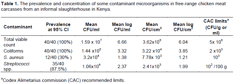
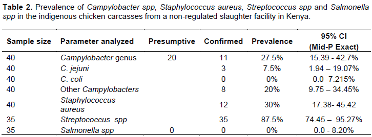
The prevalence and concentration of the contaminant bacteria, with the concentration compared to CAC-recommended concentration limits, are summarized in Table 1. Total viable counts and coliforms were recorded from all 40 carcasses. Coagulase positive Staphylococcus was isolated from 12 (30%, 95% CI: 17.38-45.42) carcasses and Streptococcus colonies from 35 (87.5%, 95% CI: 74.45-95.27%). Streptococcus Group D (Enterococcus) was isolated from 17 of the 35 samples (48.57%). Two (2) samples had mixed contamination with Groups D and F and Groups D and G, respectively. Group B was isolated from 1 sample (2.85%), Group F from 5 (14.28%), Group G from 6 (17.14), while 4 samples (11.42%) could not be classified. The concentration of all the contaminants was higher than the CAC recommended limits. Suspect Campylobacter colonies were detected in 20 (50%) of the 40 samples. DNA analyses confirmed Campylobacter genus in 11 samples (27.5%, 95% CI: 15.39-42.7); C. jejuni in three (7.5%) and C. coli in none. Primers for Campylobacter genus (C412F and C1228R) and primers for C. jejuni (ENg03F and ENg04R) generated amplicons of 812 and 773 bp, respectively. Salmonella genus was not isolated from any of the carcasses (Table 2).
DISCUSSION
CAC has set acceptable concentration limits for microorganisms in foods in order to ensure quality and safeguard human health (CAC, 1997). We observed that chicken carcasses from unregulated slaughter facilities in Nairobi have high microbial load, which include potential pathogens. The mean concentration of TVC, coliforms, coagulase positive S. aureus and Streptococcus spp. were higher than the acceptable limits. This was an indication of low hygiene standards of the slaughter facility and/or the slaughter process.
Total Viable Counts (TVC), also referred to as Aerobic Plate Count (APC) and total coliforms in a food sample informs on microbiological quality of the production process. Presence of these organisms in large numbers in raw poultry indicates unsanitary slaughter and processing practices (Svobodová et al., 2012; Vaidya et al., 2005). The presence of coliforms in the poultry meat is an indication of fecal contamination, exogenous or endogenous, and presents a public health risk of food borne diseases (Ruban and Fairoze, 2011; Temelli et al., 2011). Among the coliforms, E. coli is one of the most important cause of foodborne diarrhorea disease (WHO 2015). Specific pathogens S. aureas, Streptococcus spp. and Campylobacter spp. were identified in the meat carcass. Apart from directly causing foodborne disease, the meat may be a source of cross contamination to other carcasses and foodstuffs during processing, at retail or household level (Sirsat et al., 2014; Kennedy et al., 2011; Rasschaert et al., 2020).
S. aureus related food poisoning as the third largest cause of food related illnesses worldwide resulting from the contamination of food by preformed S. aureus enterotoxins (Kornacki, 2010; Thaker et al., 2013; Akbar and Anal, 2013). The organism is a natural flora in the skins of animals and humans and frequently contaminates raw foods of animal origin (FSANZ, 2005). However, humans are the main reservoir for S. aureus involved in human foodborne disease despite the wide-spread association of S. aureus with animals (Loir et al., 2003). The organism is also a significant cause of poultry disease and may thus contaminate carcasses and consequently other foods (Pepe et al., 2006). In this study, 30% of the chicken carcasses were contaminated with coagulase positive S. aureus and all contaminated carcasses had more than the maximum permitted limit. The high contamination rates raise public health concern since poultry meat has been linked to staphylococcal food poisoning (Loir et al., 2003; Armstong et al., 2002).
The standard provided for Streptococci in foods and packed water is 102 CFU/100 ml. In this investigation, Streptococcus spp. were isolated from 35 out of 40 carcasses (87.5%) with the mean CFU/cm2 and CFU/ml much higher than the maximum permitted limit. In the study, the Streptococci isolates were Lancefield Groups D, B, D, F, and G. Presence of Group D (Enterococcus) in carcasses was indication of feacal contamination, but may also originate from the urinary tract of humans (Poulsen et al., 2012). Molecular evidence of enterococci spread from animals to humans has been documented (Donabedian et al., 2003). Group G are part of the normal microbiota of the gastrointestinal tract and skin in animals especially cattle and potentially zoonotic. They may cause a variety of infections in humans including bacteraemia (Liao et al., 2008). Groups F and B are inhabitants of the upper respiratory mucosa and genital and gastrointestinal tracts, respectively in humans (Al-Charrakh et al., 2011; Hanna and Noor, 2020) and their presence in the carcasses may be an indication of contamination from slaughter personnel.
Prevalence of Campylobacter genus in the carcasses was 27.5% with C. jejuni contributing 7.5%, the balance being contributed by non-identified species. The public health risk is significant considering that Campylobacter spp. infections cause human gastro-enteritis more frequently than other enteric pathogens (WHO, 2015). Poultry meat accounts for approximately half of all food borne campylobacteriosis in humans (Hoffmann et al., 2017).
Negative results of Salmonella genus isolation from the meat carcasses were obtained in this study. In spite of contradictory results elsewhere (Zhao et al., 2016; Bailey and Cosby, 2005), this study alleviates fears of the chicken produced under free range production systems being common sources of human non-typhoidal infections. Indigenous free-range chicken have resistance potential to Salmonella (Msoffe et al., 2006). Further, Salmonella prevalence in poultry is age related (Beal et al., 2004) and the long duration to maturity may provide an opportunity for infection clearance ahead of slaughter.
CONCLUSION AND RECOMMENDATION
Chicken meat carcasses processed in an informal slaughterhouse were found contaminated by spoilage and pathogenic microorganism beyond the Codex Alimentarius limits. The high microbial contaminant load, which included potential human pathogens, may have been a consequence of contamination from the birds, slaughter facility or the slaughter personnel. The study recommends a need to sensitize consumers on proper handling of meat carcasses to avoid cross-contamination of other foodstuffs as well as adequate cooking. Currently, there are no regulated slaughterhouses for free-range chicken in the country. Establishment of regulated facilities accompanied by capacity building of slaughter personnel is also critically important in order to safeguard consumer health and to enable farmer’s access to high value markets.
CONFLICT OF INTERESTS
The authors have not declared any conflict of interests.
REFERENCES
|
Akbar A, Anal AK (2013) Prevalence and Antibiogram study of Salmonella and Staphylococcus aureus in poultry meat. Asian Pacific Journal of Tropical Biomedicine 3:163-168. |
|
|
Al-Charrakh AH, Al-Khafaji JKT, Al-Rubaye RHS (2011). Prevalence of β-hemolytic groups C and F streptococci in patients with acute pharyngitis. North American Journal of Medical Sciences 3:129-136. |
|
|
Antunes P, Mourão J, Campos J, .Peixe L (2016) Salmonellosis: the role of poultry meat. Clinical Microbiology and Infection 22:110-121. |
|
|
Armstong P, Peacock D, Cameron S (2002). Gastroenteritis outbreak in a sporting team linked to a barbecued chicken. Communicable Diseases Intelligence Quarterly Report 26:446-448. PMID: 12416711. |
|
|
Bailey JS, Cosby DE (2005).Salmonella prevalence in free-range and certified organic chickens. Journal of Food Protection 68:2451-2453. |
|
|
Beal RK, Wigley P, Powers C, Hulme SD, Barrow PA, Smith AL (2004). Age at primary infection with Salmonella entericaserovartyphimurium in the chicken influences persistence of infection and subsequent immunity to re-challenge. Veterinary Immunology and Immunopathology 100:151-64. |
|
|
Brichta-Harhay DM, Arthur TM, Koohmaraie M (2008). Enumeration of Salmonella from poultry Carcass rinses via direct plating methods. Letters in Applied Microbiology 46:186-91. |
|
|
Carron M, Chang Y-M, Momanyi K, Akoko J, Kiiru J, Bettridge J (2018). Campylobacter, a zoonotic pathogen of global importance: Prevalence and risk factors in the fast-evolving chicken meat system of Nairobi, Kenya. PLOS Neglected Tropical Diseases 12(8):e0006658. |
|
|
Codex Alimentarius Commission (CAC/GL 21) (1997) Principles and Guidelines for the establishment and application of Microbiological Criteria related for Foods. www.fao.org › input › download › standards. Last updated 2017. |
|
|
Donabedian SM, ThalLA, Hershberger E,Perri MB, Chow JW, Bartlett P (2003). Molecular characterization of gentamicin-resistant enterococci in the United States: evidence of spread from animals to humans through food. Journal of Clinical Microbiology 41:1109-1113. |
|
|
European Union, EU (2017). Summary report on trends and sources of zoonoses, zoonotic agents and food-borne outbreaks in 2016. European Food Safety Authority Journal 15:4634. |
|
|
Food Standards Australia New Zealand (FSANZ) (2005). Scientific assessment of the public health and safety of poultry meat in Australia. |
|
|
Hanna M, Noor A (2020). Streptococcus Group B [Updated 2020 Mar 24]. In: StatPearls [Internet]. Treasure Island (FL): StatPearls Publishing. |
|
|
Hoffmann S, Devleesschauwer B, Aspinall W, Cooke R, Corrigan T, Havelaar A, Angulo F , Gibb H, Kirk, M., Lake, R., Speybroeck N, Torgerson P, Hald T (2017) . Attribution of global foodborne disease to specific foods: Findings from a World Health Organization structured expert elicitation. PLoS ONE 12(9):e0183641. |
|
|
Ipara BO, Otieno DO, Nyikal RA, Makokha SN (2019). The role of unregulated chicken marketing practices on the frequency of Newcastle disease outbreaks in Kenya. Poultry Science 98:6356-6366. |
|
|
Kennedy J, Nolan A, Gibney S , O'Brien S, Mcmahon A, McKenzie, K Healy B, Mcdowell D, Fanning S,Wall PG (2011). Deteminants of cross-contamination during home food preparation. British Food Journal 113:280-297. |
|
|
King'ori AM, Wachira AM, Tuitoek JK (2010). Indigenous Chicken Production in Kenya: A Review. International Journal of Poultry Science 9:309-316. |
|
|
Kornacki, JL (ed.) (2010). Principles of microbiological troubleshooting in the industrial food processing environment. Food Microbiology and food safety. Springer Science+Business Media, LLC. |
|
|
Kuria JKN, Ngethe EW, Kabuage LW, GathuraPB (2018).Isolation of Campylobacter spp and Escherichia coli 0157: H7 from free-range indigenous chicken value chain in Kenya. East African Medical Journal 95:1116-1124.eISSN: 0012-835X. |
|
|
Linton D, Lawson AJ, Owen R J, Stanley J (1997). PCR Detection, Identification to species level, and fingerprinting of Campylobacter jejuni and Campylobacter coli direct from Diarrheic samples. Journal of Clinical Microbiology 35:2568-2572. |
|
|
Loir YL, Baron F, Gautier M (2003).Staphylococcus aureus and food poisoning. Genetic and Molecular Research 2:63-76. PMID: 12917803. |
|
|
Liao C-H, Liu L-C, Huang Y-T, Teng L-J, Hsueh P-R (2008). Bacteremia caused by Group G Streptococci in Taiwan. Emerging Infectious Diseases 14:837-840. |
|
|
Mkhungo MC, Oyedeji AB, Ijabadeniyi OA (2018). Food safety knowledge and microbiological hygiene of households in selected areas of Kwa-Zulu Natal, South Africa. Italian Journal of Food Safety 7:6887. |
|
|
Msoffe PLM, Minga, UM, Mtambo, MMA, Gwakisa, PS, Olsen JE (2006).Differences in resistance to Salmonella Enterica serovargallinarum infection among indigenous local chicken ecotypes in Tanzania. Avian Pathology 354:270-276. |
|
|
National Advisory Committee on Microbiology Criteria for Foods (NACMCF) (2007). Analytical utility of Campylobacter methodologies. Journal of Food Protection 70:241-250. |
|
|
Pepe O, Blaiotta G, Bucci F, Anastasio M, Aponte M, Villani F (2006) Staphylococcus aureus and staphylococcal enterotoxin A in Breaded Chicken Products: Detection and Behavior during the Cooking Process. Applied and Environmental Microbiology 72:7057-7062. |
|
|
Poulsen L, Bisgaard M, Son N, Trung N, An H, Dalsgaard A (2012). Enterococcus faecalis Clones in Poultry and in Humans with Urinary Tract Infections, Vietnam. Emerging Infectious Diseases 12:1096-1100. |
|
|
Rouger A, Tresse O, Zagorec M (2017). Bacterial contaminants of poultry meat: sources species, and dynamics. Microorganisms 5:50. |
|
|
Rasschaert G, De Zutter L, Herman L, Heyndrickx M (2020). Campylobacter contamination of broilers: the role of transport and slaughterhouse. International Journal of Food Microbiology. 322:108564. |
|
|
Ruban SW, Fairoze N (2011). Effects of processing conditions on microbiological quality of market poultry meats in Bangalore, India. Journal of Animal and Veterinary Advances 10:188-191. |
|
|
Shahdan IA, Regenstein JM, Rahman MT(2017) . Critical limits for the control points for halal poultry slaughter. Poultry Science 96:1970-1981. |
|
|
Sirsat SA, Kim K, Gibson KE, Crandall PG, Ricke SC, Neal JA (2014). Tracking microbial contamination in retail environments using fluorescent powder - A retail delicatessen environment example. Journal of Visualized Experiments 85:51402. |
|
|
Svobodová I, BoÅ™ilová G, Hulánková R, SteinhauserováI (2012). Microbiological quality of broiler carcasses during slaughter processing. Acta Veterinaria Brno 81:37-42. |
|
|
Temelli S, Kurtulus M, Sen C, Anar S (2011).Microbiological evaluation of chicken Kadinbudu meatball production stages in a poultry meat processing plant. Veterinary Journal of Ankara University 58:189-194. |
|
|
Thaker HC, Brahmbhatt MN, Nayak JB (2013). Isolation and identification of Staphylococcus aureusfrom milk and milk products and their drug resistance patterns in Anand, Gujarat. Veterinary World 6:10-13. |
|
|
Vaidya VM, Paturkar AM, Waskar VS, Zende RJ, Rawool DB (2005). Detection of indicator organisms on poultry carcass sites in an organized slaughterhouse. Journal of Muscle Foods 16:289-297. |
|
|
World Animal Health Organization, OIE (2008).Working group on animal production food safety: Animal production food safety challenges in global markets. Scientific and Technical Review of the Office International des Epizooties (Paris) 25:479-492. |
|
|
World Health Organization (WHO) (2009). Global Salm-Surv- A Salmonella surveillance and laboratory support project of the World Health Organization.Level 4 Training Course: Multiplex PCR for differentiation of C. coli and C. jejuni. Laboratory Protocols 2nd Ed. 2009. |
|
|
World Health Organization (WHO, 2015). World Health Organization estimates of the global burden of foodborne diseases: foodborne disease burden epidemiology reference group 2007-2015.ISBN 978 92 4 156516 5. |
|
|
World Health Organization (WHO, 2018). Salmonella (non-typhoidal. WHO factsheet |
|
|
Zhao X, Gao Y, Ye C, Yang L, Wang T, Weishan W (2016). Prevalence and characteristics of Salmonella isolated from free-range chickens in Shandong Province, china. BioMedical Research Internationalarticle ID 8183931. |
|
Copyright © 2024 Author(s) retain the copyright of this article.
This article is published under the terms of the Creative Commons Attribution License 4.0


