Journal of
Veterinary Medicine and Animal Health

  • Abbreviation: J. Vet. Med. Anim. Health
  • Language: English
  • ISSN: 2141-2529
  • DOI: 10.5897/JVMAH
  • Start Year: 2009
  • Published Articles: 416

Review

Risk factors, prevention and control strategies for surgical site infections in veterinary practice in Nigeria - A review

Oghenemega David Eyarefe
  • Oghenemega David Eyarefe
  • Department of Veterinary Surgery and Reproduction, Faculty of Veterinary Medicine, University of Ibadan, Nigeria.
  • Google Scholar


  •  Received: 04 November 2015
  •  Accepted: 23 May 2016
  •  Published: 31 August 2016

 ABSTRACT

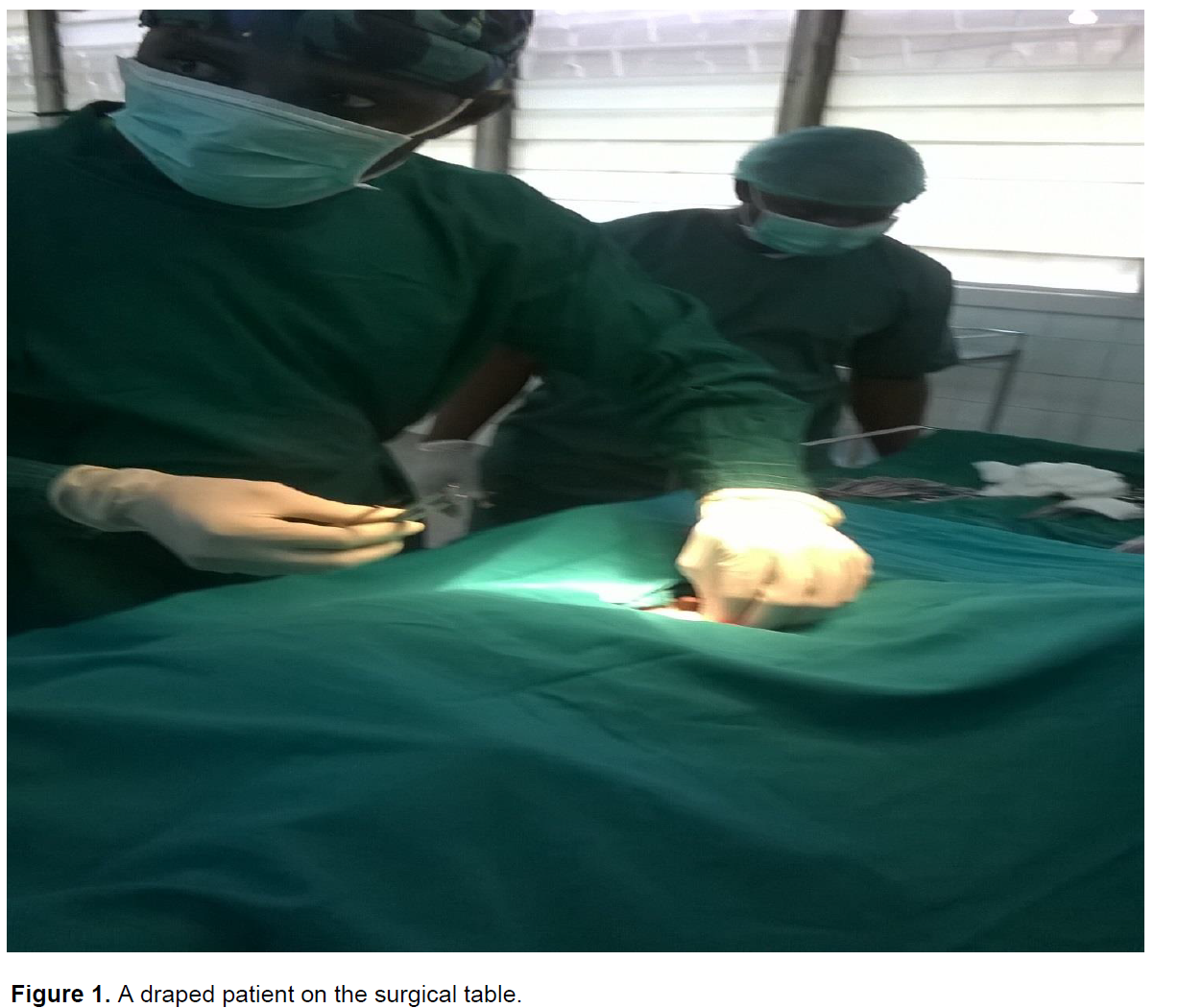
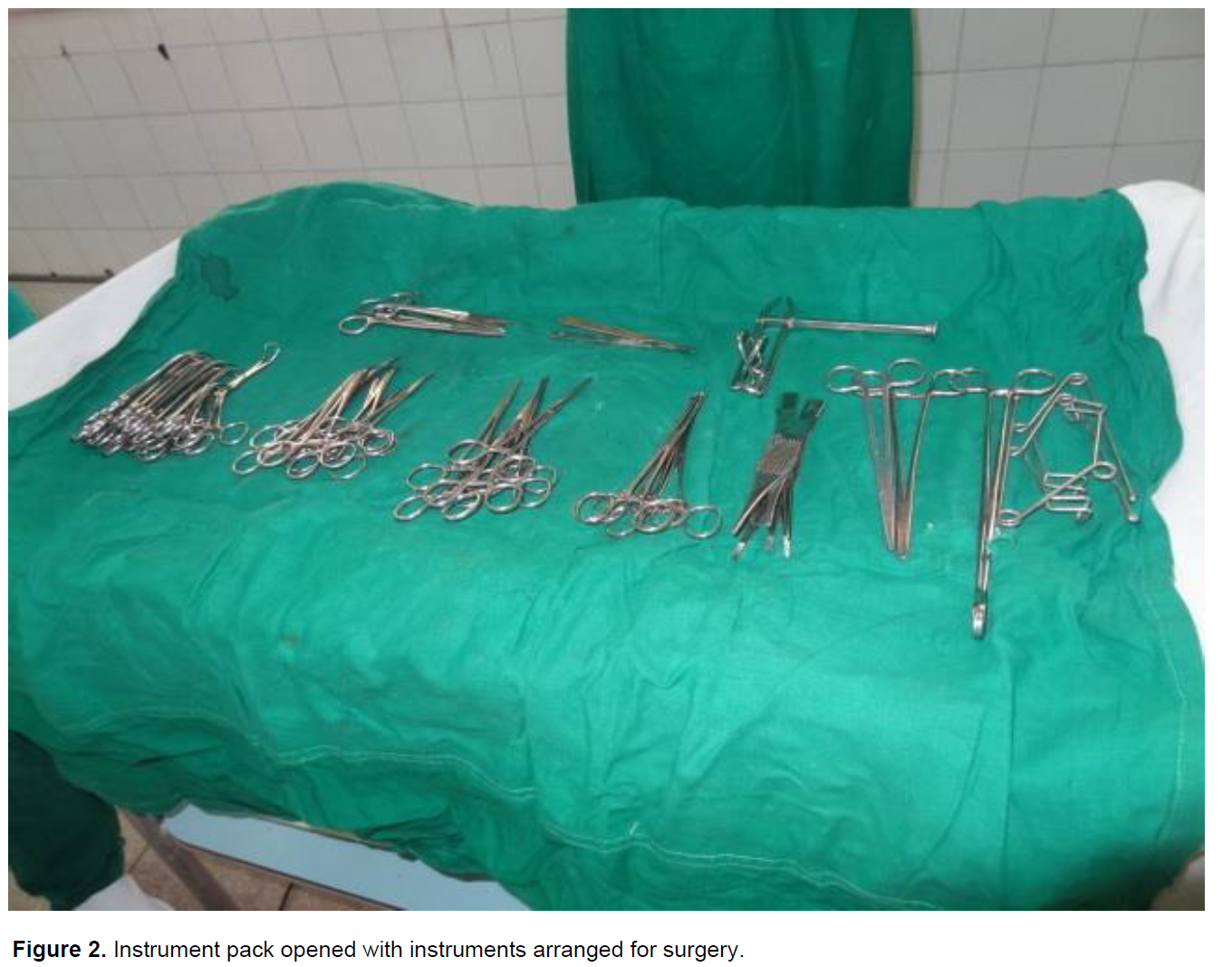
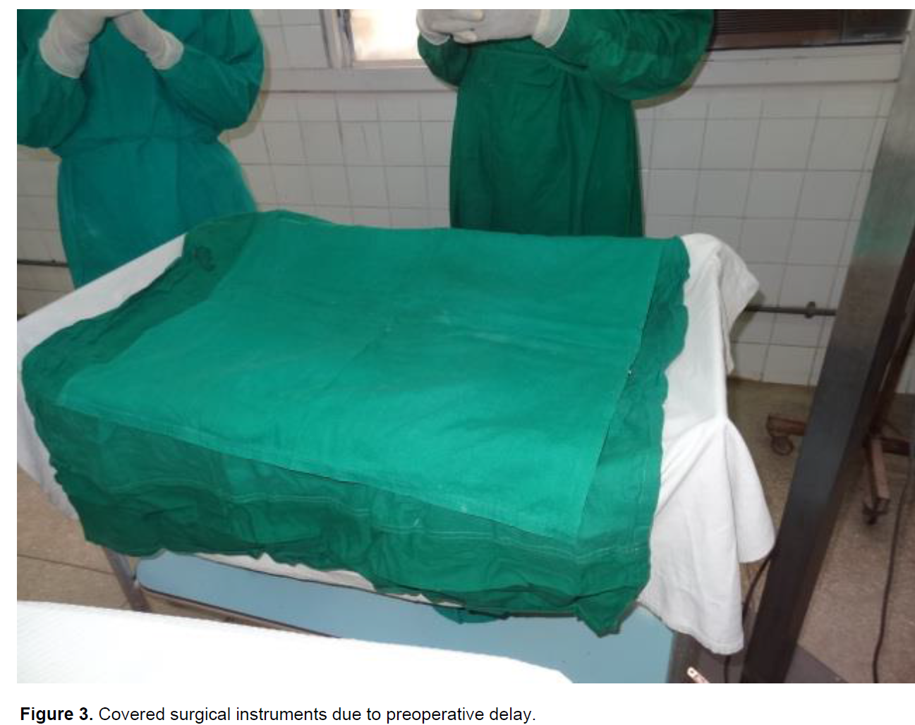
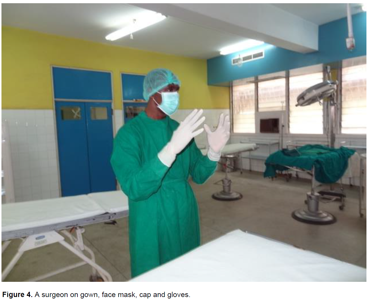
Surgical site infections (SSIs) are surgery associated nosocomial infections with multifactorial etiologies. They are adverse events that have placed heavy burden on surgery universally and have bedeviled veterinary surgery practice in Nigeria for decades, with consequent severe morbidity, mortality, financial and psychological burden on animal owners. In this paper, information on current universal trend of SSIs, including risk factors, prevention and control strategies was reviewed with emphasis on principles and practice among small animals and equine surgery practitioners. Principles guiding surgical suite design, surgical team, instruments/equipment, and patient preparation, were emphasized. It was concluded that imbibing the principles and practice of SSIs prevention strategies in Nigeria veterinary hospitals and clinics would impact positively on the veterinary health care system and the society the Veterinarian is committed to serve.

Key words: Surgical site infections, patient, surgery, veterinary.


 INTRODUCTION

Surgical site infections (SSIs) are surgery associated nosocomial infections that account for about 38% of nosocomial infections among human patients (Beldi et al., 2009), 0.8 to 18% among small animal surgery patients (Verwilghen and Singh, 2015; McMillan, 2014; Birgand et al., 2014; Weese and Halling, 2006; Eugster et al., 2004; Vasseur et al., 1985) and 0 to 50% among equine surgery patients; depending on surgical procedure and wound classification (Ahern and  Richardson, 2012; Verwilghen, 2015). The multifactorial etiology of SSIs has been associated with poor  surgery  theatre  environment, operating techniques, surgery team attitude, as well as, poor instruments, surgical team, and patient preparation (Verwilghen and Singh, 2015; McMillan, 2014; Humes and Lobo, 2009; Cheadle, 2006). The effects of SSIs is enormous on patients’ welfare and animal care-givers/owners, and include: poor surgical site cosmesis, revision of surgery (de Lissovoy et al., 2009), prolonged wound healing (Verwilghen, 2015), risk of drug side effects (AORN, 2010), increased hospital stay (Jin-joeng, 2013,), increased psychological and financial burden on dog owners (Verwilghen and Singh, 2015), emergence of multi-drug resistant (MDR) pathogenic organisms and patients’ death (Weese et al., 2007). Although SSIs cases are prevalent in veterinary facilities (Turk et al., 2015; Nazarali et al., 2014; Mayhew et al., 2012; Eugster et al., 2004; Beal et al., 2000; Whittem et al., 1999), and the need to prevent re-occurrence emphasized, the lack of SSIs surveillance programs in veterinary healthcare centres, as obtained currently in human surgery practice (Astagneau et al., 2009; Anderson et al., 2014) has led to late detection and sometimes, non-capturing of SSIs cases in many veterinary hospital record systems (Turk et al., 2015). This has made intensive control and eradication of SSIs difficult due to lack of empirical data to justify the need for worry. SSIs is prevalent in Nigeria veterinary surgery practice due to unstandardized environment for surgical procedures, poor theatre manners, poor patient and instrument preparation among others (Tsai and Caterson, 2014). Attempt at prevention had, in most cases led to antimicrobial abuse (Akinrinmade et al., 2012), and possible evolution of resistant microbial strains.
 
 
A recent growing concern for veterinary clinical practice proficiency improvement has led to the establishment of the Postgraduate College of Veterinary Surgeons, Nigeria (CVSN), as well as, the development of Veterinary Teaching Hospital (VTH) facilities across the nation. These landmark trends in the veterinary profession in Nigeria; ultimately geared toward disease prevention and control, require information complementation on current global trend in SSIs prevention and control measures. The dearth of literature on this subject in Nigerian professional and institutional journals further heightens the need for this review. This paper therefore comprehensively and systematically presents: risk factors in SSIs, as well as, time proven prevention and control measures that could be adopted to minimize or possibly eradicate SSIs occurrence in public and private surgical facilities nationwide.


 DISCUSSION

SSIs remain the third most common hospital acquired infection with costly implications for surgery in human and veterinary practice (Verwilghen, 2015; Bigand et al., 2014; Anderson et al., 2014). The problem is legendary and dates back to the beginning of practice of the surgery specialty (Milard, 2012; Clark, 1907), when the fear and risk of SSIs prevented quick surgical intervention until the patient was brought near death (Verwighen, 2015). Earlier infection control measures were implemented following Drs. Ignaz Semmelweis and Oliver Wendell Homes’ observations that contaminated hands of attending physicians served as vehicle for the spread of infections (Humes and Lobo, 2009; Adriaanse, 2000). The introduction of compulsory hand scrubbing with chlorinated lime solution before physical examination by attending physicians resulted in an impressive reduction in mortality rate (from 11.4 to 1.3% within two years) in the Vienna maternity ward (Sabbatani et al., 2014; Adriaanse, 2000), and propelled the commencement of compulsory antiseptics hand washing regimen as a means of infection control among surgeons (McMillan, 2014; Humes and Lobo, 2009). This practice became globally accepted following the publication of the Louis Pasteur germ theory in 1860, on the role of germs in infection causation, and the statement “instead of forcing ourselves to kill the microbes in wounds, would it not be more reasonable not to introduce them” (Ahern and Richardson, 2012; Verwighen et al., 2011). Infection control practice further became entrenched among communities of surgeons with Joseph Lister’s publications on anti-septic surgery concept and thesis on aseptic principles for surgeons (Hermani, 2009). The discovery of antibiotics further enhanced the curbing of SSIs. However, the current trend in microbial multi-drug resistance to antimicrobials calls for the need to identify SSIs risk factors, and strengthen the prevention and control strategies.
 
Risk factors in SSIs causation
 
Endogenous and exogenous sources of wound site bacteria contamination, and patient health status at surgery are major risk factors in the causation of SSIs among human and veterinary patient (Turk et al., 2015; Hermani, 2009). Patients’ commensal flora at the surgical site, including skin surface and body tracts (gastrointestinal and respiratory) are sources of endogenous wound site contamination and infection, while  bacteria contaminants from surgical team, the environment, surgical materials, instruments and wound dressings are exogenous sources (McMillan, 2014; Cogen et al., 2008). Canine endogenous pathogens associated with SSI have been identified and include: Staphylococcus pseudintermedius, Staphylococcus aureus Coagulase-negative staphylococci (CONS), Pseudomonas species, Enterococci and extended spectrum β-lactamase (ESBL) producing Enterobacteriaceae (E. coli, Enterobacter and Klebsiella spp) (Weese and Duijkeren, 2010). Staphylococci are the most commonly cultured bacteria from SSIs with S. pseudintermedius being the leading cause of SSIs in dogs, and S. aureus in horses (Weese et al., 2010). S. pseudintermedius is also the most isolated Staphylococci spp from small animal healthcare workers as against S. aureus in human healthcare workers (Thorup, 2014). Besides, there is an increasing concern about the multidrug resistant potentials of S. pseudintermedius which is potentially greater than those produced by methicillin resistant Staphylococcus aureus (MRSA) (Weese et al., 2010; Thorup, 2014). The bacterium also has a strong biofilm forming ability that further complicate treatment in implant associated SSIs (Thorup, 2014).
 
Zoonosis caused by S. pseudintermedius, though lowerthan that caused by S. aureus has also been reported (Weese et al., 2010). A thorough surgical patient clinical screening prior to surgery has been recommended (Centre for Disease Control (CDC), 1999). Patient health status is a major risk in SSIs causation because risk of SSI correlates directly with dose and virulence of microbial contamination and patient’s immune resistance (Owens and Stroessel, 2008). Obese patients and those with endocrinopathies such as hypothyroidism, diabetes mellitus, hyperadrenocortism, smoking, diabetes, nutritional status and consumption of certain drugs are SSIs risk in human practice (Mangram et al., 1999).
 
In animal patients, hypothermia, hypotension, surgical wound classifications and implants (Turk et al., 2015) increased body weight and endocrinopathy in intact animals (Fitzpatrick and Solano, 2010; Nicholson et al., 2002) are risk factors in SSIs causation. Other risk factors are; hair clipped at surgical site > 4 h (Mayhew et al., 2012), increased anaesthesia time (Nazarali et al., 2014), duration of surgery (Eugster et al., 2004; Nicholson et al., 2002; Vasseur et al., 1985), longer tissue manipulation, wound exposure time, noise in the theatre (Kurmann et al., 2011) and non-administration of antibacterial prophylaxis in clean contaminated and contaminated wounds (Whitten et al., 1999).
 
In equine practice, uncontrolled use of antimicrobial prophylaxis is associated with burden of multi-resistant bacteria (Damborg et al., 2012) and antimicrobial-induced colitis, especially in horses undergoing elective arthroscopic surgery (Weese and Cruz, 2009). Surgeon’s training and experience is also a risk factor as complications associated with closure of equine celiotomy incision (Wormstrand et al., 2014) and survival after colic surgery has been linked with years of experience and training of the surgeon (Wormstrand et al., 2014).  
 
Strategies for prevention and control of SSIs
 
Pre-surgical hand preparation and hand hygiene
 
Hand hygiene is a key component in prevention of SSIs (Nelson 2011; WHO, 2009). The hands of surgical staff have higher pathogenic microbial load than those of others due to their increased contact with infected wounds (Verwilghen et al., 2011; Coelho et al., 1984). Transient skin microbes acquired by contact with persons, animals and environment are known for inducing SSIs (Verwilghen et al., 2011). The isolation of zoonotic, biofilm producing, multi-drug and methicillin resistant  S. pseudintermidius from hands of small animal health care workers (Thorup, 2014), further calls for strict pre-surgical hand wash and hygiene protocol. Pre-surgical hand antisepsis is aimed at eliminating or reducing the skin microbial flora to diminish the risk of SSIs (McMillan,   2014).  
 
The practice of the correct method of pre-surgical hand preparation has been reportedly low among human and veterinary surgeons (Verwilghen et al., 2013). The common tradition is the use of antiseptic solution (chlorhexidine or povidone base soap) to scrub hand and arm with 20 to 25 scrubbing brush strokes, and hand/arm rinse over elbow or pedal controlled tap (Tanner et al., 2008). This traditional scrubbing method has been faulted due to much time involved, its inability to adequately remove resident bacteria from hand, arm and beneath finger nails, and for its compromise of the protective water-lipid layer of the superficial skin (Widmer et al., 2010; Kampf and Kramer, 2004), thus increasing the chances of pathogenic bacteria skin colonization due to impaired skin immunity (Larson et al., 1998). Besides, contact dermatitis due to skin reaction to scrubbing solutions has been reported in some individuals (Larson et al., 2006; Krautheim et al., 2004). Emphasis is currently rapidly shifting from the traditional hand scrub to hand wash and alcohol gel rubs among human surgeons (Verwilghen et al., 2013; Kampf and Kramer, 2004).  
 
Alcohol gel hand rub has the advantages of being easier and faster to use, better efficacy against hand resident microbes compared to disinfecting soap solutions and cause less skin damage with repeated use (Loffler and Kampf  2008; Kampf et al., 2003). For these reasons, it has been recommended by the World Health Organization for pre surgical antiseptic purposes (WHO, 2009). However, low compliance attitude has been observed among veterinary surgeons in shifting from the traditional hand scrub with brush and antiseptic soap solution to adjusting to alcohol hand rub despite observed advantages and WHO recommendations (Verwilghen et al., 2013; Verwilghen et al., 2011). Despite the popularity of alcohol gel hand rubs among human surgeons, cases of non-compliance with usage directive have been observed (Umit et al., 2014).
 
Surgical theatre construction and environmental hygiene practice
 
Operating theatre location and construction influence the potential for the risk of SSIs (Gastmeier et al., 2012; Hambraeus, 1988). Although veterinary literature has less information on theatre construction as correlate of SSIs, information in human literature (Sapna and Pradeep 2011) could be extrapolated and applied. It was recommended that the operating theatre be located in a blind wing, or at bottom floor, or topmost floor of hospital facility to control traffic to the area and reduce contamination (Sapna and Pradeep 2011; Lynch et al., 2009; Lidwell, 1982). A clear demarcation of the theatre into zones (the outer, restricted, and aseptic) has also been recommended to minimize contamination (Sapna et al., 2011). The use of wall and floor tiles made of polished stone or  marble  to  ease  cleaning  and  disinfection  has also been recommended (Jeong et al., 2013; Sapna and Pradeep, 2011). Also recommended are installation of a laminar air flow system for filtration and expelling of contaminated air (Tsai and Caterson, 2014; Jeong et al., 2013; Lidwell, 1982).
 
A functional surgical theatre requires constant cleaning and disinfection. All equipment within the theatre such as surgical lamps requires constant daily cleaning and instantly when fluid splashes on them during surgeries. Installation of anti-microbial copper alloys has been recommended for their bactericidal actions on touch surfaces of drip stands and chairs arms. There is also the need to treat water to reduce chances of hand contamination by water borne pathogenic microbes in surgical theatres (Tsai and Caterson, 2014; McMillan, 2014; Sapna and Pradeep 2011). Increased human traffic into and out of the operating room has been linked with increase in SSIs rate (Radcliff et al, 2013; Pokrywka and Byers, 2013; Panahi et al., 2012; Lynch et al., 2009). Rate of infection increases with the number of people in the theatre (Panahi et al., 2012; Eugster et al., 2004); as a result, restrictions should be placed on the type and number of people that can be allowed into the surgical theatre.
 
Surgical team manners
 
Surgical team members’ attitude grossly influences SSIs (Beldi et al., 2009; Lucet et al., 2012). Low attitude of team members, such as, non-compliance with hand disinfection procedures, gloving, hair and nose covering, maintaining of 50 cm distance from the surgical table by non-sterile team members, as well as, indiscriminate opening of the theatre doors and increased noise (talking) are risk factors for SSIs causation that require constant caution for prevention (Verwilghen, 2015: Kumman, 2011; Lucet et al., 2012). The presence of one or more visitors during surgical procedure constitutes SSIs risk and should be avoided (Birgand et al., 2014; Makary, 2013; Beldi et al., 2009; Boer, 2001).
 
Surgical site preparation for surgery
 
The patient’s skin microbial florae and endogenous pathogens of mucous membranes and hollow organs are risk factors in theatre wound infection (Hermani, 2009). Clipping of hair coat, scrubbing, application of antiseptic solutions, and draping prior to surgery are conventional practice to forestall SSIs (Tanner et al., 2008). In dogs removal of hair coat with clipper rather than razor blade after anaesthetic induction and immediately before surgical procedure is recommended (CDC, 1999), as coat clipping before induction is marked with incidence of SSIs (Tanner et al., 2008). The hair coat of veterinary patients harbor myriads of microbial organisms and cause contamination of the surgical  field  that  eventually leads to surgical site infection (Nelson, 2011; Cooper et al., 2000). The efficiency of hair clipping has been linked with timing of hair coat removal. Clipping of hair coat > than 4 h before surgery is fraught with high risk of SSIs and should be avoided as it gives bacteria enough time to be established on clipped skin before commencement of surgery (Mayhew et al., 2012). Surgical patients’ hair coat removal with razor blades cause more skin abrasions and compromises skin immunity compared to clippers (Mangram, 1999; Anderson et al., 2014).  
 
Skin preparation with appropriate antiseptic solution has been recommended as a preventive measure for SSIs (Nelson, 2011). Commonly used antiseptic solutions are either of alcohol or aqueous base and often contain chlorhexidine gluconate or iodophores (Loscovish, 2014; Dumville et al., 2013). A combination of 70% alcohol and 4% chlorhexidine has a faster onset, longer duration of action and broad spectrum antibacterial activities and preferred for patients’ skin preparation (Loscovish, 2014; Dumville et al., 2013; Hemani, 2009). Concentric scrubbing fashion, beginning from the proposed incision site outward has been challenged in favour of ‘back and forth motion’ with the theory  that the surgical site is inadequately sterilized with the concentric method (MacDonald et al., 2001). Draping of the surgical site is done to isolate the sterile surgical zone during surgery (Figure 1). Although draping techniques are seemingly easy, poor draping of patient increases the risk of surgical site infection (Showalter et al., 2014; Cooper, 2000). Also, blood soaked areas on drapes are potential sites for microbial multiplication and contamination of the sterile field as blood serves as good medium for bacterial growth. Disposable drapes are currently preferred to washable and re-useable fabrics as washable fabrics, over time, do not retain the water proof characteristics of an ideal draping material, and make them support infection (Showalter et al., 2014; Hopper and Moss, 2010).
 
 
Instrument preparation
 
Surgical instruments are vital to surgical procedures and their poor preparation has been associated with theatre wound infections (Hopper and Moss, 2010). Metal instruments are conventionally sterilized with moist heat using autoclave at 121°C for 13 min (Sapna, 2011). Recommended cold sterilization involves soaking of instruments in antimicrobial solutions (chlorhexidine, iodophores, and isopropyl or ethyl glycols) not lesser than 3 h prior to surgery (Cooper et al., 2000). Cold sterilization is not however advisable for instruments intended for invasive surgical procedures (Cooper et al., 2000). Gas sterilization with ethylene oxide and with gamma irradiation are also recommended where applicable (Sapna, 2011). Delay in opening of the surgical pack till time of operation is important, as pre-operative  delay  for  an extended length of time results in commencement of surgery could help prevent instrument contamination (Figures 2 and 3) (Dalstrom et al., 2008; Radcliff et al., 2013).


Pre-operative delay
 
Delay in commencement of surgery increases the chances of sterile field contamination through airborne contamination of opened surgical instruments (Hopper and Moss, 2010). Common causes of pre-operative delay include: an extended time in gaining intravenous access and anaesthestic ready time (Radcliff et al., 2013). To minimize preoperative delays, a proactive approach to anaesthesia termed “independent anaesthetic induction” has been advocated (Radclif et al., 2013). It emphasizes the need for two anaesthetic teams; one to begin induction of anaesthesia for new patients and the other to maintain anaesthesia in the current patient. This approach reduces the preoperative time and chances of surgery site infection (Radclif et al., 2013).
 
Surgical guise or theater wears
 
Surgical guise comprises conventional theatre wears (scrubs, sterile gown, a face mask, the sterile gloves, head cover, and shoe covers or boots) designed to reduce contaminants and surgical wound infections (Figure 4) (Salassa and Swiontkowski, 2014). The surgical scrub consists of the short sleeves shirt and the pant or trousers (Amirfeyz, 2007). The scrubs should not be long sleeved, and should not be worn with additional clothing like cardigans or inanimate objects like stethoscopes. It should not be worn outside the hospital or clinic facility and should not be laundered at home with other fabrics (Braswell-spruce, 2012). The human nares are colonized by the normal body flora that can become a source of infection to surgical wounds demanding the use of face mask (Salassa and Swiontkowski, 2014). Head covers help to control the spread, through aerosol, of normal hair microbes into the surgical field. The same principle holds for covering of side beards with facial mask (Fossum, 2015). Although no clear study currently shows the correlation between the use of the leg covers or booties in surgical wound infection rates, it is however inferred that street shoes could serve as sources of contamination to the theatre air and contribute to theatre wound infection (McHugh et al., 2014; Amirfeyz, 2007). Disposable sterile surgical gowns are preferred to reusable ones as their moisture proof nature prevents seep through body fluid that could contaminate wounds. A single use also prevents the discomfort associated with constant washing of the fabrics, which eventually results in wear and tear. A study has shown that the rate of theatre wound infections reduces with  disposable  gowns compared with re-usable (Moylan, 1987). Modern surgical practice therefore advocates a single use of disposable surgical gowns, face masks, head covers and surgical gloves (McHugh et al., 2014).
 
 
Breaks in surgical asepsis
 
A break in asepsis is any event that occurs that alters or compromises the aseptic attribute of the surgeons, surgical instruments or surgical field (AORN, 2010). Common breaks in surgical asepsis could include: a tear on the surgeons’ gown sterile package, torn gloves during surgery, dropped face masks; an error in an attempt at putting on the surgical guise and scrubbing by the surgeon. It also includes faulty instruments and surgical field sterilization process, errors in the patients’ positioning,  prepping and draping, as well as, dropping a contaminated instrument on a sterile instruments table, and when a sneeze or cough occurs across a sterile field (Sapna, 2011). Surgical glove puncture and contamination of sterile field is common in veterinary and human surgery (Nelson, 2011). Surgical gloves become punctured in 35% of cases after two hours of surgery, but only 20% of glove punctures are noted by the surgeon (Nelson, 2011). Double gloving has been encouraged in orthopaedic procedures where glove punctures often occurs through instruments, on the first finger, with grave SSIs consequences in implant associated procedures (McMillian, 2014). It is recommended that glove be changed after an hour of surgery, and open gloving may be done where assisted intraoperative gloving is not feasible (Duxbury et al., 2003).
 
Four classes of breaks in surgical asepsis have been identified and preventive measures suggested: Class 1: breaks which are spotted as soon as they occur and contained immediately. Class 2: breaks that are noticed a short while afterthey occurred and can still be  managed; Class 3: breaks which are almost impossible to contain because they were identified to have occurred far into the operative procedure and Class 4:  breaks that are never identified (AORN, 2010). It is important that all non-scrubbed personnel keep a distance of 12 inches from scrubbed personnel (Hopper and Moss, 2010). It is essential that a skilled perioperative nurse who has been well trained to detect lapses in aseptic techniques always be in the operating room. It is important to document common breaks in aseptic techniques that are seen to occur in surgery on a daily basis, so that control measures could be taken to prevent a re-occurrence.
 
Prolonged surgical time, techniques and tissue trauma
 
Increased stay of patient in operating room more than one hour prior to surgery increases the chances of theatre  wound  infection (Radcliff et al., 2013). Prolonged instruments contamination prior to surgery. In event of delay, covering of instruments with a sterile towel until surgical time is a major contributor to theatre wound infections (Radcliff et al, 2013; Jeong et al., 2013; Tsai and Caterson, 2014). The explanation given is that traumatized internal organs are exposed in the open for long and tissue perfusion delayed (Radcliff et al, 2013; Jeong et al., 2013; Tsai and Caterson, 2014; Cooper, 2000). A skillful surgeon is aware of the importance of adequate tissue oxygenation to the outcome of the wound healing. Supplementing oxygen intake with a ventilator is recommended in addition to maintaining the optimum body temperature (Anderson et al., 2014).
 
Temperature regulation: Maintaining normothermia is important to the outcome of theatre wounds as studies have shown a positive correlation between adverse body temperature and surgical wound infection (Hopper et al., 2009; Cheadle, 2006). Hypothermia hinders peripheral circulation and minimizes oxygen perfusion  in the wound area. Besides, an optimal temperature is needed by the body to trigger body immune responses to infection (Tsai and Caterson, 2014). Hypothermia reduces neutrophil functions and increase blood loss which would trigger  the need for blood support, a high risk factor for theatre wound infections (Anderson et al., 2014). Hypothermia could be managed by using warm fluid for intra-operative tissue flushing, warm water blanket, heating lamp and heating pads (Abelha et al., 2005). Well aerated environment using cool mists and fans, cold intravenous fluids, cooling blankets, as well as, oxygen administration are common means of managing hyperthermia (Sessler, 2009).
 
Drain placement: Postsurgical drains placement has been described as a high risk factor for theatre wound infection especially in veterinary patients (Tsai and Caterson, 2014; Jin-joeng, 2013). Drains and catheters are foreign to the body and their placement compromise the  skin immune status  in areas along which they pass through and make them prone to infection (Nakamura et al., 2012). Animal patients are more likely to distort the placed drains and catheters and may require restraint collars to prevent their removal and owners cooperation for monitoring (Tsai and Caterson, 2014).
 
Antibiotic use and mis-use: Antimicrobial prophylaxis (AMP) is indicated in human and veterinary surgical procedures depending on surgical wound classification (clean contaminated or contaminated), those with higher infection rates (implanting of prosthetic devices) and where infection could cause grave consequences (Akinrinmade, 2012) without compromising appropriate aseptic protocols (Eugster et al., 2004). The need for empirical laboratory evidence of microbial type and sensitivity could influence the choice of a narrower spectrum of antimicrobial agent to enhance preservation of patients’ normal microbial flora and reduce chances of antimicrobials resistance (Anderson et al., 2014; Carlet et al., 2014). The timing of AMP administration is important to ensure peak serum threshold at the time of incision to complement body immune system against infection. The CDC guidelines for human surgery patients (Bratzler and Houck, 2004) requires administration of selected AMP within 60 min of surgical incision, repeated every 2 half-lives to maintain therapeutic concentration, and discontinuation within 24 h post-surgery (Bratzler et al., 2005; Bratzler and Houck, 2004). This protocol has been confirmed effective in reducing SSIs in human surgery patients (Bratzler et al., 2005) and adopted in veterinary medicine (Verwilghen and Singh, 2015) with some modification and prolongation of antibiotics beyond 24 h in canine tibia plateau leveling osteotomy (TPLO) procedures (Nazarali et al., 2014; Frizpatrick and Solano, 2010). The choice of antibiotics AMP depend on the anticipated microbial challenge (Verwilghen and Singh, 2015). Methicillin-resistant Staphylococcus aureus (MRSA) is a major culprit in human and animal patients wound infection (Harper et al., 2013) except in canine species where S. pseudintermidius is a leading cause of SSIs (Weese et al., 2010). These organisms have been isolated commonly from the hair coat, nasal mucosa and palms of patients and hospital staff (Thorup, 2014) demanding the need for screening of patients and staff, and decontaminating of carriers with appropriate antibiotics prior to commencement of surgery (Tammelin et al, 2001).
 
Staff training and re-orientation: The need to train, re-train and engage all surgeons and peri-operative staff on various aseptic guidelines and practices in the hospital and surgical environment has been emphasized in the reduction of incidence of SSIs (Anderson et al., 2014; NICE, 2008). A routine training exercise will provide the forum to acquaint newly employed staff with theatre aseptic practices, refresh the memory of old staff and allowing for sustainability (Loscovich, 2014). Learning aids like brochures, videos  and  mock  exercises to test staff skills have also been recommended (Puntis et al., 1990). Feed-back measure to evaluate the impact of educational programs could help in commendation of staff for areas of success and also in spotting areas of lapses that can be improved upon (Loscovish, 2014).


 CONCLUSION

There is a universal effort at preventing or minimizing theatre wound infections. Efforts are made from design of the surgical suite to establishing principles, guidelines and work ethics within surgical facilities to prevent the occurrence of wound infection, enhance restoration to health, and reduce hospital stay and cost. Imbibing and practicing these established guidelines in veterinary hospitals and clinics in Nigeria should move veterinary patient health care efforts positively towards impacting on the society and the primary patient for which the veterinarian swore an oath of absolute commitment to total care.


 CONFLICT OF INTEREST

The author has not declared any conflict of interest. 


 ACKNOWLEDGEMENT

Drs. Oguntoye CO, Ajani OS and Afolabi JM helped proof-read and edit the manuscript. Their assistance is greatly appreciated. 


 REFERENCES

Abelha FJ, Castro MA, Neves AM, Landeiro NM, Santos CC (2005). Hypothermia in a surgical intensive care unit. BMC Anesthesiol. 5:7
Crossref


Adriaanse AH, Pel M, Bleker OP (2000). Semmelweis: the combat against puerperal fever. Eur. J. Obstet. Gynecol. Reprod. Biol. 90:153-158
Crossref


Ahern BJ, Richardson DW (2012). Surgical site infection and the use of antimicrobials. In: Equine Surgery, 4th edn. Eds: JA Auer JA Stick, Elsevier/Saunders, St Louis, Missouri. pp. 68-84.
Crossref


Akinrinmade JF, Oke BO (2012). Antibiotic prophylaxis in Gastro-intestinal surgery: An evaluation of current Veterinary Practices in Southwest Nigeria. Int. J. Anim. Vet. Adv. 4(4):256-262.


Amirfeyz R, Tasker A, Ali S, Bowker K, Blom A (2007). Theatre Shoes — A Link in the Common Pathway of Postoperative Wound Infection? Ann R Coll. Surg. Engl. 89(6):605-608.
Crossref


Anderson DJ, Podgorny K, Berríos-Torres SI, Bratzler DW, Dellinger EP, Greene L, Nyquist AC, Saiman L, Yokoe DS, Maragakis LL, Kaye KS (2014). Strategies to prevent surgical site infections in acute care hospitals: update. Infection Control Hospital Epidemiol. 35(06):605-627.
Crossref


Anderson ME, Sargeant JM, Weese JS (2014). Video observation of hand hygiene practices during routine companion animal appointments and the effect of a poster intervention on hand hygiene compliance. BMC Vet. Res. 10:106.
Crossref


AORN (2010). Recommended practices for hand hygiene in the perioperative setting. In: Perioperative standards and recommended practices. Denver: AORN Inc. pp. 75-89.


Astagneau P, L'Hériteau F, Daniel F, Parneix P, Venier AG, Malavaud S, Jarno P, Lejeune B, Savey A, Metzger MH, Bernet C (2009). Reducing surgical site infection incidence through a network: results from the French ISO-RAISIN surveillance system. J. Hosp. Infect. 72(2):127-34.
Crossref


Beal MW, Brown DC, Sholer FS (2000). The effects of perioperative hvpothermia and the duration of anesthesia on postoperative wound infection rate in clean wounds: a retrospective study. Vet. Surg. 29(2):123-127
Crossref


Beldi G, Bisch-Knaden S, Banz V, Mühlemann K, Candinas D (2009). Impact of intraoperative behavior on surgical site infections. Am. J. Surg. 198:157-162.
Crossref


Birgand G, Azevedo C, Toupet G, Pissard-Gibollet R, Grandbastien B, Fleury E, Lucet, JC (2014). Attitudes, risk of infection and behaviours in the operating room (the ARIBO Project): a prospective, crosssectional study. BMJ Open 4:e004274.
Crossref


Boer A, Gilad J, Meydan N, Riesenberg K, Schlaeffer F, Alkan M, Schlaeffer P (2001). Impact of active monitoring of infection control practices on deep sternal infection after open-heart surgery. Ann. Thorac. Surg. 72:515-20.
Crossref


Braswell ML, Spruce L (2012). Implementing AORN Recommended Practices for Surgical Attire, AORN J. (95)1:130.
Crossref


Bratzler DW, Houck PM (2004). Surgical Infection Prevention Guidelines Writers. Antimicrobial prophylaxis for surgery: an advisory statement from the National Surgical Infection Prevention Project. Clin. Infect Dis. 38(12):1706-15.
Crossref


Bratzler DW, Houck PM, Workgroup SIPGW (2004). Antimicrobial prophylaxis for surgery: an advisory statement from the National Surgical Infection Prevention Project. Clin. Infect Dis. 38(12):1706-15.
Crossref


Bratzler DW, Houck PM, Richards C, Steele L, Dellinger EP, Fry DE, Wright C, Ma A, Carr K, Red L (2005). Use of antimicrobial prophylaxis for major surgery: baseline results from the National Surgical Infection Prevention Project. Arch. Surg. 140(2):174-182.
Crossref


Carlet J, Rambaud C, Pulcini C (2014). Save Antibiotics: a call for action of the World Alliance against Antibiotic Resistance (WAAAR). BMC infect dis 14(1):436.
Crossref


Cheadle WG (2006). Risk factors for surgical site infection Surg. Infect. (Larchmt). 7(Suppl 1):S7-11 (Center of Disease Control and Prevention) CfDCaP Surgical site infection (event). 111CDC, editor. Atlanta (GA) CDC: 2014.


Cogen AL, Nizet Gallo RL (2008). Skin microbiota: a source of disease or defence? Br. J. Dermatol. 158:442-455
Crossref


Clark FC (1907). A brief history of antiseptic surgery. Med. Libr. Hist. J. 5:145-172.


Coelho JC, Lerner H, Murad I (1984). The influence of the surgical scrub on hand bacterial flora. Int. Surg. 69:305-307.


Dalstrom DJ, Venkatarayappa I, Manternach AL, Palcic MS, Heyse BA, Prayson MJ (2008). Time dependent contamination of opened sterile operating-room trays. J. Bone Joint Surg. Am. 90:1022-1025.
Crossref


Damborg P, Marskar P, Baptiste KE, Guardabassi L (2012). Faecal shedding of CTX-M-producing Escherichia coli in horses receiving broad spectrum antimicrobial prophylaxis after hospital admission. Vet. Microbiol. 154:298-304.
Crossref


Eugster S, Schawalder P, Gaschen F (2004). A prospective study of postoperative surgical site infections in dogs and cats. Vet. Surg. 33:542-550.
Crossref


Gastmeier P, Breier AC, Brandt C (2012). Influence of laminar airflow on prosthetic joint infections: a systematic review. J. Hosp. Infect. 81:73-78.
Crossref


Hambraeus A (1988). Aerobiology in the operating room–a review. J. Hosp. Infect. 11(Suppl A):68-76.
Crossref


Humes D, Lobo DM (2009). Antisepsis, asepsis and skin preparation. Surgery 27(10):441-445.
Crossref


Hemani ML, Lepor H (2009). Skin preparation for the prevention of surgical site infection: which agent is best? Rev Urol. 11(4):190-5.


Hopper WR, Moss R (2010) Common Breaks in Sterile Technique: Clinical Perspectives and Perioperative Implications. AORN J. 91(3):350-366.
Crossref


Jeong SJ, Ann HW, Kim JK, Choi H, Kim CO, Han SH, Choi JY, Peck KR, Kang CI, Yeom JS, Choi YH (2013). Incidence and Risk Factors for Surgical Site Infection after Gastric Surgery: A Multicenter Prospective Cohort Study. Infect Chemother 45(4):422-443.
Crossref


Kurmann A, Peter M, Tschan F, Muhlemann K, Candinas D, Beldi G (2011). Adverse effect of noise in the operating theatre on surgical-site infection. Br. J. Surg. 98:1021-1025.
Crossref


Krautheim AB, Jermann TH, Bircher AJ (2004). Chlorhexidine anaphylaxis: case report and review of the literature. Contact Dermatitis 50:113-116
Crossref


Kampf G, Kapella M (2003). Suitability of Sterillium Gel for surgical hand disinfection. J. Hosp. Infect 54:222-225
Crossref


Kampf G, Kramer A (2004). Epidemiologic background of hand hygiene and evaluation of the most important agents for scrubs and rubs. Clin. Microbiol. Rev. 17:863-893
Crossref


Larson EL, Hughes CA, Pyrek JD, Sparks SM, Cagatay EU, Bartkus JM (1998). Changes in bacterial flora associated with skin damage on hands of health care personnel. Am. J. Infect Control 26:513-521.
Crossref


Larson E, Girard R, Pessoa-Silva CL, Boyce J, Donaldson L, Pittet D (2006). Skin reactions related to hand hygiene and selection off hand hygiene products. Am. J. Infect Control 34:627-635.
Crossref


Lidwell OM, Lowbury EJ, Whyte W, Blowers R, Stanley SJ, Lowe D (1982). Effect of ultraclean air in operating rooms on deep sepsis in the joint after total hip or knee replacement: a randomised study. BMJ 285:10-14.
Crossref


Lissovoy G, Fraeman K, Hutchins V, Murphy D, Song D, Vaughn BB (2009). Surgical site infection: incidence and impact on hospital utilization and treatment costs. Am. J. infect. control 37(5):387-397.
Crossref


Loffler H, Kampf G (2008). Hand disinfection: how irritant are alcohols? J. Hosp. Infect 70(Suppl 1):44-48
Crossref


Loscovich A, Davidson EM, Orbach-Zinger S, Rudich Z, Ivry S, Rosen LJ, Avidan A, Ginosar Y (2014). Performance of septic technique during neuraxial analgesia for labour and after the publication of international guidelines on aseptic technique. Isr. J. Health Pol. Res. 3(1):9.
Crossref


Lucet JC, Laouenan C, Chelius G, Veziris N., Lepelletier D, Friggeri A, Abiteboul D, Bouvet E, Mentre F, Fleury E (2012). Electronic sensors for assessing interactions between healthcare workers and patients under airborne precautions. PLoS One 7(5):e37893.
Crossref


Lynch RJ, Englesbe MJ, Sturm L, Bitar A, Budhiraj K, Kolla S, Polyachenko Y, Duck MG, Campbell DA (2009). Measurement of foot traffic in the operating room: implications for infection control. Am. J. Med. Qual. 24(1):45-52.
Crossref


Makary MA (2013). The power of video recording: taking quality to the next level. JAMA 309:1591-1592.
Crossref


Mangram AJ, Horan TC, Pearson ML, Silver LC, Jarvis WR, Hospital Infection Control Practices Advisory Committee (1999). Guideline for prevention of surgical site infection 1999. Hospital Infection Control Practices Advisory Committee. Infect. Control Hosp. Epidemiol. 20:250-278.
Crossref


Mayhew PD, Freeman L, Kwan T, Brown DC (2012). Comparison of surgical site infection rates in clean and clean-contaminated wounds in dogs and cats after minimally invasive versus open surgery: 179 cases (2007–2008). J. Am. Vet. Med. Assoc. 240(2):193-8.
Crossref


McHugh SM, Corrigan MA, Hill AD, Humphreys H (2014). Surgical attire, practices and their perception in the prevention of surgical site infection. Surgeon 12(1):47-52.
Crossref


McMillan S (2014). An evidence-based approach to infection control in the operating theatre. Vet. Nurse 5(4):194-200.
Crossref


Millard C (2012). Destiny of the Republic: A Tale of Madness, Medicine and the Murder of a President. Knopf Doubleday Publishing Group.


Moylan JA, Fitzpatrick KT, Davenport KE (1987). Reducing Wound Infections: Improved Gown and Drape Barrier Performance. Arch. Surg. 122(2):152-157.
Crossref


National Institute for Health and Clinical Excellence (2008). Surgical Site Infection, Prevention and treatment of surgical site infection. NICE Clin. Guide. 74:6-28.


Nakamura RK, Tompkins E (2012). Nosocomial infections. Compendium (Yardley, PA), 34(April), E1–10; quiz E11. doi:10.1383/medc.33.3.22.61114
Crossref


Nazarali A, Singh A, Weese JS (2014). Perioperative administration of antimicrobials during tibial plateau leveling osteotomy. Vet. Surg. 43(8):966-971.
Crossref


Nicholson M, Beal M. Sholer F, Brown DC (2002). Epidemiologic evaluation of postoperative wound infection in clean-contaminated wounds, a retrospective study of 239 dogs and cats. Vet. Surg. 31(6):577-81.
Crossref


Nelson LL (2011). Surgical Site Infections in Small Animal Surgery. Vet. Clin. North Am. Small Anim. Pract. 41(5):1041-1056
Crossref


Owens CD, Stoessel K (2008). Surgical site infections: epidemiology, microbiology and prevention J. Hosp. Infect. 70(Suppl 2):3-10.
Crossref


Panahi P, Stroh M, Casper DS, Parvizi J, Austin MS (2012). Operating room traffic is a major concern during total joint arthroplasty. Clin. Orthop. Relat. Res. 470:2690-2694.
Crossref


Pokrywka M, Byers K (2013). Traffic in the operating room: a review of factors influencing air flow and surgical wound contamination. Infect. Disord. Drug Targets 13(3):156-61.
Crossref


Puntis JW, Holden CE, Smallman S, Finkel Y, George RH, Booth IW (1990). Staff training: a key factor in reducing intravascular catheter sepsis Arch. Dis. Child. 65:335-337.


Radcliff KE, Neusner AD, Millhouse PW, Harrop JD, Kepler CK, Rasouli MR, Albert TJ, Vaccaro AR (2015). What is new in the diagnosis and prevention of spine surgical site infections? Spine J. 15(2):336-347.
Crossref


Radcliff MR, Neusner A, Kepler, CK, Albert TJ, Rihn JA, Hilibrand AS, Vaccaro AR (2013). Preoperative Delay of More Than 1 Hour Increases the Risk of Surgical Site Infection. Spine 38(15):1318-1323.
Crossref


Sabbatani S, Catena F, Neri F, Vallicelli C, Ansaloni L, Sartelli M, Coccolini F, Di Saverio S, Catena R, Lazzareschi D, Tarasconi A, Abongwa HK, De Simone B, Pinna A (2014). TheBolognese surgeon Giuseppe Ruggi: how and why the aseptic surgery was introduced in Bologna in the middle half of the XIX century. J. Surg. Res. 192:555-563.
Crossref


Salassa TE, Swiontkowski MF (2014). Surgical attire and the operating room: role in infection prevention. J. Bone Joint Surg. Am. 96(17):1485-1492
Crossref


Sapna SM, Pradeep V (2011). The operating theatre: basic architecture. Delhi J. Ophthalmol. 21(3):9-14.


Sessler DI (2009). Thermoregulatory defense mechanisms. Critical care Med. 37(7):S203-S210.
Crossref


Showalter BM, Crantford JC, Russell GB, Marks MW, DeFranzo AJ, Thompson JT, Pestana IA, David LR (2014). The effect of reusable versus disposable draping material on infection rates in implant-based breast reconstruction: a prospective randomized trial. Ann. Plast. Surg. 72(6):S165-169.
Crossref


Tammelin A, Hambraeus A, Stahle E (2001). Routes and sources of Staphylococcus aureus transmitted to the surgical wound during cardiothoracic surgery: possibility of preventing wound contamination by use of special scrub suits. Infect. Control Hosp. Epidemiol. 22(6):338-346.
Crossref


Tammelin A, Hambraeus A, Stahle E (2001). Source and route of methicillin-resistant Staphylococcus epidermidis transmitted to the surgical wound during cardio-thoracic surgery. Possibility of preventing wound contamination by use of special scrub suits. J. Hosp. Infect. 47(4):266-276.
Crossref


Tanner J, Swarbrook S, Stuart J (2008). Surgical hand antisepsis to reduce surgical site infection. Cochrane Database Syst. Rev. (1):CD004288
Crossref


Thorup S (2014). An investigation into bacterial type and load in on veterinary compared to human healthcare workers. Copenhagen (Denmark) Department of Large Animal Sciences, University of Copenhagen.


Tsai DM, Caterson EJ (2014). Current preventive measures for health-care associated surgical site infections: a review. Patient safety surg. 8(1):1.
Crossref


Turk R, Singh A, Weese JS (2015). Prospective surgical site infection surveillance in dogs. Vet. Surg. 44:2-8.


Vasseur PB, Paul HA, Enos LR, Hirsh DC (1985). Infection rate in clean surgical procedures. A comparism of ampicillin prophylaxis vs a placebo. J. Am. Vet. Assoc. 187(8):825-827.


Verwilghen D (2015). Surgical site infections: What do we know? Equine Vet. J. 47:753-755.
Crossref


Verwilghen D, Singh A (2015). Fighting surgical site infections in small animals are we getting anywhere? Vet. Clin. North Am. Small Anim. Pract. 45:243-276.
Crossref


Verwilghen D, Findji S, Weese JS, Singh A, Dupre G, Catry B, Van Galen G (2013). Evidence based hand hygiene in veterinary surgery: what is holding us back? In: Annual Symposium of the American College of Veterinary Surgeons, ACVS, San Antonio, Texas.


Verwilghen D, Grulke S, Kampf G (2011). Presurgical Hand Antisepsis: Concepts and Current Habits of Veterinary Surgeons. Veterinary Surgery 40:515-521.
Crossref


Weese JS, Cruz A (2009). Retrospective study of perioperative antimicrobial use practices in horses undergoing elective arthroscopic surgery at a veterinary teaching hospital. Can. Vet. J. 50:185-188.


Weese JS, van Duijkeren E (2010). Methicillin-resistant Staphylococcus aureus and Staphylococcus pseudintermedius in veterinary medicine. Vet. Microbiol. 140(3-4):418-429
Crossref


Weese JS, Halling KB (2006). Perioperative administration of antimicrobrals associated with elective surgery for cranial eructate ligament rupture in dogs: 83 cases (2003-2005). J. Am. Vet. Med. Assoc. 229(1):92-95.
Crossref


Whittern TL, Johnson AL, Smith CW, Schaeffer DJ, Coolman BR, Averill SM, Cooper TK, Merkin GR (1999). Effect of perioperative prophylactic antimicrobial treatment in dogs undergoing elective orthopedic surgery. J. Am. Vet. Med. Assoc. 215(2):212-216.


Widmer AF, Rotter M, Voss A, Nthumba P, Allegranzi B, Boyce J, Pittet D (2010). Surgical hand preparation: state-of-the-art. J. Hosp. Infect 74(2):112-22.
Crossref


Wormstrand BH, Ihler CF, Diesen R, Krontveit RI (2014). Surgical treatment of equine colic-a retrospective study of 297 surgeriesin Norway 2005–2011. Acta Vet. Scand. 56:38.
Crossref


World Health Organization (WHO) (2009): WHO Guidelines on Hand Hygiene in Health Care. Geneva, Switzerland, WHO. 270 p.


          */?>