ABSTRACT
In order to determine the prevalence of caprine paratuberculosis and the possible risk factors associated a cross-sectional epidemiological study (February to June, 2009) was carried out in dairy goat intensive production units in Guanajuto, Mexico. Blood (n=821) and fecal (n=240) were sampled from goats (older than one year of age) were randomly selected from thirteen dairy goat intensive production units. Serum samples were analyzed using immunodiffusion serological test (AGIT) and fecal samples were used for confirmatory diagnosis by IS 900 Nested PCR. Analysis of results was carried out with STATA 7® software. Paratuberculosis prevalence in the studied population was 9.87% (range 1 to 32%), females had 10.25% and male 6.24%; for body condition were 14.02% emaciated, 7.94% thin and 8.7% good. By Nested PCR analysis detected 64 (26.67%) fecal samples as positive. Risk factor analysis indicated that alpine breed had a OR=2.1 (95%, CI 0.76 to 7.65), females in theirs second parturition showed OR= 2.94 (95%,CI 1.04 to 8.3), from three to fifth parturition had OR= 5.88 (95%, CI 4.3 to14.7) and with more than six parturitions had an OR=7 (95%, CI 2.79 to 18.21)., Animals that presented pasty to liquid feces had OR=2.10 (95%, CI 0.84 to5.25). The results in this study suggest that there is an increased risk for paratuberculosis as the number of parturitions increases and is also related to the consistency and handling of feces in the flocks.
Key words: Paratuberculosis, caprine, epidemiology, nested, polymerase chain reaction (PCR), immunodiffusion, intensive production.
Paratuberculosis is a chronic infectious disease caused by the facultative intracellular bacteria Mycobacterium avium subspecies paratuberculosis (Map) that affects the gastrointestinal tract causing granulomatous enteritis of a large variety of animals, among which are domestic ruminants such as bovines, sheep and goats (Kennedy
and Benedictus, 2001; Amoldi et al., 1983; Ramírez et al., 1983).
The main elimination pathway of Map is through feces which contaminate water and feed. Animals are infected in the first weeks of life through water, colostrum or milk that has been contaminated with feces of the animals infected with paratuberculosis. The animals that are most susceptible to be infected are newborns all the way up to 6 months of age (Gilardoni et al., 2012; Martínez et al., 2012). Feces of goats and sheep infected with paratuberculosis lose their normal consistency and become pasty with intermittent diarrhea accompanied with progressive loss of body condition and death (Gumber et al., 2006). It is thought that stressing conditions such as nutritional deficiencies, concurrent diseases, crowding, parturition and high production pressure contribute to the start of clinical signs (Merkal et al., 1975). Unlike cattle, background on ovine and caprine paratuberculosis prevalence data are scarce and much of case is known, It assumes that is similar to cattle, paratuberculosis has a worldwide distribution, the prevalence in some countries is high as 45%, as is the case for the United States of America (Pithua and Kollias, 2012), similar rates are reported in Canada (Bauman et al., 2016, Sorensen et al., 2003). In cattle, it is endemic in The Netherlands, Austria, and Belgium, where the prevalence is 54, 7 and 41%, respectively (Singh et al., 2008). In Australia, infection rates fluctuate between 9 and 22% respectively. In Argentina reports prevalence rates is 18.8% in dairy farms and 6.8% in beef farms (Gilardoni et al., 2012). In Rio de Janeiro, Brazil, the prevalence is 33%. In Venezuela, 72% of the herds is infected (Sánchez-Villalobos et al., 2009), in Chile report prevalence of 9.3% (Kruze et al., 2007; Pinto et al., 2002).
Paratuberculosis in Mexico is widely distributed and studies indicate that the prevalence rates between 5 and 30% mainly in cattle, goats, sheep and fighting bulls (Córdova et al., 2010). The main economic losses incurred by infection with paratuberculosis are due to lower milk production, premature culling and/or seizure of carcasses at slaughterhouse (Harris and Barletta; 2011).
Diagnosis of paratuberculosis can be done based on clinical and epidemiological aspects, through the study of lesions present, direct identification of Map in clinical samples and indirectly by measuring the immune response of the animal against the infection. Serological diagnosis of paratuberculosis in sheep and goat can be done using agar-gel immunodiffusion test (AGIT) which has a sensitivity between 70 and 80% and 100% specificity (Ferreira et al., 2003). Confirmatory diagnosis of paratuberculosis is done by bacteriological culture although the limiting factor is that the incubation period for a Map culture is between 8 and 16 weeks. A further diagnosis option is to use a PCR test (polymerase chain reaction), with primers designed to detect the IS900 insertion sequence that is specific to Map, using ADN from bacterial cultures, tissue samples from granulomatous lesions (mesenteric lymph nodes and small intestine), feces and milk, which has a sensitivity and specificity above 90% and the result is obtained in one to two weeks (Erume et al., 2011; Garrido et al., 2000). Paratuberculosis causes important economic losses to livestock production and as such it is important to determine the possible pathways by which production units and herds become infected and develop the clinical phase of the disease.
The purpose of this study was to determine the prevalence and possible risk factors associated to paratuberculosis in dairy goat intensive production units.
Selection of sample size and study population
A cross-sectional epidemiological study was carried out on the dairy goat population from thirteen intensive dairy goat production units located in the State of Guanajuato, Mexico. The study was carried out between February and June 2009. Sample size was determined using a livestock census of 3,500 heads which was the total present in the livestock production units (LPU), goat farmers that were invited to participate; they signed an informed consent letter. In order to determine the sample size, the proportions formula (Levy and Lemeshow, 1980) was used with an estimated prevalence of 0.03 and a 0.1 estimated error; 10% was added to the calculated “n” to account for losses in field and/or in the laboratory. Animals older than one year were randomly selected from each production unit taking into account the existing animals present at the time of visit following the sample size calculation formula (Cannon and Roe, 1982), which provides the probability of detecting at least one sick animal considering the expected prevalence. Information was collected using questionnaires for each LPU and individual charts for each animal selected.
Epidemiological information
All information collected was blind-coded in order to ensure the confidentiality of the participants. Questionnaires of each LPU included location, type of handling, feeding variables and time as goat farmer. The individual charts consisted of age, gender, physiological status, breed, body condition, and type of animal, place of origin, treatment of diarrhea and physical characteristics of the feces sample. All information, together with the corresponding diagnostic test results, was transferred into two electronic databases (one for the LPU data and one with the chart data) for their epidemiological analysis using the STATA 7® software, descriptive analysis and odds ratios using logistic regression models was calculated.
Blood and fecal samples
Blood samples were collected from the jugular vein in sterile tubes in order to obtain sera, and fecal samples were collected directly from the rectum using a polyethylene bag. Samples were individually tagged for their latter analysis in the laboratory.
Serological diagnosis
Sera samples were evaluated by duplicate throughout the agar-gel immunodiffusion test (AGIT) which was done following the method described in Hernandez (2007). The antigen used in the test was a protoplasmic antigen of the 3065 strain of Mycobacterium avium subspecies paratuberculosis.
Nested polymerase chain reaction test (Nested PCR)
For the nested PCR diagnostic test, selected samples that were positive to the AGIT test and also randomly negative samples from each LPU were selected, up to a total of 240 samples. DNA was extracted following the methods described in Jaimes et al. (2008) and Garrido et al. (2000).
Nested PCR was carried out following the methods described in Erume et al. (2001), using the following primers for IS 900. The first PCR: ptb1 (5’ TGA TCT GGA CAA TGA CGG TTA CGG A 3’) and ptb4 (5’CGC GGC ACG GCT CTT GTT 3’). The amplification product was to 563 base pairs (bp) and used for the second reaction using the primers: ptb2 (5’ GCC GCG CTG CTG GAG TTA A 3’) and ptb3 (5’ AGC GTC TTT GGC GTC GGT CTT G 3’), amplification product was 210 bp. Amplification products were visualized in 2% agarose gels stained with ethidium bromide.
Serological diagnosis
Serological diagnosis for caprine paratuberculosis was done by the agar-gel immunodiffusion tests (AGIT) to determine the presence of antibodies against Map in this study in which 81 of 821 of evaluated animals were AGIT positive.
IS900 Map nested PCR analysis
A 210 bp amplification product of the IS900 Map was observed in 64 (26.67%) of 240 samples evaluated and at least one animal of each farm was positive nested PCR test.
Descriptive analysis
Goat farmers had between 4 and 38 years of being involved in goat production with a mean of 20 years. Of the LPUs, 69.2% (9 of 13) had animals born within the same LPU, while the remaining 30.7% (4 of 13) had animals that were purchased from outside the LPU.
All of the LPUs had an animal health plan that encompassed cleaning of pens, feces handling. Regarding cleaning of pens and feces handling, each farm had their own activities schedule with 45.15% (6 of 13) doing them daily, 7.69% (1 of 13) every month and 15.38% (2 of 13) every six months. Each LPU had their own feces handling procedures of which 69.23% (9 of 13) incorporated feces into soil as organic fertilizer, 15.28% (2 of 13) donate them to brick manufacturing entities, 7.69% (1 of 13) used them for compost and the remaining LPU did not provide specific information.
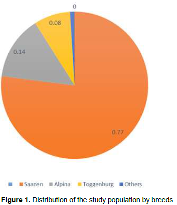
The total sample size was 821 goats composed of 89.16% (732) females and 10.84 % (89) males . The predominant breed was Saanen (77.1%), followed by Alpine (14%) and Toggenburg (8.2%). The remaining goats were grouped into a category that included Lamancha, Nubian and Boer breeds (Figure 1).
Caprine paratuberculosis prevalence
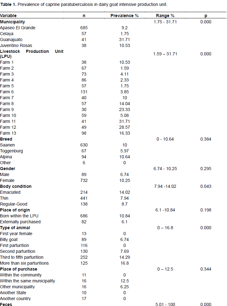
The general prevalence of caprine paratuberculosis was 9.87% (range 1to 32 %). Females had 10.25% and male 6.24%; the prevalence estimated by municipality was for Guanajuato 31.7%, Juventino Rosas 10.53%, Apaseo el grande 9.2% and Celaya 1.75%. For each LPU the prevalence varied, and the rates between 1.59 and 31.71%.
For dairy breads alpine, saanen and togenburg prevalence obtained was 10.64, 10.00 and 5.97%, respectively.
For body condition the prevalence in emaciated animals had 14.2%, 7.94% in thin animals and 8.7% for animals with good body condition. The animal purchased within the same municipality had 12.5%, while the animals purchase in other municipality had 6.25% of the prevalence (Table 1).
Risk factors analysis
In order to identify risk factors associated with prevalence, a logistic regression analysis was performed. Risk factor analysis for breed, gender, place of origin and body condition did not have statistical significance, but alpine breed had an OR=2.1 (95%, CI 0.76 to 7.65). For animal type, females in theirs second parturition showed OR=2.94 (95%, CI 1.04 to 8.3) from three to fifth parturition had OR=5.88 (95%, CI 4.3 to 14.7) and with more than six parturitions had an OR=7 (95%, CI 2.79 to 18.21). For municipality Guanajuato had OR=26 (95%, CI 3.24 to 208.93), Juventino Rosas OR=6.58 (95%, CI 071 to 61.41) and Apaseo el Grande OR=5.67 (95%, CI 0.77 to 41.67). Animals that presented pasty to liquid feces had OR=2.10 (95%, CI 0.84 to 5.25) (Table 2).
The prevalence obtained in this study was 9.87%; results agree with those obtained from studies in caprine production units in several states of the Mexican Republic as Puebla 28% (Gallaga, 2001), Coahuila 18.36% (Toledo et al., 2010) and Guerrero 3.4% (Villalobos, 2011)
indicating that the prevalence of the disease had wide distribution between caprine production units in Mexico.
In Chile 2500 sera samples were evaluated for detection of antibodies anti-Map by ELISA (Pinto et al., 2002), the prevalence showed in the intensive production system was 9.3%, and It could be because of the animals stay for long time in the flocks. In México, it is very common for goat farmers to have for long periods time their animals and the average life span of a goat in this system is about six or more years. Serological diagnosis of paratuberculosis in sheep and goat is done using the AGIT methodology with sensitivities ranging from 70 to 80% and 100% specificity. This methodology is simple and quick for testing whole herds and in this study it was considered an efficient test to determine the presence of antibodies against Map. The main disadvantage of this test is that it detects the presence of the disease once the clinical phase has been initiated (Hernandez, 2007; Gumber et al., 2006; Ferreira et al., 2003).
Kruze et al. (2007) evaluated 383 caprine fecal samples for bacteriological culture and IS900 Map PCR, in which was found 50% of flocks and 9.1% of the animals tested were positive from all flocks with intensive management and presence of imported breeds. This study focused on intensive production systems and indeed all thirteen production units had at least one animal positive to paratuberculosis diagnosed by Nested PCR. The Nested PCR detected 26.67% of animals as positive confirming that the disease is present in the flocks. The used of the Nested PCR as confirmatory test, allowed the validation of the results obtained by AGIT screening test. The Nested PCR has the advantage of detecting animals that are sheding in feces low quantities of bacilli and are at an early infection stage of the disease which has a low humoral immune response. It is recommended that for integral diagnosis of
paratuberculosis both a serological test and a confirmatory test such as nested PCR are carried out (Erume et al., 2011; Jaimes et al., 2008).
As risk factors, gender, breed and place of origin, there were no statistical differences but with respect to place of origin; the introduction of infected animals with Map, is the main route of spread diseases among flocks. This practice was also common for all infected flocks, since it was found that farmers, directly or indirectly, had imported animals selected for dairy genetic fitness of countries whose paratuberculosis infection status is widely recognized (Kennedy and Benedictus, 2001).
In this study 30.7% of the farmers goat have a common practice to buy or trade animals in the same region, generally the animal`s health status is unknown, so there is a risk of introducing infected goats in the flocks, so the health status should be emphasize in relation to paratuberculosis at the time of goats acquisition. Body condition is considered as an indicator of the presence of the disease, and even though the design of this study did
not allow the determination of causality or the start of the disease due to the temporal structure of the data, the“emaciated” body condition category had higher seroprevalence of the disease; these animals are considered to be in the clinical phase of the disease and therefore are the main infection source for the rest of the flock.
The main dairy goat breeds evaluated in this study were alpina, toggenburg , and saanen, had a high prevelence, possible causes of these may be due to various management measures each farm. Dairy goats are subjected to stress conditions of productive, reproductive rate, and genetic predisposition, makes them more susceptible and predispose the animals infected with Map (Pithua and Kollias, 2012).
In Korea the apparent regional prevalence of caprine paratuberculosis was estimated at 21.1, 18.2 and 38.2% for Northern, central and southern regions respectively (Lee et al., 2006). They conclude that possible risk factor for caprine paratuberculosis were all associated with intensive management. In addition a flock size more than 100 heads and presence of foreign specialized dairy goat breeds are associated with prevalence. Guanajuato in Mexico is one of the states with the largest goat production, and high prevalence observed by municipalities, can be due to the fact that ten years ago, the introduction of specialized goat breeds is increased, without knowing their paratuberculosis health status and the mobilization of animals between farms was carried out without restrictions.
Handling of feces constituted a risk factor with an OR = 2.10, which agrees with Villalobos (2012); that reported that when pen cleaning and feces removal was not carried out regularly this had an OR= 3.6 for the presence of the disease into the flocks. These management practices should be considered as an important part of the measures disease control.
Results in this study showed that the dairy goat production units had a 9.87% prevalence of the disease (considering the AGIT results) and, since paratuberculosis is a flock problem and the detection of a single animal can implicate that up to 25% of the herd could be infected, there could be many more infected animals. These results point to handling practices within and between production units that are obligatory but are nevertheless dependent upon the degree of cooperation and expressed interest for goat farmers, in reducing the risk of infection. Such practices include slaughter of positive animals, and the limitation of animal movement between production units, especially towards the LPUs that were negative to paratuberculosis. Also, it must be considered that the diagnosis of this disease should be done before a recently purchased animal enters the herd, and also a regular monitoring program should be in place.
Paratuberculosis is a chronic infection that affects dairy goat herds that are under intensive production systems and further studies should be carried out in order to determine the epidemiological distribution of the disease and quantify its economic impact on production.
The authors have not declared any conflict of interests.
REFERENCES
|
Amoldi JM, Hurley SS, Lesar S (1983). Johne's disease in Wisconsin cattle-a survey of cull cows. In Proc. Int. Colloq. Paratuberculosis 1:16-21.
|
|
|
|
Bauman CA, Jones-Bitton A, Menzies P, Toft N, Jansen J, Kelton D (2016). Prevalence of paratuberculosis in the dairy goat and dairy sheep industries in Ontario, Canada. Can. Vet. J. 57:169-175.
|
|
|
|
|
Cannon RM, Roe RT (1982). Livestock disease surveys: a field manual for veterinarians. Australian Government Publishing Service. pp. 125-135.
|
|
|
|
|
Córdova LD, Guzmán RCC, Santillán FMA, Favila HLC, Urrutia MJ, Gámez V (2010). La paratuberculosis en la ganadería bovina de San Luis Potosí, México. XXXIV Congreso Nacional de Buiatría. (Trabajo completo). Monterrey N. L. México.1:35-40.
|
|
|
|
|
Erume J, Spergser J, Rosengarten R (2001). Rapid detection of Mycobacterium avium subspecies paratuberculosis from cattle and zoo animals by Nested PCR. Afr. Health Sci. 1:83-89.
|
|
|
|
|
Ferreira F, Fonseca LS, Lilembaum W (2003). Agar gel immunodiffusion test (AGID) evaluation for detection of bovine paratuberculosis in Rio de Janeiro, Brazil. Lett. Appl. Microbiol. 35(3):173-175.
Crossref
|
|
|
|
|
Gallaga MEP (2011). Determinación de la Seroprevalencia y factores de riesgo de la paratuberculosis, en las regiones caprinas de libres y la Mixteca en el Estado de Puebla. (Tesis de Maestría). México DF: Universidad Nacional Autonóma de México.
|
|
|
|
|
Garrido JM, Cortabarria N, Oguiza JA, Aduriz G, Juste RA (2000). Use of PCR method on fecal samples for diagnosis of sheep paratuberculosis. Vet. Microbiol. 77:379-386.
Crossref
|
|
|
|
|
Gilardoni LR, Paolicchi FA,Mundo SI (2012). Bovine paratuberculosis: a review of the advantages and disadvantages of different diagnostic tests. Rev. Arg. Microbiol. 44:201-215.
|
|
|
|
|
Gumber S, Eamens G, Whittington RJ (2006). Evaluation of a pourquier ELISA kit in relation to agar gel immunodiffusion (AGID) test for assessmente of the humoral immune response in sheep and goats with and without Mycobacterium paratuberculosis. Infect. Vet. Microbiol. 115:91-101.
Crossref
|
|
|
|
|
Harris NB, Barletta RG (2001). Mycobacterium avium subspecie paratuberculosis in Veterinary Medicine. Clin. Microbiol. Rev. 14(3):489-512.
Crossref
|
|
|
|
|
Hernández COA (2007). Obtención de un antígeno protoplasmático de Mycobacterium avium subespecie paratuberculosis para el diagnóstico de la enfermedad de Johne en ovinos. (tesis de Licenciatura), Mexico: Universidad Autonóma de México. Cite author name in body of work.
|
|
|
|
|
Jaimes NG, Santillán FMA, Hernández COA, Córdova LD, Guzmán RCC, Arellano RB, Díaz AE, Tenorio GVR, Cuellar OA (2008). Detección de Mycobacterium avium subespecie paratuberculosis, por medio de PCR-anidada a partir de muestras de heces de ovino. Vet. Méx. 39(4):377-386.
|
|
|
|
|
Kennedy D, G Benedictus. 2001. Control of Mycobacterium paratuberculosis in agricultural species. Rev. Sci. Tech. 20(1):151-79.
Crossref
|
|
|
|
|
Kruze J, Salgado M, Collin MT (2007). Paratuberculosis en reba-os caprinos chilenos. Arch. Med. Vet. 39(2):147-152.
Crossref
|
|
|
|
|
Lee KW,Jung BY, Moon OK, Yang DK, Lee SH, Kim JY, Kweon CH (2006). Seroprevalence of Mycobacterium avium subespecie paratuberculosis in Korean Goat Black (Capra hircus aegragus). J. Vet. Med. Sci. 68(12):1379-1381.
Crossref
|
|
|
|
|
Levy PS, Lemeshow S (1980). Sampling for Health Professionals Lifetime Learning Publications. Belmont, California.
|
|
|
|
|
Martínez CAG, Santillán FMA, Guzmán RCC, Favila HLC, Córdova LD, Díaz AE, Hernández AL, Blanco OM (2012). Desarrollo de un Inmuno Ensayo-Enzimático (ELISA), para el diagnóstico de paratuberculosis en bovinos. Rev. Mex. Cienc. Pecu. 3(1):1-18.
|
|
|
|
|
Merkal RS, Larsen AB, Booth GD (1975). Analysis of the effects of in apparent bovine paratuberculosis. Am. J. Vet. Res. 36:837-842.
|
|
|
|
|
Pinto J, Maier L, Ríos C, Martínez R (2002). Prevalencia y factores de riesgo de paratuberculosis en sistemas productivos intensivos caprinos de la zona central de Chile. Resúmenes del XII Congreso Nacional de Medicina Veterinaria, Chillán, Chile.
|
|
|
|
|
Pithua P, Kollias NS (2012). Estimated Prevalence of Caprine Paratuberculosis in Boer Goat Herds in Missouri, USA. Vet. Med. Int.10:1-5.
Crossref
|
|
|
|
|
Ramírez PC, Ramírez CC, Valero EG, Trigo TE (1983). Paratuberculosis en cabras en México. Téc. Pecu. Méx. 44:104-109.
|
|
|
|
|
Sánchez-Villalobos A, Arráiz-Rodríguez N, Becerra-Ramírez L, Faria N, Montero-Urdaneta M, Oviedo-Bustos A, Pino-Ramírez D (2009). Infección por Mycobacterium avium subspecie paratuberculosis en un reba-o criollo limonero. Rev. Cient. 19(6):555-565.
|
|
|
|
|
Singh SV, Singh AV, Singh R, Sharma S, Shukla N, Misra S, Sandhu KS (2008). Sero-prevalence of Bovine Johne's disease in buffaloes and cattle population of North India using indigenous ELISA kit based on native Mycobacterium avium subspecies paratuberculosis 'Bisontype'genotype of goat origin. Comp. Immunol. Microbiol. 31(5):419-433.
Crossref
|
|
|
|
|
Sorensen O, Rawluk S, Wu J, Manninen K, Ollis G (2003). Mycobacterium paratuberculosis in dairy herds in Alberta. Can. Vet. J. 44(3):221.
|
|
|
|
|
Toledo OA, Favila HLC, Díaz AE, Santillán FMA, Córdova LD, Isidro RLM, Pastor LFJ (2010). Seroprevalencia de paratuberculosis caprina en la Región Lagunera. Resultados preliminares. XLI Reunión Nacional de Investigación Pecuaria. Campeche México.
|
|
|
|
|
Villalobos SI (2011). Estudio Epidemiológico de brucellosis y paratuberculosis caprina en el Estado de Guerrero. (Tesis de Licenciatura) México DF: Universidad Nacional Autonóma de México.
|
|