Full Length Research Paper
ABSTRACT
Bovine tuberculosis (BTB) is a chronic contagious disease of cattle which has economical, public health and veterinary importance worldwide. So far, test and slaughter is the most feasible method to control the disease but identification of lesions from slaughtered animals does not always coincide with ante-mortem test results. Fifty four animals which were found positive by comparative intradermal tuberculin test (CIDT) or INF-γ release assay or both were slaughtered and post mortem examination (PME) was conducted to identify tuberculous (TB) lesions. The animals were composed of four age groups (≤ 3; 4-6; 7-9 and ≥ 10 years) the two sexes and two breeds (Boran, Boran-Friesian cross). Out of all the slaughtered animals, lesions were detected from 70.3% (38/54) of them. No significant difference was observed among the four age groups and two sexes of both breeds of animals on the level of TB lesion detection. Higher proportion of the lesion was observed by mediastinal (40.4%) followed by bronchial (34.8%), retropharyngeal (17.9%) and lastly miliary form and mesenteric lymph nodes in equal proportion (3.3%). Among the CIDT positive animals, 59.2% of them had lesions. Similarly, 46.2% of INF-γ release assay positive animals had lesions during postmortem examination. Of the total CIDT (+) and INF-γ (-) animals, postmortem lesions were detected from 22.2% of them, and in the vice-versa, of the CIDT (-) and INF-γ (+) animals, 9.2% of them contain TB lesions. Using the PME as reference test, sensitivity of CIDT was 84.2% and the specificity was 6.2%. Similarly, the sensitivity and specificity of the INF-γ release assay was 65.7 and 37.5% respectively. It is concluded that, both tests predict the development of TB lesions. However, in our condition, CIDT was found more predictable for detection of TB lesions as compared to INF-γ release assay.
Key words: Bovine tuberculosis (BTB), comparative intradermal tuberculin test (CIDT), interferon gamma (INF-γ) release, post mortem examination (PME), tuberculous (TB) lesion
Abbreviation: CIDT, Comparative intradermal tuberculin test; INF-γ, interferon gamma; BTB, bovine tuberculosis; PPD, purified protein derivative; PME, postmortem examination.
INTRODUCTION
Bovine tuberculosis is a chronic contagious zoonotic disease caused by Mycobacterium bovis, member of the Mycobacterium tuberculosis complex (MTBC), affecting cattle, other domestic animals, humans and a wide range of wild life species. Infected cattle can become infectious long before they elicit any clinical sign or lesion and concurrent infections are also seen (Gupta et al., 2009). Even after careful examination, the clinical signs in cattle are not pathognomonic (la Rau-Domenech et al., 2006). Unlike many other diseases where antibody (humoral immunity) is the main immune defense mechanism, tuberculosis is caused by intra-cellular pathogen, thus, the immune response rely on cell mediated immunity (Ritacco et al., 1991). So, the common ante-mortem tests rely on this principle. The comparative intradermal tuberculin test or simply ‘skin test’ is the most common diagnostic test and has been implemented for decades since it was first reported in 1982 ( Sinder, 1982). Another test which targets quantifying the amount of the most stable cytokine (INF γ) produced in response to TB antigen exposure came into application since the first report in 1991 (Wood et al., 1991). Post slaughter diagnosis of TB mainly targets the protection of the community from the disease however it has advantage on predicting the extent of the disease but not the real prevalence (Biffa et al., 2010).
The ability of ante mortem examinations to predict the level and degree of postmortem lesion development could provide information to take the necessary measure during examination of the carcass, judgment or disposal. Although there are few studies with the objective of comparing antemortem tests and postmortem diagnosis of TB (Ameni et al., 2006), differences due to breed, management practice and geographical locations are yet to be studied. Thus, the objective of this work is to evaluate the ability of comparative intradermal tuberculin and interferon gamma tests to predict TB lesions development independently or in combination on cattle at different age range, breeds and sexes.
MATERIALS AND METHODS
Study animals
A cross-sectional study was conducted at a governmental farm located in Holetta, central Ethiopia. A total of 502 cattle were tested for BTB by CIDT and INF-γ release assay. 125 animals were found positive by both or either of the two tests. Among them, 54 of them were selected for slaughter in order to clean the farm from infection. The other half were kept in quarantine for further study. The selected animals were from both sexes (male and female), breed (Boran and Holstein –boran cross) and any of the four age categories (≤ 3; 4-6; 7-9 and ≥ 10 years).
Comparative intradermal tuberculin test
Two sites, twelve centimeter apart, on the right neck of the animal were shaved at equal distance from the cervical lymph node, and skin thickness was measured with a caliper and recorded. One site was injected with an aliquot of 0.1 ml of 2,500-IU/ml bovine PPD (Veterinary Laboratories Agency, UK), into the dermis, and the other was similarly injected with 0.1 ml of 2,500-IU/ml avian PPD (Veterinary Laboratories Agency, UK). After 72 h, the skin thickness at the injection sites was measured. In brief, increase in skin thickness only at avian PPD injected site indicate the animal was positive for avian tuberculosis but not either of Mycobacterium tuberculosis or M. bovis (the mammalian tuberculosis), however, if there is increment of skin thickness for both injections (avian and bovine), the increase for Bovine PPD in mm was deducted from the increase of avian PPD to make decision. If the difference is above 4 mm, it will be considered as positive for BTB (OIE, 2000).
Whole-blood culture and IFN-γ release assay
Blood samples were collected from the jugular vein into heparinized vacutainers and dispensed at 250 µl volume into 96-well flat-bottom culture plates. Antigens (each avian and bovine PPD) were added in 25 µl aliquots to give ï¬nal assay concentrations of 10 g/ml and 25 µl of saline were used as negative control for each sample. All the samples were duplicated to minimize pipetting error. Cultures were incubated at 37°C, in a humid, 5% CO2 atmosphere for 48 h, and supernatants were harvested and frozen. Levels of IFN-γ in the supernatants were measured by an enzyme-linked immunosorbent assay by using the bovine IFN-γ test kit (BOVIGAMTM test kit) (Commonwealth Serum Laboratories, Australia) in accordance with the manufacturer’s instructions.
Detailed post-mortem examination
Detailed post-mortem examination was conducted on the lymph nodes of the head, the lungs, and the intestine namely mandibular; medial retro-pharyngeal; cranial and caudal mediastinal; left and right bronchial and mesenteric lymph nodes. Each lymph node was sliced at 2 mm thickness in order to find the typical calcified or caseous tuberculous lesion. Body cavities and visceral organs were inspected to detect miliary type of lesions.
Statistical analysis
Chi-square (χ2) was used to analyze the prevalence of BTB among different age groups, breeds and sexes by both tests identifying the differences as significant if the P-value is less than 5% (P<0.05). Multivariate logistic regression was used to estimate the risk of exposure to the infection quantified by the odds ratio. Bivariate correlation analysis was conducted to understand the relationship between test score values and number of TB lesions identified per examined animals. All the statistics were computed by SPSS statistics version 17.0.
RESULTS
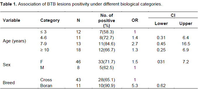
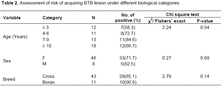
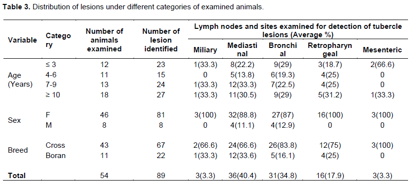
Among the 54 animals, 47 were confirmed for BTB by CIDT and 35 by using BOVIGAMTM test kit (INF-γ release assay). Out of all the slaughtered animals, tuberculous lesion was detected from 70.3% (38/54) of them examined through detailed postmortem examination. No significant difference was observed among the four age groups and two sexes of both breeds of animals on the frequency of tuberculous lesion development. Tuberculous lesion was found nearly three times higher on animals at 7-9 years of age as compared to animals at three or lesser years of age. The frequency of the tuberculous lesions was found more than five times higher in Boran as compared to cross breed animals (Tables 1 and 2). Higher proportion of the lesion was observed on mediastinal (40.4%) followed by bronchial (34.8%), retropharyngeal (17.9%) and lastly miliary form and mesenteric lymph nodes in equal proportion (3.3%) (Table 3).
Among the CIDT positive animals, 59.2% of them had lesions. Similarly, 46.2% INF-γ release assay positive animals contained lesions during postmortem examination (Table 4). Lesions were detected from a total of 61 sites of all animals diagnosed by INF-γ release assay and the distribution of the lesion was 1.69 lesion/head of post mortem examined animals. Similarly, a total of 78 lesions were detected from different lymph nodes of all 47 CIDT positive animals and the lesion were distributed with 1.65 lesion/head of post mortem examined animals. Out of all animals examined, three (5.5%) miliary types of lesion were detected which were confirmed by both tests.
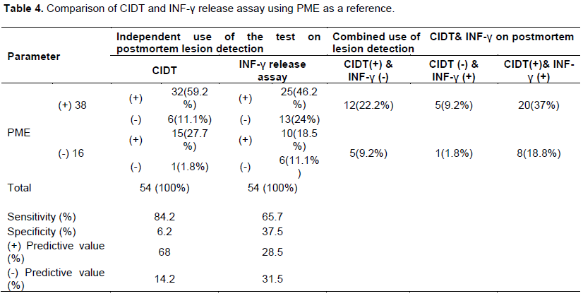
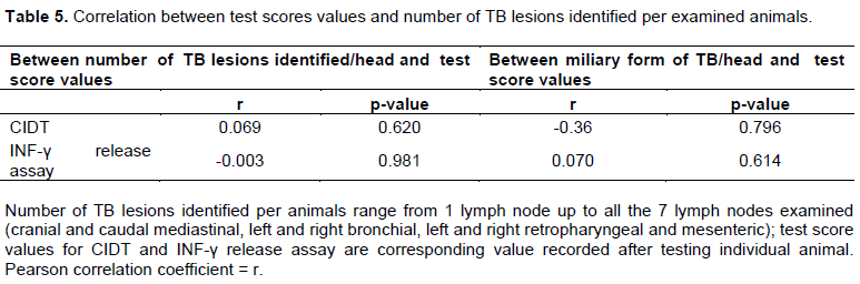
Of the total CIDT positive and INF-γ negative animals, postmortem lesions were detected from 22.2% of them, and in the vice-versa, of the CIDT negative and INF-γ positive animals, 9.2% them had TB lesions. However, the two tests share the highest proportion of individuals having postmortem lesion rather than exclusion of one from the other (37%). The sensitivity of CIDT was 84.2% and the specificity was 6.2%. Similarly, the sensitivity and specificity of Bovigam was 65.7 and 37.5% respectively (Table 4). No significant correlation between scores of both CIDT and INF-γ release assay and the distribution of post mortem lesion was detected. However, the values scored by INF-γ release assay and the number of lesions detected show slightly negative correlation. Again, no correlation was observed between the scores of the two tests and occurrence of military tuberculosis. However, when the CIDT scores decrease, the occurrence of miliary tuberculosis increase (Table 5).
DISCUSSION
Although the current study was not intended to study prevalence of BTB, higher proportion of the slaughtered animals had at least one lesion (67.8%). The distribution of the lesion was not differing significantly among the age groups but in younger animals (≤ 3 years of age) animals, TB lesions were detected almost three times less likely than animals at the age range of 7 to 9 years. Since TB is a chronic disease, the tuberculous lesion might develop long after the infection time which coincides with the age of the animal (Radostits et al., 2000); however, in this study, two animals which were less than three years of age were observed, one having miliary type of lesions and the other having lesions almost in all of the diagnosed lymphnodes (data not shown). Guirado and Schlesinger (2013) explained that T-cell functions such as cytokine production (IL-2 and IFNγ), cytotoxic activity, and T cell proliferation decrease when the age increases. This implies the increased risk of acquiring the disease while exposure in aged individuals. However, poor nutrition and immunosuppression have impact on development of sufficient immune response besides the age factor in M. tb infection in human cases (Peggy and Ellen, 2008). The inconsistency observed in this study in respect to lesion development in relation to aging could align with such and other unknown factors like co-infection with immune-suppressive diseases or infection by very high dose of the pathogen.
Higher proportion of the lesions were detected from pulmonary LN (mediastinal and bronchial) (78%) followed by LN of the head (Mandibular) (18%). Similarly, Tigre et al. (2012) identified 63.3% of the total lesions from the thoracic cavity. Since aerosol is the most common route of infection, it is expected to identify lesion on the primary site of replication.
Another interesting finding in this study is that majority of the mesenteric LN lesions were detected from younger animals (16.7%). Oral route of infection from contaminated milk during suckling could be the most common route of infection in young animals since the animals are mostly kept indoors (Regassa et al., 2010). However, the animals may acquire the infection through grazing and lesions can be detected at GIT system as described earlier by Ameni et al. (2006) who compared animals kept indoors with the ones in pasture. In another study by Ayana et al. (2013), it was explained that, although route of infection could be the primary site to develop the granulomatous lesion, reactivation and dissemination could spread the bacteria throughout the body and lesions might appear far from their site of infection. Similarly, Guirado and Schlesinger (2013) discussed reactivation of TB that occurs 80% in the lung which could be noticed when the lung associated lymph nodes contain considerable lesions. Hence, lesions that develop extra-pulmonary sites might be due to escaping bacilli from pulmonary tubercle.
Miliary type of tuberculosis is the most hazardous form of tuberculosis among the rest localized lesions in terms of public health significance, especially in countries where consumption of raw meat is common due to cultural reasons. In the current study, 5.5% of the examined animals had miliary tuberculosis which was very alarming. Similarly, a10 years retrospective study (from 1992-2001) of animals slaughtered at Addis Ababa abattoir indicate whole carcass condemnation due to generalized tuberculosis was 0.024% with annual increment rate of 0.34% (Asseged et al., 2004).
In terms of distribution, the mean number of lymph nodes with TB lesions per skin test reactive and INF-γ positive animals were 1.65 and 1.69, respectively. Similarly, Liebana et al. (2008) has identified the mean number of lymph nodes with tuberculosis TB-like lesions per TB-confirmed animal was 1.7. Lesser ratio of mean number of lesions was observed by Asseged et al. (2004) which was 1.5.
In the current study, of all the CIDT positive animals, TB lesions were detected from 59.2% of them which is closer to the finding in an earlier study by Costello et al. (1997) who illustrated that out of all tuberculin reactive animals, 65.6% of them contain TB-like lesion at least in one of their lymph nodes. But, it is higher than another study by Llamazares et al. (1999) who showed that, lesions were detected only from 50% of tubercline reactive animals. However, high percentage of overlaps between skin test and postmortem lesions were illustrated by Ameni et al. (2006) who detected lesions from 91% of the reactors. After a year, the same author found even higher proportion of intersection between the skin test and most mortem examination which was 95% (Ameni and Erkihun, 2007). It was reviewed that, there is wide range (50-80%) of missing detectable TB lesion from skin test reactive animals (la Rau-Domenech et al., 2006). O’Hagan et al. (2015) found lesions only from 43% of the total 24,923 reactor animals in Ireland. Failure to detect visible gross TB lesions while conducting postmortem examination might not infer freedom from TB infection. This is probably due to technical gaps to identify the typical lesion, early infection which does not develop detectable tubercle lesion yet, time spent during diagnosis, proper lymph nodes slicing distance and latent infection of M. bovis (Biffa et al., 2010; Corner, 1994; la Rau-Domench et al., 2006).
On the other hand, we found that TB lesions were detected from 46.2% of animals positive for INF-γ release assay. In New Zealand, TB lesions were identified in 37.2% of INF-γ reactive animals (Sincliar et al., 2016). Breed, environment and management practices might produce the difference on the level of INF production upon TB exposure.
Different proportion of exclusion was observed when the two tests combined rather than their independent application considering postmortem lesions as a reference. Of the total CIDT positive and INF-γ negative animals, postmortem lesions were detected from 22.2% of them, and in the vice-versa, of the CIDT negative and INF-γ positive animals, 9.2% had TB lesions. However the two tests share the highest proportion of individuals having postmortem lesion rather than exclusion of one from the other (37%). According to a study by Clegg et al. (2017), out of the total skin test reactive but INF-γ negative animals, lesions were detected from 11.8% of them. Again, in their study, lesions were detected from 18.9% of SICCT negative but INF-γ positive animals. Despite the ability of INF-γ test to detect early infections in comparison with skin test (Wood and Jones, 2001), in the current study, the INF-γ assay was found less sensitive to detect lesions unlike what was seen in the work of Clegg et al. (2017). The explanation for this phenomenon is not well understood, but the shift on immune mechanism from cellular to humoral during the later period of the infection can more likely affect the single cytokine INF-γ dynamics rather than the skin test since the second one elicits more general inflammatory reaction than the first (Clegg et al., 2017).
Using postmortem lesion development as a reference, the sensitivity of CIDT and INF-γ release assay was 84.2 and 65.7% respectively. On the other hand, the specificity of the two tests was 6.2 and 37.5% in the same order. The higher sensitivity of the skin test and the higher specificity of the INF-γ release assay coincide with the nature of the two tests. The chance of getting lesions from CIDT positive animals seems higher than INF-γ release assay positive animals and this might be because the first one has higher probability of detecting cross-reactive animals for other mycobacterial infection. However, the change in the cytokine INF-γ level is monitored in highly controlled environment (in vitro) and measurement was done using the very sensitive type of ELISA which is the sandwich ELISA (Bovigam kit).
In the current study, no significant correlation was observed among any of the tests and/or pathological conditions. However, the correlation tend towards negative (-0.003) when the INF-γ release score related with the extent of distribution of TB lesion. Similarly, a negative correlation trend between INF-γ responses and the pathology scores was observed by Ameni et al. (2006) in animals kept indoors but not those kept in pasture.
Conversely, negative correlation between the occurrences of generalized tuberculosis (miliary TB) and skin test scores was observed, that is, the chance to detect miliary tuberculosis is high in skin test low scoring animals as compared to those which scored high. Focus was given to miliary tuberculosis because of the assumption that this form of BTB affects more the systemic immunological response as compared to the other forms. In the present study, animals at stage of miliary tuberculosis could be justified as a shift from the T-cell mediated immune response towards the B-cell response via production of antibodies discussed under different studies since,cellular mechanisms of control of the bacilli start to exhaust (Buddle et al., 2005; Cassidy et al., 2001; Cooper, 2009). Earlier study by Ritacco et al. (1991) also demonstrated that there is inverse relationship between cellular and humoral responses to tubercle bacilli in cattle with natural infection. In conclusion, both CIDT and INF-γ release assay predict the presence of TB lesions post slaughter. CIDT was found to be more predictable for detection of postmortem lesions as compared to INF-γ release assay in our condition. However, combined use of the two tests predicts the existence of lesions from considerable proportion of the examined animals. Abattoir workers should focus more while examining animals with positive TB history.
CONFLICT OF INTERESTS
The authors have not declared any conflict of interest.
REFERENCES
|
Ameni G,Aseffa A, EngersH,Young D,Hewinson G, Vordermeier M (2006). Cattle husbandry in Ethiopia is a predominant factor affecting the pathology of bovine tuberculosis and gamma interferon responses to mycobacterial Antigens. Clinical and Vaccine Immunology, |
|
|
Ameni G, Erkihun A (2007). Bovine tuberculosis on small-scale dairy farms in Adama Town, Central Ethiopia, and farmer awareness of the disease. Revue Scientifique et Technique-Office International des Epizooties 26 (3):711-719. |
|
|
Asseged B, Woldesenbet Z, Yimer E, Lemma E (2004). Evaluation of abattoir inspection for the diagnosis of Mycobacterium bovis Infection in cattle at Addis Ababa Abattoir. Tropical Animal Health and Production 36:537-546. |
|
|
Ayana T, Ketema T, Mamo G, Tesfaye ST, and Ameni G (2013).Isolation and molecular characterization of non tuberculosis Mycobacteria from skin positive reactors and pathological lesions of cattle at Bako, Western Ethiopia. African Journal of Microbiology Research, |
|
|
Biffa D, Bogale A, Skjerve E (2010). Diagnostic efficiency of abattoir meat inspection service in Ethiopia to detect carcasses infected with Mycobacterium bovis: Implications for Public Health. BMC Public Health 10(1):462. |
|
|
Buddle MB, Wedlock DN, Denis M, Skinner MA (2005). Identification of immune response correlates for protection against bovine tuberculosis. Veterinary Immunology and Immunopathology 108:1-2 |
|
|
Cassidy JP, Bryson DG, Cancela MMG, Forster F, Pollock JM, Neill SD (2001). Lymphocyte subtypes in experimentally induced early-stage bovine tuberculous lesions. Journal of Comparative Pathology 124(1):46- |
|
|
Clegg TA, Good M, Doyle M, Duignan A, More SJ, Gormley E (2017). The Performance of the interferon gamma assay when used as a diagnostic or quality assurance test in Mycobacterium bovis infected herds. Preventive Veterinary Medicine 140:116-121. |
|
|
Cooper MA (2009). Cell-mediated immune responses in tuberculosis. Annual Review of Immunology 27:393-422. |
|
|
Corner LA (1994). Post mortem diagnosis of Mycobacterium bovis infection in cattle. Veterinary Microbiology 40:53-63. |
|
|
Costello E, Egan JW, Quigley FC, O'Reilly PF (1997).Performance of the single intradermal comparative tuberculin test in identifying cattle with tuberculous lesions in Irish herds. Veterinary Record 141(9):222-224. |
|
|
Guirado E, Schlesinger LS (2013).Modeling the Mycobacterium Tuberculosis Granuloma - the Critical Battlefield in Host Immunity and Disease. Frontiers in Immunology 4:98. |
|
|
Gupta MP, Kumar H, Singla LD (2009). Trypanosomosis concurrent to tuberculosis in black bucks. The Indian Veterinary Journal 86:727-728. |
|
|
la Rau-Domenech Rde, Goodchild AT, Vordermeier HM, Hewinson RG, Christiansen KH, Clifton-Hadley RS (2006). Ante mortem diagnosis of tuberculosis in cattle: A review of the tuberculin tests, γ-Interferon assay and other ancillary diagnostic techniques. Research in Veterinary Science 81(2):190-210., |
|
|
Liebana E, Johnson L, Gough J, Durr P, Jahans K, Clifton-Hadley R, Spencer Y, Hewinson RG, Downs SH(2008). Pathology of naturally occurring bovine tuberculosis in England and Wales. The Veterinary Journal 176(3):354-360. |
|
|
Llamazares ORG, Martín CBG, Martín AA, Criado EL, Rodríguez LD, Ferri EFR (1999). Comparison of different methods for diagnosis of bovine tuberculosis from tuberculin- or interferon-γ-reacting cattle in Spain .Journal of Applied Microbiology 87:465-471 |
|
|
O'Hagan MJH, Courcier EA, Drewe JA, Gordon AW, McNair J, Abernethy DA (2015). Risk factors for visible lesions or positive laboratory tests in bovine tuberculosis reactor cattle in Northern Ireland. |
|
|
OIE (2000). Principles of prevention and control of aquatic animal diseases. Conf. OIE, pp. 27-49. |
|
|
Peggy P, Ellen P (2008). Nutrition and tuberculosis: A review of the literature and considerations for TB control programs. United States Agency for International Development, Africa's Health 2010 Project, 1. |
|
|
Radostits OM, Gay CC, Hinechliff KW (2000).Veterinary medicine, A Textbook of the diseases of cattle, sheep, pigs, goat and horses. 9th ed W. B. Saunders Company Ltd., London. |
|
|
Regassa A, Tassew A, Amenu K, Megersa B, Abunna F, Mekibib B, Macrotty T, Ameni G (2010). A cross-sectional study on bovine tuberculosis in Hawassa Town and its surroundings, Southern Ethiopia. Tropical Animal Health and Production 42(5):915-920. |
|
|
Ritacco V, Lopez B, Kantor IND, Barrera L, Errico F, Nader A (1991). Reciprocal cellular and humoral immune responses in bovine tuberculosis. Research in Veterinary Science 50:365-367. |
|
|
Sincliar JA, Dawson KL,Buddle BM (2016). The effectiveness of parallel gamma-interferon testing in New Zealand's bovine tuberculosis eradication programme. Preventive Veterinary Medicine 127:94-99. |
|
|
Sinder D (1982). The tuberculin skin test. American Review of Respiratory Disease 125(3P2):108-118. |
|
|
Tigre W, Alemayehu G, Abetu T, Ameni G (2012). Preliminary study on the epidemiology of bovine tuberculosis in Jimma Town and its surroundings, Southwestern Ethiopia. African Journal of Microbiology Research 6(11):2591-7. |
|
|
Wood PR, Corner LA, Rothel JS, Baldock C, Jones SL, Cousins DB, McCormick BS, Francis BR, Creeper J, Tweddle NET(1991). Field comparison of the interferon-gamma assay and the intradermal tuberculin test for the diagnosis of bovine tuberculosis. Australian Veterinary Journal 8(9):286-290. |
|
|
Wood PR, Jones SL (2001). "BOVIGAMTM: An in vitro cellular diagnostic test for bovine tuberculosis. Tuberculosis 81(1-2):147-155. |
|
Copyright © 2024 Author(s) retain the copyright of this article.
This article is published under the terms of the Creative Commons Attribution License 4.0


