Full Length Research Paper

ABSTRACT
Mycobacterium avium subspecies paratuberculosis (Map), is the etiological agent of paratuberculosis, a disease that affects cattle and causes economic losses to the animal husbandry industry. Its opportune diagnosis, in herds, is part of the control measures of the disease; therefore, the objective of this work was to compare paratuberculosis detection of infected bovines with the Fluorescence Polarization Assay (FPA) and Enzyme-linked Immunosorbent Assay (ELISA). Six-hundred and three sera and feces samples, from bovines older than 2 years old were studied. The sera were assessed with the FPA technique using as antigen a protein fraction of 35 kDa obtained from the raw extract of the Map strain 3065, and for ELISA the protoplasmic antigen of the same strain was used. DNA was obtained from the feces and assessed by nested PCR. The correlation of results was established by Kappa Test. The FPA test had sensitivity (Se) of 88.50% and specificity (Sp) of 91.42% (p ≤0.000); for ELISA Se 83.86% and Sp 89.87% (p ≤0.000) were obtained. Concordance (K) between tests was 0.6742%, and when compared with nested polymerase chain reaction (PCR), the FPA test had K = 0.7314%, while for ELISA it was 0.5771%. The FPA technique using as antigen the protein fraction of 35 kDa showed a higher sensitivity and specificity, moreover it was a simple technique for the determination of the antigen-antibody interaction, and therefore it becomes an alternative diagnostic tool to detect paratuberculosis infected bovines.
Key words: Paratuberculosis, diagnosis, fluorescence polarization assay (FPA), enzime-linked immunoabsorbent assay (ELISA).
INTRODUCTION
Paratuberculosis or Johne's disease is an infectious contagious disease that is characterized by the production of chronic granulomatous enteritis. The etiological agent is Mycobacterium avium subsp. paratuberculosis (Map) (Arsenault et al., 2014). This serious economically important disease has a worldwide distribution with prevalence rate variation from five to 30% (Mortier et al., 2015).
Animals younger than 6 months of age are infected by ingesting bacilli through feed and water, as well as, sucking on teats contaminated with infected feces (Castellanos, 2010).
Affected bovines suffer chronic enteritis, the signs of which are diarrhea, submandibular edema, weight loss, and low body condition that leads eventually to death (Cirone et al., 2007; Speer et al., 2006). Clinical signs are only observed in adults between 18 and 24 months of age. Appetite and general behavior of the animal remain normal during the early stages, but milk production and body condition worsen due to nutrient malabsorption. As the disease advances, there is lethargy, depression, bristly hair, hypoproteinemia and submandibular edema (Mortier et al., 2015).
Tests based on the humoral immune response are the most frequently used for the diagnosis of the infection. One of the more evaluated tests has been the ELISA which is considered a good option for diagnosis, since there are several brands of commercial kits; it has a sensitivity of 50 to 83% and a specificity of 70 to 89% (Martinez et al., 2012).
Another technique that allows the determination of the antigen-antibody interaction is the FPA. The principle of this test is based on the property that antibodies have a high affinity and specificity to join a particular antigen; this property is used to recognize a specific analyte of interest, and by different mechanisms, generate a light signal that may be measured. In the case of FPA, the signal which is being measured is polarized fluorescent light. The size of the molecule is the main factor that influences the rotation speed. For example, a small molecule rotates at a higher speed than a larger molecule. These molecules move and rotate freely in the solution, and the result that is obtained is going to depend on how much the molecule has been able to rotate during the time the excited fluorescence state lasts. The smaller the molecule, the faster it shall rotate, and its polarization of fluorescence shall be lower (Nielsen and Gall, 2001; Marcelo et al., 2017).
If a molecule is marked with a fluorochrome, the rotation time through a 68.5° angle can be established by measuring the intensity of polarized light in vertical or horizontal planes. A large molecule emits more light in a single plane (more polarized) than a small one that rotates faster and emits more depolarized light (Nielsen and Gall, 2001; Marcelo et al., 2017). Polarized fluorescence has been used for the diagnosis of bovine brucellosis and tuberculosis, demonstrating a sensitivity of 95.5% and specificity of 99.0% for brucellosis and 92.9 and 98.3%, respectively for tuberculosis (Surujballi et al., 2002; Jolley et al., 2007; Marcelo et al., 2017).
Paratuberculosis diagnostic methods must be based on their capacity to detect infected animals; the specificity and reliability of serodiagnosis tests is determined by the type of mycobacterial antigens that are used, hence the identification of the structural antigenic components must be well characterized and specific for each mycobacterial species to be able to provide the means to improve specificity and sensitivity of immunodiagnostic assays.The economic losses caused by paratuberculosis on the animal husbandry industry are a serious problem, and therefore it is necessary to have quick tests for diagnostic of the disease. The objective of this work was to compare FPA and ELISA to detect paratuberculosis infected bovines.
MATERIALS AND METHODS
Samples
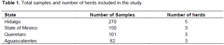
The sampling was carried out in dairy herds, with and without prior paratuberculosis diagnosis, which belonged to cooperating cattlemen. The dairy herds were located in the States of Mexico, Hidalgo, Queretaro and Aguascalientes (Table 1). To estimate the sample size, the cattle census of each State of the Mexican Republic included in the study were taken into consideration, as well as a paratuberculosis prevalence of 10% with a confidence level of 95% and 5% error. Six-hundred and three serum and feces samples were taken from bovines, older than two years of age, most of them from the Holstein breed. The blood was taken from the caudal vein in vacutainer tubes and centrifuged at 180 xg, for ten minutes to obtain the serum; the feces samples were directly taken from the rectum with palpation gloves and were kept at 4°C until their use.
Fluorescence polarization assay (FPA)
The protocol described by Torres (2015), was followed to obtain and mark with fluorochrome the protein fraction of 35 kDa from Map 3065 strain.
The sera were diluted 1:100 in borosilicate tubes with Tris buffer (pH 7.2, 0.01 M), then incubated for 30 minutes at room temperature.
The initial reading of the diluted sera was carried out in an FPA Sentry 2000 reader; 10 µl of fluorescein marked antigen was incubated for 5 min at room temperature and a second reading was carried out. The results were expressed in milli-polarization units (mP), which is the difference between both readings. The samples were processed in duplicate with positive and negative serum controls of Map (Allied & Monitor Inc). Results were considered positive when they were equal to or greater than 126 mP (Torres, 2015).
Enzyme-linked immunosorbent assay (ELISA)
The protocol described by Martinez et al. (2012), was followed to fix the protoplasmic antigen of Map from strain 3065 to the microplates. A dilution 1:160 was obtained with the problem sera using a 0.02% M. phlei, 0.1% gel and 0.05% Tween 80 solution. The diluted sera were incubated for an hour at room temperature. Map positive and negative sera controls were used (Allied Monitor & Inc). Each well received 100 µl of each, suspected serum, negative and positive controls; then incubated at room temperature for 30 min, and then washed four times with 300 µl PBS-Tween 20 (1X). 100 µl of a 1:10 000 dilution of Anti-IgG bovine HRP was added to each well and incubated at room temperature for 30 min. The plate content was discarded and washed with PBS-Tween 20 (1X), four times; 100 µl of the substrate solution 2’2 azino-bis-(3-ethyl-benzothiazoline-sulfone-6) -(diamonium) (ABTS, AMPRESCO) was added to each well and incubated for 30 min at room temperature and 1.5 mM sodium nitrate (Sigma Aldrich) in 0.1M citric acid (Sigma Aldrich) was used as stop solution. Plate reading was carried out at 650 nm in a spectrophotometer (ELx800 Biotek).
Sera with results equal to or greater than 0.22 optic density (OD) were considered positive to the ELISA test.
Nested polymerase chain reaction (n-PCR)
DNA extraction from feces was carried out according to the protocol described by Jaimes et al. (2008).
The primers described by Erume et al. (2001), were used for the first PCR reaction: Paratb1 (5´ TGA TCT GGA CAA TGA CGG TTA CGG A 3´) and Paratb 4 (5´ CGC GGC ACG GCT CTT GTT 3´) and the product that was obtained had 563 bp; for the second reaction, Paratb 2 (5´ GCC GCG CTG CTG GAG TTA A 3´) and Paratb 3 (5´ AGC GTC TTT GGC GTC GGT CTT G 3´) were used, obtaining a final product of 210 bp.
For the first reaction, 2 µl (15 ng/ µl) of DNA, obtained from bovine feces were used, with the following reagent conditions: for 48 µl of premix solution, 5 µl were used of Reaction Buffer 10X (67 mM/µl), 4 µl MgCl 2 30 mM 20 X, 1 µl DNTP (200mM), 1 µl Paratb 1 (25 pMol), 1 µl Paratb 4 (25 pMol), 0.25 µl Polimerase 500 U and 35.75 µl double-distilled water. Amplification was carried out in an Axigene thermocycler using the following program: an initial denaturing at 98°C/3 min, followed by 35 cycles of denaturing at 98°C/30 s, annealing at 65 °C/30 s, and extension at 72 °C/30 s, followed by a final extension at 72°C/3 min and then 4°C /three min. For the second reaction, 3 µl of the first reaction were taken as a DNA template and were transferred into PCR microtubes containing the same amount and concentration of reagents previously described with the exception that the Paratb1 and Paratb4 primers were substituted by primers Paratb2 (25 pMol) and Paratb3 (25 pMol). The same thermocycler program was used. For the visualization of the amplification products, electrophoresis was carried out on 2% agarose gels and stained with ethidium bromide.
Establishing test sensitivity and specificity
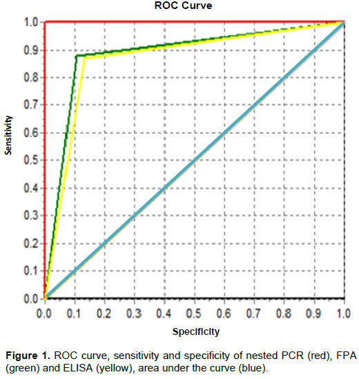
Receiver operational characteristics (ROC) analysis was carried out, which is a graphic representation of sensitivity and specificity of a binary system according to the discrimination threshold variation. The Kappa Test or Concordance Index was estimated to measure the association with the results obtained in FPA, ELISA and nested PCR. The statistical analysis was carried out with the Intercooled Stata 7.0 and Epidat 3.1 software packages.
RESULTS
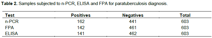
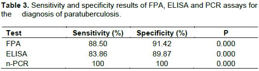
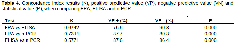
The results obtained with the FPA (142/603) and ELISA (141/603) tests were similar; the FPA test identified only one more serum positive (Table 2). The sensitivity and specificity of the FPA technique were 88.50 and 91.42%, respectively, when compared with ELISA and n-PCR. The FPA assay shows high sensitivity and specificity (Table 3, Figure 1). Concordance between tests showed a good correlation of FPA when compared to ELISA (0.6742%) and n-PCR (0.7314%); the positive predictive value of FPA was 75.6% when compared with ELISA and 87.7% compared with polymerase chain reaction (PCR). Likewise, the negative predictive values were 90.8 % and 89.3% respectively, which indicates that the test has agreater specificity (Table 4).
DISCUSSION
Currently, research lines in mycobacteriosis are focusing on the assessment of low molecular weight protein type antigens. These antigens, since they are highly specific, are the most viable candidates to be used in diagnostic tests to detect animals infected with tuberculosis and/or paratuberculosis. The low molecular weight antigens have been previously used in serological diagnostic techniques such as ELISA and FPA, where the sensitivity and specificity has been demonstrated to be higher in both techniques (Beck et al., 2005; Chaubey et al., 2016). In this work, the FPA and ELISA techniques were evaluated using as a confirming test n-PCR. FPA had an 88.5% sensitivity and 91.42% specificity, while ELISA had 83.86% and 89.87% respectively; when the concordance analysis was carried out, an acceptable result of (K=0.6742) was obtained. Therefore, both tests are a good option to determine the presence of anti-Map antibodies in cattle.
With n-PCR, 162 positive samples were detected while with FPA and ELISA 142 and 141 positives were detected, respectively. It is possible to find samples that are positive in n-PCR but negative in the serology tests, because in the subclinical stage of paratuberculosis the humoral immune response decreases and the cellular immune response increases. Another reason for alteration of antibody production can be the loss of immune response to the infection. This suppressing activity is known as anergy and occurs in immunocompromised animals such as old animals or that are in the final stages of the disease. Nevertheless, it is considered that these animals may be eliminating the bacilli that are not detected by serological diagnosis but are detected by n-PCR (Martinez et al., 2012; Chaubey et al., 2016). Nielsen and Gall (2001), mentioned that the FPA test is more specific than sensitive; albeit Jolley et al. (2007), when using the FPA test with the 22 kDa protein MPB70 from an M. bovis strain for the diagnosis of bovine tuberculosis, obtained a 99.9% specificity. Also, Surujballi et al. (2009), used FPA for diagnosis of tuberculosis in cervids and obtained a sensitivity and specificity above 81.00%.
FPA has been used in the diagnosis of bovine brucellosis, and the sensitivity and specificity has been reported at 99.00 and 95.50%, respectively (Marcelo et al., 2017). Even though the FPA technique is simple and has several advantages over other serological techniques, up until now there had not been studies on its use and application to the diagnosis of paratuberculosis. The results obtained in this work show that the use of FPA might contribute to and be an alternative for the diagnosis of bovine paratuberculosis.
The Map 3065 strain protoplasmic antigen that was used for ELISA, has the characteristic of being a complex preparation of lipids, carbohydrates, proteins and nucleic acids that contain antigenic determinants in common with most of the Map strains, with which an acceptable sensitivity and specificity has been obtained (Martinez et al., 2012). In comparison, the antigen that is used in the FPA test is a protein fraction of 35 kDa, and as such has more specificity. Proteins below 50 kDa are involved in the humoral and cellular response to intracellular microorganism infections, and therefore they are candidates to be used as antigens in the FPA test since they allow for more specificity of the assay (Nielsen and Gall, 2001; Franco et al., 2005; Mon et al., 2012; Chaubey et al., 2016).
Bauman et al. (2019) evaluated Bulk tank milk (BTM) to determine Map's presence at the herd level in production, using fecal- culture, PCR, mELISA paratuberculosis tests in 29 dairy goat herds and 21 dairy sheep flocks; theirs results showed that mELISA was more sensitive for identifying farms with affected animals (those with detectable circulating antibodies in their serum or milk). Increasing sensitivities to 87.50% (serum ELISA as reference test) and 71.40% (milk ELISA as reference test) in dairy goats and 72.70% (serum ELISA as reference test) and 87.50% (milk ELISA as reference test) in dairy sheep; While the sensitivity was 50% and speficivity 83.00%; when used PCR of the BTM and fecal culture. BTM has been evaluated in dairy cattle as a potential herd-level paratuberculosis testing strategy so inclusion of FPA for the detection of anti-map antibodies in milk, is an alternative that would allow to have a diagnostic test with greater sensitivity and specify, as well as determine the health status of paratuberculosis of the herd in production.
Control of paratuberculosis in herds is based on the opportune identification and elimination of infected animals. Nevertheless, the low sensibility that diagnostic tests have and the subclinical presentation of the infection, make the diagnosis of the disease difficult, especially in young animals (Chaubey et al., 2016).
CONCLUSION
The FPA test using the 35 kDa antigen may be considered as an alternative that allows better sensitivity and specificity to diagnose paratuberculosis.
CONFLICT OF INTERESTS
The authors have not declared any conflict of interests.
ACKNOWLEDGEMENT
The authors are grateful to INIFAP SIGI 12484419581 for the financial resources provided.
REFERENCES
|
Arsenault RJ, Maattanen P, Daigle J, Potter A, Griebel P, Napper S, (2014). From mouth to macrophage: mechanisms of innate immune subversion by Mycobacterium avium subsp. paratuberculosis. Veterinary Research 45(1):54. |
|
|
Bauman CA, Jones-Bitton A, Jansen J, Kelton D, Menzies P (2019). Evaluation of bulk tank milk PCR and bulk tank milk modified ELISA tests for the detection of paratuberculosis at the herd level in goat and sheep dairies in Ontario, Canada. Journal of Dairy Science 102(1):511-520. |
|
|
Beck ST, Leite OM, Arruda RS, Ferreira AW (2005). Humoral response to low molecular weight antigens of Mycobacterium tuberculosis by tuberculosis patients and contacts. Brazilian Journal of Medical and Biological Research 38:587-596. |
|
|
Castellanos RE (2010). Caracterización molecular de aislados de Mycobacterium avium subespecie paratuberculosis. Mapa epidemiológico en España (2010). Tesis doctoral. Universidad Complutense de Madrid, Facultad de Veterinaria. España. |
|
|
Chaubey KK, Gupta RD, Gupta S, Singh SV, Bhatia AK, Jayaraman S, Kumar N, Goel A, Rathore AS, Sahzad, Sohal JS, Stephen BJ, Singh M, Goyal M, Dhama K, Derakhshandeh A (2016). Trends and advances in the diagnosis and control of paratuberculosis in domestic livestock. The Veterinary Quarterly 36(4):203-227. |
|
|
Cirone K, Morsella C, Romano M, Paolicchi F (2007). Mycobacterium avium subsp. paratuberculosis: presencia en los alimentos y su relación con la enfermedad de Crohn. Revista Argentina de Microbiología 39:57-68. |
|
|
Erume J, Spergser J, Rosengarten R (2001). Rapid detection of Mycobacterium avium subsp paratuberculosis from cattle and zoo animals by nested PCR. African Health Sciences 1(2):83-89. |
|
|
Franco J, Camarena JJ, Noriega JM, Blanuer R, Ruiz MJ, Marin J (2005). Serological response (Western blot) to fractions of Mycobacterium tuberculosis sonicate antigen in tuberculosis patients and contacts. International Journal of Tuberculosis and Lung Disease 5(10):958-962. |
|
|
Jaimes NG, Santillán FMA, Hernández COA, Córdova LD, Guzmán RCC, Arellano RB, Díaz AE, Tenorio GVR, Cuéllar OJA (2008). Detección de Mycobacterium avium subespecie paratuberculosis, por medio de PCR-anidada a partir de muestras de heces de ovinos. Vet. Méx. 39(4):377-386. |
|
|
Jolley ME, Nasir MS, Surujballi OP, Romanowska A, Renteria TB, De la Mora A, Lim A, Bolin SR, Michel AL, Kostovic M, Corrigan EC (2007). Fluorescence polarization assay for the detection of antibodies to Mycobacterium bovis in bovine sera. Veterinary Microbiology 120(1-2):113-121. |
|
|
Marcelo I, Hernan B, Ruth S, Milena G, Judith G, Julio P, David H, Jorge M, Martin C, Byron P (2017). Determining a diagnostic cut-off on fluorescence polarization assay (FPA) for bovine brucellosis in Carchi, Ecuador. Open Journal of Animal Sciences 7:425-432. |
|
|
Martinez CAG, Santillán FMA, Guzmán RCC Favila HLC, Córdova LD, Díaz AE, Hernández AL, Blanco OM (2012). Desarrollo de un Inmuno-Ensayo Enzimático (ELISA) para el diagnóstico de paratuberculosis en bovinos. Revista Mexicana de Ciencias Pecuarias 3(1):1-18. |
|
|
Mon ML, Viale M, Baschetti G, Alvarado Pinedo F, Gioffre A, Travería, G, Willemsen P, Bakker D, Romano MI (2012). Search for Mycobacterium avium subspecies paratuberculosis antigens for the diagnosis of paratuberculosis. Veterinary Medicine International 2012.doi:10.1155/2012/860362. |
|
|
Mortier RA, Barkema HW, De Buck J (2015). Susceptibility to and diagnosis of Mycobacterium avium subspecies paratuberculosis infection in dairy calves: A review. Preventive Veterinary Medicine 121(3-4):189-98. |
|
|
Nielsen K, Gall D (2001). Fluorescence polarization assay for the diagnosis of brucellosis. Journal of Immunoassay and Immunochemistry 22(3):183-201. |
|
|
Speer CA, Scott MC, Bannantine JP, Waters WR, Mori Y. Whitlock RH, Eda S (2006). A novel enzyme-linked immunosorbent assay for diagnosis of Mycobacterium avium subsp. paratuberculosis infection (Johne's Disease) in cattle. Clinical and Vaccine Immunology 13(5):535-540. |
|
|
Surujballi O, Romanowska A, Sugden EA, Turcotte C, Jolley ME (2002). A fluorescence polarization assay for the detection of antibodies to Mycobacterium bovis in cattle sera. Veterinary Microbiology 87(2):149-157. |
|
|
Surujballi OM, Lutze-Wallace C, Turcotte C, Savic M, Stevenson D, Romanowska A, Monagle W, Berlie-Surujballi G, Tangorra E (2009). Sensitive diagnosis of bovine tuberculosis in a farmed cervid herd with use of an MPB70 protein fluorescence polarization assay. Canadian Journal of Veterinary Research 73(3):171-176. |
|
|
Torres VR (2015). Obtención y evaluación de proteínas de bajo peso molecular a partir de un extracto crudo de Mycobacterium avium subsp. paratuberculosis para el diagnóstico de paratuberculosis por fluorescencia polarizada. Tesis de Maestría. Posgrado-FMVZ UNAM. México. |
|
Copyright © 2024 Author(s) retain the copyright of this article.
This article is published under the terms of the Creative Commons Attribution License 4.0


