Review

ABSTRACT
The objective of this review is to generate information about the current status of contagious caprine pleuropneumonia (CCPP) which is recognized as highly infectious, and devastating disease of goats for about 140 years. However, current reports indicated that sheep can be affected. CCPP is caused by Mycoplasma capricolum subspecies capripneumoniae (Mccp) which is severe and dramatic mycoplasmas, challenging to isolate and properly identify. Coughing, respiratory distress and very high morbidity and mortality are the main clinical signs that described the disease. Transmission of CCPP occurs through aerosol while animals are in nearby contact. There are a number of approaches for the rapid detection and identification of Mccp based on a PCR. In Africa, Asia and Middle East morbidity and mortality can reach about 100% which causes huge economic loss. Presence of CCPP in Ethiopia reported in 1983 and later confirmed from an outbreak in 1990 in Ogaden, Eastern Ethiopia. Since then CCPP has been considered as endemic disease in Ethiopia. Prevention and control of CCPP is undertaken through vaccination, quarantine, restriction of movement, culling of infected and exposed animals and keeping the hygiene of premises. However, it remains one of the standing problems of goat production. Therefore, more research, awareness creation about CCPP, transmission path way, prevention and control methods, in small ruminant rearing areas to reduce the impact of the disease.
Key words: CCPP, goat, Mccp, Ethiopia.
INTRODUCTION
Ethiopia is endowed with huge number of livestock that have not yet been optimally exploited mainly due to technical and institutional constraints (Shapiro et al., 2017). The sector has huge support to Ethiopia’s national economy and livelihoods the community and has a promise to the economic development of the country (Alemayehu et al., 2015).
Based on Central Statistical Agency of Ethiopia (CSA, 2015), Ethiopia is one of the country having largest livestock population. Ethiopia has over 29.11 and 29.33 million heads of goats and sheep, respectively. Now a days, demand for meat and meat products rising (Narrod et al., 2011), small ruminants are considered as a main asset for livestock farmers in East Africa (Armson et al., 2021) and playing crucial economic and cultural roles in Ethiopia (Gobena, 2016; Wodajo et al., 2020).
Sheep and goats are very important to the household economy of Borana community in terms of providing a source of convenient amount of cash on a more frequent basis. This can partially substitute for sales of cattle (Coppok, 1994). At farm level small ruminants serve as investment and insurance due to their short generation interval and high frequency of multiple births (Asmare et al., 2016) as well as small feed requirement and adaptability to harsh environmental conditions (Urgessa et al., 2012).
Despite their huge social and economic roles the services and revenue obtained from small ruminants is sub-optimal due to several constraints. The most important constraints to small ruminant productions are widespread endemic diseases including parasitic infestation, viral and bacterial diseases (Singh et al., 2017; Pawar et al., 2019). Among various diseases, Contagious Caprine Pleuropneumonia (CCPP) is a major threat to goats (Teshome et al., 2018) which causes major economic losses (Asmare et al., 2016; Teshome et al., 2018).
CCPP has become one of major concern in some African countries because of high morbidity and mortality. Often uncontrolled movements of animals in search of water and feeds and informal trading systems as well as communal grazing are considered important drivers for the dynamics of CCPP in sub-Saharan Africa (Mbyuzi et al., 2014). In goats CCPP is devastating disease causing high morbidity and mortality occurring in Africa, Asia and Middle East (Teshome et al., 2018). It is a classical trans-boundary animal disease (Shahzad et al., 2016; Teshome et al., 2018). Moreover, the disease is included in the list of notifiable diseases of World Organization for Animal Health (OIE) as it threatens a significant number of goat populations throughout the world and causes a significant socioeconomic impact in infected territories (Atim et al., 2016; Teshome et al., 2018). Though the disease is confined to goats, subclinical cases were reported in sheep and some wild ruminant species (Asmare et al., 2016).
Contagious caprine pleuropneumonia is clinically characterized by coughing, respiratory distress and very high morbidity and mortality (OIE, 2018). The causative agent, Mycoplasma capricolum subspecies capripneumoniae, is one of the most serious and dramatic mycoplasmas, which is very difficult to isolate and correctly identify (Sherif et al., 2012). But there are a number of methods for the rapid detection and identification of Mccp based on a PCR system by which a segment of the 16S rRNA gene from all members of the Mycoplasmamycoides cluster can be amplified. The PCR product is then analyzed by restriction enzyme cleavage for the identification of Mccp DNA (Noah et al., 2011).
In Ethiopia CCPP occurs in most extensive goat rearing areas namely, Afar, Borana, Omo valley, West Gojjam and in the lowlands of Tigray (Yigezu et al., 2004). Reviewing different studies are very important in designing control and prevention methods. Therefore, in this paper the authors reviewed the current knowledge on CCPP and finally gave special emphasis to the Ethiopian situation.
LITERATURE REVIEW
Mycoplasma
Mycoplasma belong to the class Mollicutes, which are notable bacteria for inflicting a number of infections in animals and humans (Awan et al., 2009). They are the smallest free-living fastidious bacteria. They are about 300 nm in diameter, bound by a triple layered membrane and unlike conventional bacteria they don’t have a rigid cell wall containing murein (peptidoglycans) (Samiullah, 2013). Members of the genus Mycoplasma are associated with pneumonia, conjunctivitis, arthritis, abortion and infertility (Fischer et al., 2012). In small ruminants, they have been known to cause genital disease and mastitis in addition to the above mentioned diseases (Kumar et al., 2011).
The mycoplasmas belonging to the “mycoides cluster” are classified under class Mollicutes, order Mycoplasmatales, family Mycoplasmataceae, and the genus Mycoplasma. Mycoplasma mycoides cluster is made up of 6 species, subspecies or group of strains that are pathogenic for ruminants (Younis et al., 2015). It includes Mycoplasma mycoides subspecies mycoides (MmmSC), Mycoplasma mycoides subspecies mycoides (MmmLC), Bovine sero-group 7, Mycoplasma mycoides subspecies capri (Mmc), Mycoplasma capricolum subspecies capripneumoniae (Mccp) and Mycoplasma capricolum subspecies capricolum (Mcc) (Santos, et al., 2013; Fischer et al., 2015). Most of the members of the Mycoplasma mycoides cluster are significant pathogens of small ruminants including Mccp, Mmc and Mcc all of which can infect lungs of small ruminant inducing respiratory disease (Ejaz et al., 2015).
Mycoplasma capricolum subspecies capripneumoniae
The emergence of Mycoplasma mycoides clusters was dated back to about 10,000 years. The emergence and spread of the cluster has been suggested to coincide with the domestication of small ruminants (Prats-van der Ham et al., 2015). The organism; as a causal agent of CCPP was first isolated in 1976 in Kenya and was shown to cause CCPP and later, it was isolated in many countries of Africa and Asia (Thiaucourt et al., 2000). It was given its species name “M. capricolum subspecies capripneumoniae” in 1993 (Peyraud et al., 2014). Strain F38 is the type strain of Mccp described after its identification in Kenya as a cause of CCPP. Different strains were isolated from different countries in Africa and Asia (Pettersson et al., 1996). Molecular characterization of strains revealed valuable information used to classify them into many groups and help in understanding the epidemiology of the diseases (Li et al., 2020). The taxonomic status of F38 was unclear until in 1993 when it became a subspecies of M. capricolum and was named M. capricolum subspecies capripneumoniae (Nicholas, 2012).
Contagious Caprine Pleuropneumonia (CCPP)
Historical aspect
Contagious caprine pleuropneumonia has been known as a clinical disease of goats for over 140 years. CCPP was first described in 1873 in Algeria. It was not initially recognized as contagious because the disease was endemic in most of the regions examined; climate conditions were instead blamed for disease outbreaks. In 1881, there was a major outbreak in South Africa following the introduction of diseased goats coming from Turkey, and this led to the conclusion that CCPP was highly infectious. The burden and distribution of this disease, however, remains largely unknown (Atim et al., 2016). CCPP is highly infectious and devastating respiratory disease affecting goats (OIE, 2015; Atim et al., 2016). It is extremely contagious and frequently fatal; in some naive flocks, the morbidity and mortality may reach 100% and causes major economic losses in Africa, Asia and the Middle East, where goat husbandry plays significant role in the livelihood of the community (OIE, 2015).
Despite being an OIE notifialbe disease, there have been very few declarations of outbreaks of the disease to the OIE during the last 15 years, due to lack of awareness of the disease and possible confusion with other diseases, such as peste des petits ruminants (PPR) or pasteurellosis (Peyraud et al., 2014).
Epidemiology
Affected animals: Previously, CCPP has been reported only in domestic goats regardless of sex and age (Thiaucourt and Bolske, 1996). However, various wild animal families or orders among ungulates are quoted as susceptible too (OIE, 2017). The wild goat (Capra aegagrus), Nubian ibex (Capra ibex nubiana), Laristan mouflon (Ovis orientalis laristanica) and Gerenuk (Litocranius walleri) were affected (Arif et al., 2007; AU-IBAR, 2013). Similarly reports from the United Arab Emirates claim that CCPP occurred in captive Rhim and Dumani Gazelles and other deer species (Nicholas et al., 2008). Moreover reports show that members of the hippotraginae subfamily, the Arabian oryx, can be affected by CCPP (Prats-van der Ham et al., 2015) and outbreak of contagious caprine pleuropneumonia in endangered Tibetan antelope (Pantholops hodgsonii) in China (Yu et al., 2013).
McMartin et al. (1980) reported that Mccp does not cause disease in sheep, either spontaneously or experimentally. However, there are some reports describing the isolation of Mcpp from sheep with respiratory disease returning to Eritrea with refugees from Sudan (Houshaymi et al., 2002) and from healthy sheep in Kenya that have been in contact with goat herds affected by CCPP (Litamoi et al., 1990). Isolation of Mccp has been also reported from sick sheep mixed with goats in Uganda (Bolske et al., 1995).The occurrence of CCPP was 32.5% and 5% in goats and sheep, respectively, while case fatality was 30 and 8% in goats and sheep, respectively reported by Abd-Elrahman et al. (2019).
In a serological survey for CCPP in Ethiopia, sheep tested were found to be sero-positive (Hadush et al., 2009; Teshome et al., 2018). Isolation of Mccp from cattle with mastitis has also been reported (Kumar and Garg, 1991) and these reports confound the perceived host specificity of this pathogen. In addition to serological, molecularly occurrence and case fatality of CCPP in sheep were a new record for Mccp in Egypt by (Abd-Elrahman et al., 2019).
Distribution of CCPP in the world: CCPP affects goats in more than 40 countries of the world thereby posing a serious threat to goat farming around the globe (Iqbal Yatoo et al., 2019). The first incidence of CCPP was reported in 1873 in Algeria. In 1881, the disease was then spread to South Africa by a shipment of Angora goats to the cape colony of South Africa (Sadique, 2010). Mccp has been isolated in a number of African, Asian and Middle East countries (AU-IBAR, 2013). Although a precise description of the distribution of CCPP is not available, the clinical disease has been reported in 30 countries mainly in Africa, Asia and Middle East (AU-IBAR, 2013; OIE, 2014).
The African countries where Mccp has been isolated are Chad (Lefevre et al., 1987), Eritrea, Ethiopia, Kenya, Mauritius, Niger, Sudan, Tanzania , Tunisia , and Uganda (AU-IBAR, 2013). In Asia and the Mediterranean region, isolations have been reported in China (Li et al., 2007), Oman (Jones and Wood, 1988), Turkey (Jones and Wood, 1988), the United Arab Emirates, and Yemen (Rurangirwa et al., 1987). In Mali, goats have been suspected of infection based on serological evidence (Rurangirwa et al., 1990) and in Pakistan based on molecular tests (AU-IBAR, 2013; Awan et al., 2010).
Different sero-prevalence from different countries have been reported: 33.5% Southern Darfuor state, 30% Khartoum state, 38.5% Kassala state and 36% Red Sea state, by Abbass (2006) in Sudan; 8.52%, by Shahzad et al. (2016) in Pakistan; 3.91%, by Wazir et al. (2016) in Pakistan; 45.73%, by Shahzad et al. (2012) in Beetal, Pakistan; 52.1 and 35.5% by Mbyuzi et al. (2014) in Tanzania; 64.4%, by Nyanja et al. (2013) in Tanzania; 47.2% by Kipronoh et al. (2016) in Kenya; 20.8 % by Atim et al. (2016) in Uganda and 37.5% by Cetinkaya et al. (2009) in Turkey.
Transmission of the disease
Transmission of CCPP from infected to susceptible animals occurs through aerosol droplets produced during coughing when animals are in close contact (Wazir et al., 2016). Transmission of the disease within and between flocks occurs from direct and repeated contacts between sick and healthy animals, and the principal route of infection is by the inhalation of infective droplets from active or carrier animals to healthy animals. Factors such as overcrowding especially during confinement at night enclosures, stress due to extreme weather or weather change, lack of vaccination against Mccp, poor management and concurrent infections contribute to the occurrence and spread of the disease (OIE, 2008; Swai et al., 2013; Shahzad et al., 2016).
In Africa where extensive and traditional husbandry is dominant, pathogens spread when animals meet at watering points and grazing areas (Mekuria and Asmare, 2010; AU-IBAR, 2013). Infective Mccp are believed to persist in chronic, latent carrier goats which have recovered from clinical CCPP although isolation efforts are not successful. These are considered to be responsible for the perpetuation of the disease in endemic areas (Thiaucourt et al., 1996). Experimental studies revealed that transmission of the disease is via intranasal, intratracheal, endobronchial routes and as well as by contact infection (Abbass, 2006).
Clinical signs
The clinical signs of CCPP may be confused with infections caused by other Mycoplasma species or pasteurellosis (Liljander et al., 2015). The classical disease caused by Mycoplasma capricolum subspecies capripneumoniae is restricted respiratory system (Thiacourt et al., 1996). Typical cases of CCPP are characterized by extreme fever (41–43ºC), and high morbidity and mortality in susceptible herds affecting all ages (AU-IBAR, 2015; Teshome et al., 2018). Anorexia, weakness, emaciation, dullness, exercise intolerance, and respiratory signs including dyspnea, polypnea, coughing, and nasal discharges are common signs (Radostitis et al., 2007; OIE, 2014; Shahzad et al., 2016; Tharwat and Al-Sobayil, 2017). In some goats abortion can occur (Wazir et al., 2016). Coughing is irregular and nasal discharge is often absent initially (OIE, 2009), however, frothy nasal discharge and stirringly salivation often seen shortly before death (Thiacourt et al., 1996).
The acute disease is more noticeable in naive populations in newly affected areas with high mortality and morbidity. The incubation period generally lasts on average 10 days but may vary between 2 and 28 days. The first signs of CCPP are reluctance to walk and the onset of fever. Respiration accelerates and becomes painful with violent bouts of coughing. Affected animals stand with limbs abducted and neck extended. In the terminal stages, the goats are unable to move and death follows quickly. In sub-acute or chronic forms, signs are milder with coughing usually noticeable only following exercise (Nicholas, 2012).
Pathological lesions
Mccp causes lesions specifically in thoracic cavity (Wazir et al., 2016) and fibrinous pleuropneumonia is the typical gross pathological lesion observed at necropsy (Nicholas, 2002; Teshome et al., 2018). Macroscopic lesions of pleuropneumonia are often unilateral (Liljander et al., 2015) with rare cases involving both lungs or an entire lobe may become consolidated (Amare, 2012). Lesions in classical CCPP are confined to the thoracic cavity. Pea-sized yellowish nodules are seen in the lungs in early cases, whereas in more established cases there is marked congestion around the nodules (Liljander et al., 2015). The pulmonary pleurae become thickened, sometimes covered by deposit of fibrin and pleural adhesion to the thorax observed (Smith and Sherman, 2009; Sadique et al., 2012; Kumar et al., 2015). Lung hepatisation, serofibrinous, pleuritis, accumulation of straw-colored pleural fluid in the pleural cavity and a varying degree of lung consolidation or necrosis with marble appearance is common (Ettorre et al., 2007; OIE, 2008; Goncalves et al., 2010; Shahzad et al., 2016). In addition bronchial and mediastinal lymph nodes are swollen, edematous with areas of congestion in acute cases. In some circumstances pericardial sacs are filled with a sero-haemorragic fluid. The liver and kidneys are enlarged with hemorrhages and diffused necrotic foci (Gelagay et al., 2007). In severe and advanced cases tracheal congestion and in some cases, hepatisation and abscessation of lungs (Kumar et al., 2015) as a consequence of secondary bacterial infection are encountered (Thiaucourt and Bolske, 1996).
Histological examination of the lung tissues have shown acute serofibrinous to chronic fibrino-necrotic pleuropneumonia with serofribinous fluids and inflammatory cells (dominated by neutrophils) in the alveoli, bronchioles, interstitial septae and sub-pleural connective tissue (Msami et al., 2001; Nicholas and Churchward, 2012). Pulmonary fibrosis, peribronchiolar mononuclear cuffing has also been observed (Hussain et al., 2012). Intralobular edema is more prominent, but interlobular edema has also occasionally been reported. Peribronchial and peribronchiolar lymphoid hyperplasia with mononuclear cell infiltration is also described (Srivastava et al., 2010; Nicholas and Churchward, 2012; AU-IBAR, 2013).
Diagnosis
An accurate and reliable diagnostic technique is essentially required for rapid detection and confirmation of infected animals (Rurangirwa et al., 1987). Prompt diagnosis is crucial for effective disease control and monitoring. However, the spread and burden of CCPP remain largely unknown, mainly due to inadequately funded veterinary services, an absence of infrastructure enabling swift sample transport, and the lack of rapid, inexpensive, sensitive, and specific diagnostic tests applicable for field use (Liljander et al., 2015).
Tentative diagnosis of CCPP can be given based on clinical signs but confirmation of the disease is difficult for reasons that, from clinical point of view, CCPP cannot be differentiated from a number of diseases presenting similar respiratory signs in small ruminants, such as Peste des petits ruminants and pasteurellosis, thus laboratory confirmation is required for differential diagnosis with other diseases (AU-IBAR, 2015). However, being fastidious organisms it is very difficult to isolate Mycoplasmas on ordinary media in in vitro culture. In fact, it has been observed that a negative bacteriological result does not indicate the absence of infection (Sadique et al., 2012).
Confirmatory diagnosis is based on the isolation of Mccp from clinical samples of lung or fluids (Nicholas and Churchward, 2012). Whole genome sequencing has become the gold standard for high resolution typing method, which supersedes all previous phenotypic or genotypic methods, which could be applied for major public health pathogens (Loire et al., 2020).
Culturing
The isolation of Mccp is difficult because of its highly fastidious nature (Nicholas and Churchwardm, 2012). It has been successfully grown and isolated from infected lungs through culturing on Hayflick medium (H25P) reported by Balikci et al. (2008), Cetinkaya et al. (2009) and Noah et al. (2011). Similarly modified Hayflicks media have been used for the growth and isolation of Mccp organisms (Manso-Silvan et al., 2011). Other than Mccp (five to seven days in vitro growth), all members of the Mycoplasma mycoides cluster grow within 24-48 h, producing colonies that are 1–3 mm in diameter (Thiaucourt et al., 1996). On agar media, under inverted microscope Mccp colonies have the typical fried egg appearance observed in other mycoplasmas (Daniel et al., 2015).
As previously discussed, the sample of choice from affected animals is the pleural fluid, which contains high numbers of mycoplasmas and sections of hepatized lung preferably at the interface between normal and diseased tissue. Samples must be sent quickly in a cool condition but will become of little value if journey time is longer than 2 days. Sending frozen samples is recommended but not always practical (Nicholas and Churchward, 2012). During the investigation of CCPP in Eritrea, excellent isolation rates of Mccp were achieved from lyophilized lung samples even though isolation was carried out only several weeks after arrival (Houshaymi et al., 2000). Choice of medium is also critical, and best results were obtained during the same investigation using a commercial medium (Mycoplasma Experience, Reigate) (Houshaymi et al., 2002; Nicholas, 2012).
Biochemical tests
Nowadays biochemical tests for identification of bacteria have been replaced by more specific molecular techniques (OIE, 2014).
Serological tests
Classical methods of isolation and identification of mycoplasmas is laborious and time consuming and are complicated by serological cross-reactions between the closely related organisms (Awan et al., 2009); thus cross-reactivity between members of the M. mycoides cluster, together with similarities in the clinical diseases that they cause, all play a part in hindering accurate diagnosis of CCPP (March et al., 2000). A number of serological tests currently exist, but most are difficult to use in situ, due to lack of specificity, or require resources unavailable in many countries affected by the disease (March et al., 2000). There are quite a few serological tests that are available to be used in the field for the confirmatory diagnosis of CCPP (Samiullah, 2013). These serological tests include the following.
Complement fixation test (CFT)
CFT has been used to detect the antibody response of goats to Mccp (DaMassa et al., 1992). CFT though recommended as a designated test for international trade and one of the diagnostic tests for endemic region but sensitivity and/or specificity of CFT for CCPP diagnosis is not known yet (Radostitis et al., 2009; OIE 2014; Asmare et al., 2016) and is believed to be low as it shows cross-reaction due to use of a crude antigen (Iqbal Yatoo et al., 2019).
Latex agglutination test (LAT): LAT has been used for detecting antibodies in CCPP-infected goats using whole blood or serum (Thiaucourt et al., 1994; March et al., 2000, El-Manakhly and Tharwat, 2016; Sheikh et al., 2016, and Tharwat and Al-Sobayil, 2017)and it is more sensitive than CFT and has been reported to be very convenient in the field conditions with a quick result. Latex beads sensitized with the polysaccharide produced by Mccp and present in culture supernatant have been used in a slide agglutination test. It is a very useful test in an outbreak because it can be performed at the pen side using a drop of whole blood. This test is sensitive at an early stage of the disease as long as IgM persists in the serum. Its specificity is not well characterized. Cross-reactions may occur as Mccp polysaccharides are similar to those produced by M. leachii and may be found in other bacteria (OIE, 2014).
Enzyme-Linked Immunosorbent Assay (ELISA): Recently, a competitive enzyme-linked immunoassay (cELISA) was modified to produce a heat- stable laboratory diagnostic kit suitable for prevalence and vaccine efficacy screening (Peyraud et al., 2014) and it is a newly developed test, which permits the specific detection of antibodies in animals, which have been affected by CCPP. This test is based on the use of a monoclonal antibody (MAb), which is competing with goat antibodies to bind to the antigen that is coated on the plates. The specificity of the test depends on the epitope that is recognized by the MAb. The introduction of the cELISA for CCPP will permit the implementation of serological studies on a large scale (Younis et al., 2015). However, it is not suitable for detecting acute disease in the field; because sero-conversion appears 2-3 weeks after infection and acute cases lead to death before insurgence of antibody response (Peyraud et al., 2014).
Direct and indirect fluorescent antibody tests: These are the simple, reliable and rapid serological methods applied to clinical samples for the identification of most Mycoplasma species (Thiaucourt et al., 1996). Among many, the indirect fluorescent antibody (IFA) test is the most commonly used and is applied to unfixed Mycoplasma colonies on agar (OIE, 2008).
Growth inhibition test (GIT): GIT is the least sensitive and simplest of the tests available for CCPP diagnosis (OIE, 2008). It depends on the direct inhibition of growth of Mycoplasma on solid media by specific hyper immune serum, and detects primary surface antigens. The GIT is particularly useful in identifying Mccp because they appear to be serologically homogeneous, and antiserum to the type strain produces wide inhibition zones (OIE, 2014).
Molecular diagnosis: Due to antigenic and genetic similarities among the member of the mycoides cluster, the best and most accurate diagnostic method for identification of Mccp is molecular typing (Woubit et al., 2004). Recently a field-applicable recombinase polymerase amplification assay for rapid detection of Mccp has been developed (Liljander et al., 2015). All members of the M. mycoidescluster have two rRNA operons and there are differences in the sequence of 16S rRNA genes of the two operons. Many of the members of Mycoplasma share genomic and antigenic structures that often cause immunological cross-reactions. However, different species can be distinguished from each other by using different molecular techniques (Sadique, 2010; Younis et al., 2015).
Two polymerase chain reaction (PCR) assays for the specific identification of Mccp have been published. The first one (Bascunana et al., 1994) is based on the amplification of the 16S rRNA gene of the mycoides cluster. The PCR product is then analyzed by restriction enzyme cleavage for the identification of the Mccp amplicon. The second one (Woubit et al., 2004) is based on a specific amplification of Mccp using primers sequences specific to this species (OIE, 2014). Molecular techniques have been reported to greatly improve detection of Mccp (Nicholas and Churchward, 2012) and reverse DNA sequence analysis of Mccp has been successfully used to study genetic diversity of Mccp strains (Heldtander et al., 2001). Species identification based on PCR targeting 16S rRNA genes and restriction at positions where unique differences occur between the two operons has been demonstrated previously for Mccp (Bascunana et al., 1994). An improved resolution method, MLSA (multi-locus sequence analysis) based on the analysis of several genetic markers has also been used for the identification of Mccp (Manso-Silvan et al., 2011). Sequence-based genotyping methods for bacterial typing are technically simple, objective oriented and portable (Van Belkum et al., 2007). Moreover they allow direct amplification and sequencing of the organism from clinical materials (Manso-Silvan et al., 2011).
In recent years, with the improvement of sequencing technologies, several complete Mycoplasma genomes have become publicly available. The first complete genome sequence of Mccpstrain M1601, isolated from clinically infected animals in China, was described by Chu et al., (2011). Recently, three additional complete and annotated Mccp genome sequences have been published: Mccpstrain 9231-Abomsa (Dupuy and Thiaucourt, 2014), the type strain F38 and the field strain ILRI181, isolated during outbreaks in Kenya (Falquet et al., 2014). The development of simple, accurate and cheaper diagnostic tools will be helpful for efficient surveillance, detection and effective control of CCPP.
Prevention and control
Prevention and control of CCPP is undertaken through vaccination, quarantine, movement controls, slaughter of infected and exposed animals and cleaning and disinfection of premises (AU- IBAR, 2015).
Treatment: As a result, good quality vaccines are almost never used in regions where CCPP is highly prevalent, for technical as well as financial reasons and goat owners resort frequently to antibiotic treatments in case of an outbreak (Loire et al., 2020). Various therapeutic trials were reported by different authors who used different antibiotics of variable efficacy like streptomycin and long-acting oxytetracycline. Streptomycin treated goats suffering from natural and experimental CCPP recovered on the third day of treatment and became completely immune to re-infection with Mccp (Rurangirwa and McGuire, 2016). Administration of long-acting oxytetracycline stopped morbidity and mortality, and controlled further CCPP spread (Giadinis et al., 2008). Some antibiotics, such as tetracyclines, fluoroquinolones (e.g., danofloxacin) and the macrolide family, can be effective if given early (OIE, 2008). Among them tylosin in naturally infected goats and danofloxacin in experimentally infected goats showed successful results (Kumar et al., 2015). Tylosin is a macrolide bacteriostatic antibiotic used in sheep, goats, cattle and swine for treatment of local and systemic infections caused by Mycoplasma. The goats treated with tylosin show positive response within 2-3 days and show recovery from clinical disease after 3rd day (Kumar et al., 2015). Danofloxacin was found to be highly effective in the treatment of clinical CCPP in goats (Ozdemir et al., 2006), however, complete elimination of Mycoplasma is reported to be rare, and treated animals may be potential carriers. The degree of risk from treated animals spreading Mccp is still uncertain (OIE, 2008).
Vaccination: Vaccination is the most cost effective technique in the control of CCPP than any other control measures. Inactivated mycoplasma protein based vaccine obtained by centrifugation has been in use since many years (Tesgera et al., 2017). An inactivated vaccine with saponin as an adjuvant was produced in Kenya following a series of trials; the vaccine was proved to be effective in the field and protects animals for approximately one year (OIE, 2017). According to Gelagay et al. (2007) inactivated CCPP vaccine adjuvated with saponin and ISA 50 significantly reduce morbidity and mortality of goats due to CCPP and also indicated the importance of utilization of ISA 50 as alternative adjuvant to minimize post-vaccinal reactions associated with the use of saponin. Moreover, their study revealed the possibility of combining anthrax vaccine with CCPP vaccine in areas where vaccination is practiced for the two diseases which showed better protection. Currently study focusing on evaluation of the safety and immunogenicity of inactivated whole culture of CCPP vaccine developed at National Veterinary Institute (NVI) showed that it is as immunogenic as the protein based vaccine, which could be used for mass vaccination if field immunogenicity trials show good results (Gelagay et al., 2007; Tesgera et al., 2017). The standing problem regarding the production and widespread use of vaccination to control CCPP at present is the lack of technological advances that enable production and packaging of sufficient doses of the purified protein vaccine.
Economic importance of CCPP
“Goats are important commodities to a large segment of the world's population as a source of meat, milk, and skin. CCPP is a disease of major economic importance in Africa and Asia, posing a major constraint to goat production. The direct losses of the disease result from its high mortality, reduced milk and meat yield, cost of treatment, control, disease diagnosis and surveillance. Moreover, there are indirect losses due to the imposition of trade restrictions’’ (Au-ibar.org, 2013).
Status of CCPP in Ethiopia
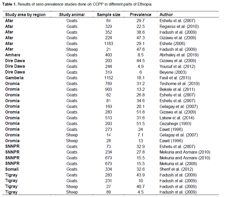
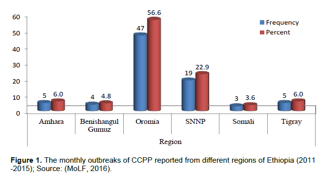
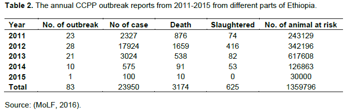
In Ethiopia the presence of CCPP has been suspected since 1983. It was confirmed later in 1990 by isolation and identification of Mccp from an outbreak of the disease in Ogaden, Eastern Ethiopia by Thiaucourt et al. (1992). Since then the disease has been known to be endemic in different regions of the country (Lakew et al., 2014). Limited availability of vaccine, shortage of antibiotics, high cost of treatment and scarce veterinary services aggravated the situation in the remotest parts of the country (Mekuria and Asmare, 2010). CCPP has been reported from almost all regions of Ethiopia (Asmare et al., 2016). The disease is endemic in most extensive goat rearing areas of Ethiopia (Gelagay et al., 2007); mainly in the arid and semi-arid low lands of rift valley, Borana rangelands, South Omo, Afar and other pastoral areas of Ethiopia where about 70% the national goat population are found (Amare, 2012).Different sero-prevalence has been reported in goats and sheep by various authors as shown in (Table 1). In addition to the different sero-prevalence studies conducted, there have been monthly reports of disease outbreaks to the Ministry of Livestock and Fisheries (MoLF), veterinary department from different regions of the country as indicated below in Figure 1 and the annual outbreak reports from 2011-2015 obtained from Ministry of Livestock and Fisheries (MoLF) are shown in Table 2.
CONCLUSION
This reviewed literature revealed that outbreaks of CCPP have been occurring throughout Africa and Asia. It has considerable effects on the livelihoods of poor farmers where goat production plays important role. The status of the disease is not well known and people have little or no awareness on the potential risk of the disease. Besides, the lack of affordable, easy to use and reliable diagnostic tools and control measures hampers the efforts to prevent the effects of the disease on the economy of thesector. Hence, emphasis is needed in the following areas.
RECOMMENDATIONS
1) Small ruminant rearing countries should promote further research focusing on the understanding of its geo-spatial epidemiological status so as to design a feasible control strategy
2) Improvement in the production of vaccine and vaccination coverage
3) Development of new diagnostic tools 4) Awareness creation about CCPP, transmission path way, prevention and control methods, for small ruminant farmers
5) Promotion of regional and international collaborations for development of efficient control strategies.
CONFLICT OF INTERESTS
The authors have not declared any conflicts of interests.
REFERENCES
|
Abbass S (2006). A study on contagious caprine pleuropneumonia in four selected states in the Sudan. MSc Thesis, University of Khartoum, Faculty of Veterinary Science, Khartoum, Sudan, pp. 1-88. |
|
|
Abd-Elrahman AH, Khafaga AF, Abas OM (2019). The first identification of contagious caprine pleuropneumonia (CCPP) in sheep and goats |
|
|
in Egypt: molecular and pathological characterization. Tropical Animal Health and Production 52(3):1179-1186. |
|
|
Abrhaley A, Ejo M, Fentie T (2019). Seroprevalence and Risk Factors Associated with Contagious Caprine Pleuropneumonia in Western Amhara, Northwest Ethiopia. Journal of Veterinary Medicine 1-7. |
|
|
Ahaduzzaman M (2020). Contagious caprine pleuropneumonia (CCPP): A systematic review and metaanalysis of the prevalence in sheep and goats. Transboundary and Emerging Diseases 00:1-13. |
|
|
Alemayehu G, Hailu B, Seid N (2015). Quality Constraints in the Market Chains for Export of Small Ruminants from Afar Pastoral and Agro-Pastoral Areas. Animal and Veterinary Sciences 3(2):51. |
|
|
Amare G (2012). Sero-prevalence and participatory study of contagious caprine plueropneumonia in Gulina Wereda, Afar National Regional State, Ethiopia. DVM. Thesis, FVM. AAU, Debrezeit, Ethiopia. |
|
|
Arif A, Schulz J, Thiaucourt F, Taha A, Hammer S (2007). Contagious caprine pleura pneumonia outbreak in captive wild ungulates at Al Wabra wildlife preservation, state of Qatar. Journal of Zoo and Wildlife Medicine 38(1):93-96. |
|
|
Armson B, Ekiri AB, Alafiatayo R, Cook AJ (2021). Small ruminant production in Tanzania Uganda, and Ethiopia: A systematic review of constraints and potential solutions. Veterinary Science 8(5):1-13. |
|
|
Asmare K, Abayneh T, Mekuriaa S, Ayelet G, Sibhat B, Skjerve E, Szonyi B, Wieland B (2016). A meta-analysis of contagious caprine pleuropneumonia (CCPP) in Ethiopia. Acta Tropica 158:231-239. |
|
|
Atim S, Ayebazibwe C, Mwiine F, Erume J, Tweyongyere R (2016). A Survey for contagious caprine pleuropneumonia in Agago and Otuke districts in Northern Uganda. Open Journal of Veterinary Medicine 6(1):9-14. |
|
|
AU-IBAR (2013). Contagious caprine pleuropneumonia. African union Inter African bureau for animal resources. Www/AU-IBAR Org. |
|
|
AU-IBAR (2015). Standard methods and procedures (SMPs): for contagious caprine pleuro pneumonia (CCPP) in the Greater Horn of Africa, Nairobi. |
|
|
Awan MA, Abbas F, Yasinzai M, Nicholas RAJ, Babar S, Ayling RD, Attique MA, Ahmad Z (2009): Prevalence of Mycoplasma capricolum subspecies capricolum and Mycoplasma putrefaciens in goats in Pishin district of Balochistan. Pakistan Veterinary Journal 29(4):179-185. |
|
|
Awan MA, Abbas F, Yasinzai M, Nicholas RAJ, Babar S, Ayling RD, Attique MA, Ahmed Z, Wadood A, Khan FA (2010). First report on the molecular prevalence of Mycoplasma capricolum subspecies capripneumoniae (Mccp) in goats the cause of contagious caprine pleuropneumonia (CCPP) in Balochistan province of Pakistan. Molecular Biology Reports 37(7):3401-3406. |
|
|
Balikci E, Kizil O, Karapinar T, Karahan M, Ozdemir H, Dabak M (2008): Efficacy of marbofloxacin for natu- rally occurring contagious caprine pleuropneumonia. Small Ruminant Research 77(1):75-79. |
|
|
Bascunana CR, Mattsson JG, Bolske G, Johansson KE (1994): Characterization of the 16S rRNA genes from Mycoplasma species strain F38 and development of an identification system based on PCR. Journal of Bacteriology 176(9):2577-2586. |
|
|
Bekele T, Asfaw Y, GebreEgziabeher B, Getachew Abebe G (2011). Sero-prevalence of contagious caprine pleuropneumonia in Borana and Guji lowlands, Southern Ethiopia. Ethiopia Veterinary Journal 15(2):69-76. |
|
|
Belachew H (2004). Livestock marketing and animal health in Ethiopia.In proceedings of the 18th Annual Conference of the Ethiopian Veterinary Association (EVA), Addis Ababa. Ethiopia P 52. |
|
|
Beyene N (2003). Epidemiology and Sero-surveillance of contagious caprine pleuropneumonia in Dire Dawa Administrative Region, Ethiopia. DVM Thesis, AAU, faculty of Veterinary medicine, Debrezeit, Ethiopia pp. 1-23. |
|
|
Bolske G, Johansson K, Heinonen R, Panvuga P, Twinamasiko E (1995). Contagious caprine pleuropneumonia in Uganda and isolation of Mycoplasma capricolum subsp. capripneumoniae from goats and sheep. Veterinary Record 137(23):594. |
|
|
Bradbury JM (1983). Methods in Mycoplasmology, Mycoplasma characterization. New York, USA: Academic Press pp. 363-366. |
|
|
Cetinkaya B, Kalin R, Karahan M, Atil E, Manso-Silván L, Thiaucourt F (2009). Detection of contagious caprine pleuropneumonia in East Turkey. |
|
|
Chaber A, Lignereux L, Qassimi M, Saegerman C, Manso-Silva'n L, Dupuy V, Thiaucourt F (2014). Fatal transmission of contagious caprine pleuropneumonia to an Arabian oryx (Oryx leucoryx). Veterinary Microbiology 173(1):156-159. |
|
|
Chu Y, Gao P, Zhao P, He Y, Liao N, Jackman S, Zhao Y, Birol I, Duan X, Lu Z (2011). Genome sequence of Mycoplasma capricolum subsp. capripneumoniae strain M1601. Journal of Bacteriology 193(21):6098-6099. |
|
|
Coppock D (1994). The Borana plateau of southern Ethiopia: Synthesis of pastoral research development and change, 1980-91. ILCA (International Livestock Center for Africa), Addis Ababa, Ethiopia 5:150-160. |
|
|
CSA (2015). Agricultural Sample Survey. Volume II report on livestock and livestock characteristics (private peasant holdings). Central Statistical Agency (CSA): Addis Ababa, Ethiopia. |
|
|
DaMassa A, Wakenell P, Dale Brooks D (1992). Review article Mycoplasmas of goats and sheep. Journal of Veterinary Diagnostic Investigation 4(1):101-113. |
|
|
Dawit K (1996). Serological and microbiological studies of major respiratory mycoplasmoses of goats in Yabello (Range lands of Borena), Ethiopia. DVM Thesis, Addis Ababa University, faculty of Veterinary medicine, Debre-zeit, Ethiopia pp. 1-58. |
|
|
Dupuy V, Thiaucourt F (2014). Complete genome sequence of Mycoplasma capricolum subspecies. capripneumoniae strain 9231-Abomsa. Genome Announcements 2(5):e01067-14. |
|
|
Ejaz H, Hashmi H, Awan MA, Kakar MA, Naeem M, Ameen S, Bukhari FA, Hameed T, Tariq MM (2015). 'Molecular study on the prevalence of respiratory Mycoplasma Species in small ruminants of Kuchlak , District Quetta and Khanozai , District Pishin, Balochistan' 47(2):473-478. |
|
|
El-Manakhly EM, Tharwat MA (2016). Correlation between latex agglutination test positivity and contagious caprine pleuropneumonia chronicity. Journal of Agriculture and Veterinary Science 9(2):121-135. |
|
|
Eshete G (2006). Serological and participatory epidemiological survey of CCPP in Afar pastoral areas, North East Ethiopia. In: MSc Thesis. Addis Ababa University, CVMA, Debre Zeit. |
|
|
Eshetu L, Yigezu L, Asfaw Y (2007). A study on contagious caprine pleuropneumonia (CCPP) in goats at an export oriented abattoir, Debrezeit, Ethiopia. Tropical Animal Health and Production 39(6):427-432. |
|
|
Ettorre C, Sacchini F, Scacchia M, Salda LD (2007). Pneumonia of lambs in the Abruzzo region of Italy: anatomopathological and histopathological studies and localisation of Mycoplasma ovipneumoniae. Veterinary Italian 43(1):149-155. |
|
|
Falquet L, Liljander A, Schieck E, Gluecks I, Frey J, Jores J (2014): Complete genome sequences of virulent Mycoplasma capricolum subsp. capripneumoniae strains F38 and ILRI181. Genome Announcement 2(5):e01041-14. |
|
|
Food and Agriculture Organization (FAO) (2012). Online retrieved from: www. Disaster risk reduction. Net/ FAO disease cards CCPP. Doc. |
|
|
Fasil A, Yilkal A, Getnet A (2015). Epidemiological study of contagious caprinepleuropneumonia (CCPP) in selected districts of Gambella Region, Western Ethiopia. African Journal of Agricultural Research 10(24):2470-2479. |
|
|
Fischer A, Santana-cruz I, Hegerman J, Gourlé H, Schieck E, Lambert M, Nadendla S, Wesonga H, Miller RA, Vashee S, Weber J, Meens J (2015). High quality draft genomes of the Mycoplasma mycoides subsp. mycoides challenge strains Afadé and B237. Standards in Genomic Sciences 10(89):1-11. |
|
|
Fischer A, Shapiro B, Muriuki C, Heller M, Schnee C, Bongcam-Rudloff E, Vilei E, Frey J, Jores J (2012). The Origin of the 'Mycoplasma mycoides cluster' coincides with domestication of ruminants. PLoS One 7(4):3-8. |
|
|
Gelagay A, Teshale S, Amsalu W, Esayas G (2007). Prevalence of contagious caprine pleuropneumonia in the Borana pastoral areas of Ethiopia. Small Ruminant Research 70(2):131-135. |
|
|
Gezahegn M (1993). Preliminary study of contagious caprine pleuropneumonia (CCPP) in selected sites of East Shewa. DVM Thesis.AAU/FVM pp. 1-62. |
|
|
Giadinis ND, Petridou EJ, Sofianidis G, Filioussis G, Psychas V, Hatzopoulous E, Haratzias H (2008). Mortality in adult goats attributed to Mycoplasma capricolum subspecies capricolum. Veterinary Record 163(9):278-279. |
|
|
Gizawu D, GebreEgziabher B, Ayelet G, Asmare K (2009). Investigation of mycoplasma infection in goats slaughtered at ELFORA export abattoir, Ethiopia. Ethiopian Veterinary Journal 13(1):41-58. |
|
|
Gobena MM (2016). Production Performance, Challenges and Opportunity of Goat Production in Ethiopia. Advances in Life Science and Technology 50:26-35. |
|
|
Goncalves R, Mariano I, Nunez A, Branco S, Fairfoul G, Nicholas R (2010). A typical non-progressive pneumonia in goats. Veterinary Journal 183:219-221. |
|
|
Hadush B, Eshetu L, Mengistu W, Hailesilassie M (2009). Sero-prevalence of contagious caprine pleuropneumonia in Kefta Humera, Alamata (Tigray) and Abaala (Afar), Northern Ethiopia. Tropical Animal Health and Production 41(5):803-806. |
|
|
Harbi MSMA, El-Tahir MS (1981). Mycoplasma strain F38 and contagious caprine pleuropneumonia in the Sudan. Veterinary Record 108(12):261. |
|
|
Heldtander M, Wesonga H, Bolske G, Pettersson B, Johansson K (2001). Genetic diversity and evolution of Mycoplasma capricolum subsp. capripneumoniae strains from eastern Africa assessed by 16S rDNA sequence analysis. Veterinary Microbiology 78(1):13-28. |
|
|
Houshaymi BM, Miles RJ, Nicholas RAJ (2000). Studies on strains of M. capricolum subsp. capripneumoniae isolated from outbreaks of contagious caprine pleuropneumonia. Small Ruminant Research 45:139-143. |
|
|
Houshaymi B, Tekleghiorghis T, Wilsmore AJ, Miles RJ, Nicholas RAJ (2002). Investigations of outbreaks of contagious caprine pleuropneumonia in Eritrea. Tropical Animal Health and Production 34(5):383-389. |
|
|
Hussain R, Auon M, Khan A, Khan MZ, Mahmood F, UR-rehman S (2012). Caprine pleuropneumonia in Beetal goats. Tropical Animal Health and Production 44(3):675. |
|
|
Iqbal Yatoo M, Raffiq Parray O, Tauseef Bashir S, Muheet Ahmed Bhat R, Gopalakrishnan A, Karthik K, Dhama K, Vir Singh S (2019). Contagious caprine pleuropneumonia-a comprehensive review. Veterinary Quarterly 39(1):1-25. |
|
|
Jones GE, Wood AR (1988). Microbiological and serological studies on caprine pneumonias in Oman. Research in Veterinary Science 44(1):125-131. |
|
|
Kamani J, Baneth G, Shimon Harrus S (2019). An annotated checklist of tick-borne pathogens of dogs in Nigeria. Veterinary Parasitology: Regional Studies and Reports 15:100255. |
|
|
Kipronoh K, Ombuib J, Binepald Y, Wesongae H, Gitongae E, Thuranirad E, Kiarac H (2016). Risk factors associated with contagious caprine pleuro-pneumonia ingoats in pastoral areas in the Rift Valley region of Kenya. Preventive Veterinary Medicine 132:107-112. |
|
|
Kumar A, Garg DN (1991). Isolation of mycoplasma F-38 from the milk of mastitic cows. Veterinary Record 128(18):429. |
|
|
Kumar CP, Goud KS, Sundar NS, Prasad VD (2015). Diagnosis and management of contagious caprine pleuropneumonia. Intas Polivet 16(2):404-406. |
|
|
Kumar P, Roy A, Bhanderi BB, Pal BC (2011): Isolation, identification and molecular characterization of Mycoplasma isolates from goats of Gujarat State, India' 81(4):443-458. |
|
|
Kusiluka LJ, Semuguruka WD, Kazwala RR (2000). Demonstration of Mycoplasma capricolum subsp. capripneumoniae and Mycoplasma mycoides subsp. mycoides, Small Colony Type in Outbreaks of Caprine Pleuropneumonia in Eastern Tanzania. Acta Veterinaria Scandinavica 41(3):311-319. |
|
|
Lakew M, Sisay T, Ayelet G, Eshetu E, Dawit G, Tadele T (2014). Sero-prevalence of contagious caprine pleuropneumonia and field performance of inactivated vaccine in Borana pastoral area, southern Ethiopia. African Journal of. Microbiology Research 8(24):2344-2351. |
|
|
Leach RH, Erno H, MacOwan KJ (1993). Proposal for designation of F38-type caprine Mycoplasmas as Mycoplasma capricolum subsp. capripneumoniae subsp. nov. and consequent obligatory relegation of strains currently classified as M. capricolum (Tully, Barile, Edward, Theodore, and Erno, 1974) to an additional new subspecies, M. capricolum subsp. capricolum subsp. nov. Journal of Systematic Bacteriology 43(3):603-605. |
|
|
Lefevre PC, Breard A, Farouk Ial, Buron S (1987). Mycoplasma species F38 isolated in Chad. Veterinary Record 121(24):575-576. |
|
|
Li Y, Wang R, Sun W, Song Z, Bai F, Zheng H, Xin J (2020). Comparative genomics analysis of Mycoplasma capricolum subsp. capripneumoniae 87001. Genomics 112:615-620. |
|
|
Li Y, Zhang JH, Hu SP, Wang L, Xin JQ (2007). Reclassification of the four China isolated strains of the pathogen for contagious caprine pleuropneumonia. Wei Sheng Wu Xue Bao 47(5):769-773. |
|
|
Liljander A, YuM O,'Brien E, Heller M, Nepper JF, Weibel DB, Gluecks I, Younan M, Frey J, Falquet L, Jores J (2015). Field applicable recombinase polymerase amplification assay for rapid detection of Mycoplasma capricolum subspecies capripneumoniae. Journal of Clinical Microbiology 53(9):2810-2815. |
|
|
Litamoi JK, Wonyangu S, Siman P (1990). Isolation of mycoplasma biotype F38 from sheep in Kenya. Tropical Animal Health and Production 22(4):260-262. |
|
|
Loire E, Ibrahim AI, Manso-Silván L, Lignereux L, Thiaucourt FA (2020). Whole-genome worldwide molecular epidemiology approach for contagious caprine pleuropneumonia. Heliyon 6(10). |
|
|
MacOwan KJ, Minette JE (1976). A mycoplasma from acute contagious caprine Pleuropneumonia in Kenya. Tropical Animal Health and Production 8(2):91-95. |
|
|
Manso-Silvan L, Dupuy V, Chu Y, Thiaucourt F (2011). Multi locus sequence analysis of Mycoplasma capricolum subsp. capripneumoniae for the molecular epidemiology of contagious caprine pleuropneumonia. Veterinary Research 42(1):86. |
|
|
March J, Gammack C, Nicholas R (2000). Rapid detection of contagious caprine pleuropneumonia using a Mycoplasma capricolum subsp. capripneumoniae capsular polysaccharide-specific antigen detection latex agglutination test. Journal of Clinical Microbiology 38(11):4152-4159. |
|
|
Mbyuzi A, Erick V, Komba E, Kimera S, Kambarage D (2014). Sero-prevalence and associated risk factors of peste des petitsruminants and contagious caprine pleuropneumonia in goats and sheep in the Southern Zone of Tanzania. Preventive Veterinary Medicine 116(1):138-144. |
|
|
McMartin D, MacOwan K, Swift L (1980). A century of classical contagious caprine pleuropneumonia: from original description to aetiology. British Veterinary Journal 136:507-515. |
|
|
Mekuria S, Asmare K (2010): Cross sectional study on contagious caprine pleuropneumonia in selected districts of sedentary and pastoral production systems in Southern Ethiopia. Tropical Animal Health and Production 42(1):65-72. |
|
|
Mekuria S, Zerihun A, Gebre-Egziabher B, Tibbo M (2008). Participatory investigation of contagious caprine pleuropneumonia (CCPP) in goats in the Hammer and Benna Tsemay Districts of Southern Ethiopia. Tropical Animal Health and Production 40(8):571-582. |
|
|
MoLF (2016): Monthly disease outbreak report. Ministry of Livestock and Fish. Addis Ababa, Ethiopia. |
|
|
Monnerat MP, Thiaucourt F, Nicolet J, Frey J (1999). Comparative analysis of the lppA locus in Mycoplasma capricolum subsp. capricolum and Mycoplasma capricolum subsp. capripneumoniae. Veterinary Microbiology 69(3):157-72. |
|
|
Msami HM, kapaga AM, Heldtander M, Bolske G (2001). Contagious caprinepleuropneumonia in Tanzania. Veterinary Record 148(1):22-23. |
|
|
Muthomi EK, Rurangirwa FR (1983). Passive haemagglutination and complement-fixation as diagnostic tests for contagious caprine pleuropneumonia caused by F-38 strain of mycoplasma. Research in Veterinary Science 35:1-4. |
|
|
Narrod, C., Tiongco, M. and Scott, R. (2011). Current and predicted trends in the production, consumption and trade of live animals and their products. OIE Revue Scientifique et Technique 30(1):31-49. |
|
|
Nicholas R, Churchward C (2012). Contagious Caprine Pleuropneumonia: New Aspects of an Old Disease. Transboundary and Emerging Diseases 59(3):189-196. |
|
|
Nicholas RAJ (2002). Nicholas RAJ. Improvements in the diagnosis and control of diseases of small ruminants caused by mycoplasmas. Small Ruminant Research 45(2):145-149. |
|
|
Niclholas R, Ayling R, McAuliffe L (2008). contagious caprine pleuropneumonia. In: Mycoplasma diseases of ruminants. |
|
|
Wallingford, UK: CAB International pp. 114-131. |
|
|
Noah EY, Kusiluka LJM, Wambura P, Kimera SI, Veterinary C, Salaam E (2011): Field Isolation of Mycoplasma capripneumoniae in Central Zone of Tanzania 3(6):434-442. |
|
|
Nyanja P, Kusiluka L, Mellau S (2013). Prevalence of contagious caprine pleuropneumonia in goats in Musoma District of Mara Region, Tanzania. Tanzania Veterinary Journal 28(1). |
|
|
OIE (2008). Contagious caprinepleuropneumonia. Manual of standards for diagnostic tests and vaccines.Office of International Epizootics, Paris pp. 1000-1012. |
|
|
OIE (2009). World Animal Health Information Database - Version: 1.4. World Animal Health Information Database. Paris, France. |
|
|
OIE (2014). Contagious caprine pleuropneumonia, OIE Tersterial Animal health code. |
|
|
OIE (2015). Animal Health in the World. OIE Listed Diseases. (Accessed at http: //www. oie. nt/ on 9/2/2016). |
|
|
Ozdemir U, Loria GR, Godinho KS, Samson R, Rowan TG, Churchward C, Ayling RD, Nicholas RAJ (2006). Effect of danofloxacin (Advocin A180) on goats affected with contagious caprine pleuropneumonia. Tropical Animal Health and Production 38(7):533-540. |
|
|
Pawar PD, Singla LD, Kaur P, Bal MS, Javed M (2019). Evaluation and correlation of multiple anthelmintic resistance to gastrointestinal nematodes using different faecal egg count reduction methods in small ruminants of Punjab, India. Acta Parasitologica 64(3):456-463. |
|
|
Perreau P, Breard A, Goff C le (1984). Experimental infection of goats with type F.38 mycoplasma strains (CCPP). Annales de Microbiology 135(1):119-124. |
|
|
Pettersson B, Leitner T, Ronaghi M, Bolske G, Uhlen M, Johansson KE (1996). Phylogeny of the Mycoplasma mycoides cluster as determined by sequence analysis of the 16S rRNA genes from the two rRNA operons. Journal of Bacteriology 178(14):4131-4142. |
|
|
Peyraud A, Poumarat F, Tardy F, Manso-Silván L, Hamroev K, Tilloev T, Amirbekov M, Tounkara K, Bodjo C, Wesonga H, Nkando IG, Jenberie S, Yami M, Cardinale E, Meenowa D, Jaumally MR, Yaqub T, Shabbir MZ, Mukhtar N, Halimi M., Ziay GM, Schauwers W, Noori H, Rajabi AM, Ostrowski S, Thiaucourt F (2014). An international collaborative study to determine the prevalence of contagious caprine pleuropneumonia by monoclonal antibody-based cELISA. BMC Veterinary Research 10(1):48-57. |
|
|
Prats-van der Ham M, Amores Fedela C, Paterna J, Tatay-Dualde A, Gomez-Martin Á (2015). Contagious caprine pleuropneumonia (CCPP) and other emergent mycoplasmal diseases affecting small ruminants in arid landsJournal of Arid Environments 119:9-15. |
|
|
Prats-van der ham M, De la Fe C, Amores J, Paterna A, Tatay-dualde J, Gómez-Martín A. (2015). Contagious caprine pleuropneumonia (CCPP) and other emergent mycoplasmal diseases affecting small ruminants in arid lands. Journal of Arid Environments 119:9-15. |
|
|
Radostitis OM, Blood DC, Cray CC (2007). Veterinary Medicine: A textbook of the disease of cattle, sheep, goats, pigs and horses. 8th eds, Baillier Tindall. London, p. 1214. |
|
|
Regassa F, Netsere M, Tsertse T (2010). Sero-prevalence of contagious caprine pleuropneumonia in goat at selected Woredas of Afar Region. Ethiopian Veterinary Journal 14(1):83-89. |
|
|
Rurangirwa FR, McGuire TC (2016). Contagious caprine pleuropneumonia: Diagnosis and control. Online retrieved from http:// www.fao.org/ wairdocs/ilri/ x5473b/ x5473b11. |
|
|
Rurangirwa FR. (1996). Contagious caprine pleuropneumonia. In: Manual of Standards for Diagnostic Tests and Vaccines. Office. International des Epizooties, pp. 374-383. |
|
|
Rurangirwa FR, Kouyate B, Niang M, McGuire TC (1990). CCPP antibodies to F38 polysaccharide in Mali goats. Veterinary Record 127(14):353. |
|
|
Rurangirwa FR, McGuire TC, Mbai L, Ndung'u L, Wambugu A (1991): Preliminary Field Test of Lyophilised Contagious Caprine Pleuropneumonia Vaccine. Research in veterinary science 50(2):240-241. |
|
|
Rurangirwa F, McGuire T, Kibor A, Chema S. (1987). An inactivated vaccine for contagious caprine pleuropneumonia. Veterinary Record 121(17):397-400. |
|
|
Sadique U (2010). Studies on Pathogenesis and Molecular Characterization of Contagious Caprine Pleuropneumonia in Small Ruminants. MSc Thesis, Department Of Pathology, University of Veterinary and Animal Sciences, Lahore, Pakistan, pp. 1-184. |
|
|
Sadique U, Chaudhry ZI, Younas M, Anjum AA, Hassan ZU, Idrees M, Mushtaq M, Sajid A, Sabtain M, Jhang AS, Sciences A (2012). Molecular Characterization of Contagious Caprine Pleuropneumonia (CCPP) in Small Ruminants of Khyber Pakhtunkhwa. Pakistan Materials and Methods 22(2):33-37. |
|
|
Samiullah S (2013). Contagious caprine pleuropneumonia and its current picture in pakistan: A review', Veterinarni Medicina 58(8).389-398. |
|
|
Shahzad W, Munir R, Khan MS, Ahmad MUD, Khan MA, Ijaz M, Shakil M, Iqbal M, Ahamd R (2012). Characterization, Molecular Diagnosis and Prevalence of Caprine Mycoplasmosis in Different Areas of Pakistan. Pakistan Journal of Zoology 44(2):559-568. |
|
|
Shahzad W, Yaqoob T, Mukhtar N, Munir R, Ahmad R, Khan M, Hussain A (2016). Sero-prevalence of Mycoplasma capricolum subsp. capripneumoniae in goats through cELISA in different districts of Punjab, Pakistan. Journal of Animal and Plant Sciences 26(4):931-937. |
|
|
Shapiro BI, Gebru G, Desta S, Negassa A, Nigussie K, Aboset G, Mechale H (2017). Ethiopia livestock sector analysis. ILRI Project Report. Nairobi, Kenya: International Livestock Research Institute (ILRI). |
|
|
Sheikh FD, Wani SA, Fatima K, Masoodi TH. (2016). Contagious caprine pleuropneumonia: a major threat to pashmina farming in Ladakh pp. 1-8. |
|
|
Sherif M, Addis M, Tefera M (2012). Contagious caprine pleuropneumonia: serological survey in selected districts of Jijiga Zone, Ethiopia. Asian Journal of Animal Sciences 6(6):309-315. |
|
|
Singh E, Kaur P, Singla LD, Bal MS (2017). Prevalence of gastrointestinal parasitism in small ruminants in western zone of Punjab, India. Veterinary World 10(1):61-66. |
|
|
Smith MC, Sherman DM (2009). Liver and pancreas. In: Goat Medicine, 2nd Ed, Wiley-Blackwell, State Avenue, Lowa USA pp. 513-515. |
|
|
Srivastava A, Meenowa D, Barden G, Salguero F, Cherchward C, Nicholas R (2010). Contagious caprine pleuropneumonia in Mauritius. Veterinary Record 167(8):304-305. |
|
|
Swai ES, Kaaya JE, Noah EY (2013). Antibody response to Mycoplasma capricolum subsp. capripneumoniae bacterium in small holder dairy goats in Tanzania. Tropical animal Health and Production 45(7):1603-1608. |
|
|
Tesgera T, Sori H, Yami M, Mamo B (2017). Evaluation of safety and immunogenicity of inactivated whole culture contagious caprine pleuropneumonia trial vaccine in National Veterinary Institute, Ethiopia. African Journal of Microbiology Research 11(11):466-473. |
|
|
Teshome D, Sori T, Sacchini F, Wieland B (2019). Epidemiological investigations of contagious caprine pleuropneumonia in selected districts of Borana zone, Southern Oromia, Ethiopia. Tropical Animal Health and Production 51(3):703-711. |
|
|
Tharwat M, Al-Sobayil F. (2017). Ultrasonographic findings in goats with contagious caprine pleuropneumonia caused by Mycoplasma capricolum subsp. capripneumoniae. BMC Veterinary Research 13(1):263. |
|
|
Thiacourt F, Libbeau G, LeFoff C, Lefevere PC (1994). The use of monoclonal antibody in diagnosis of contagious caprine pleuropneumonia. Veterinary Microbiology 41(3):191-2003. |
|
|
Thiaucourt F, Bolske G (1996): Contagious caprine pleuropneumonia and other pulmonary mycoplasmoses of sheep and goats. Revue Scientifique et Technique-Office International des Epizooties 15(4):1397-1414. |
|
|
Thiaucourt F, Bolske G, Leneguersh B, Smith D, Wesonga H. (1996). Diagnosis and control of contagious caprine pleuropneumonia. Revue Scientifique et Technique-Office International des Epizooties15(4):1415-1429. |
|
|
Thiaucourt F, Lorenzon S, David, A. and Breard, A. (2000). Phylogeny of the Mycoplasma mycoides cluster as shown by sequencing of a putative membrane protein gene. Veterinary Microbiology 72(3):251-268. |
|
|
Thiaucourt F, Pible O, Miotello G, Nwankpa N, Armengaud J,(2018.Improving quality control of Contagious Caprine Pleuropneumonia vaccine with tandem mass spectrometry.Proteomics 1800088. |
|
|
Urgessa D, Duguma B, Demeka S, Tolamariam T (2012). Sheep and goat production systems in Iluabbabora zone of Oromia regional state, Ethiopia: Feeding and management strategies. Global Veterinaria 9(4):421-429. |
|
|
Van Belkum A, Tassios PT, Dijkshoorn L, Haeggman S, Cookson B, Fry NK, Fussing V, Green J, Feil E, Gerner Smidt P, Brisse S, Struelens M (2007). European Society of Clinical Microbiology and Infectious Diseases (ESCMID). Study Group on Epidemiological Markers (ESGEM). Clinical Microbiology and Infectious Disease 13(3):1-46. |
|
|
Wamwayi HM, Wafula JS, Litamoi JK, Nandokha EN (1989). Detection of antibody to mycoplasma F38 in goat sera by an enzyme-linked immunosorbent assay. Tropical Animal Health and Production 21(1):43-49. |
|
|
Wazir I, Hussain I, Khan M, Ali M, Rahman H, Ashraf F, Khan S, Khan B, Ullah S, Ullah Q, khyber K, Khan A (2016). Seroepidemiological analysis of contagious caprine pleuropneumonia through cELISA in selected districts of Khyber Pakhtunkhwa, Pakistan. ASRJETS 26(3):274-281. |
|
|
Wodajo HD, Gemeda BA, Kinati W, Mulem AA, van Eerdewijk A, Wieland B (2020). Contribution of small ruminants to food security for Ethiopian smallholder farmers. Small Ruminant Research 184(106064):1-10. |
|
|
Woubit S, Lorenzon S, Peyraud A, Manso-Silvan L, Thiaucourt F (2004). A specific PCR for the identification of Mycoplasma capricolum subsp. capripneumoniae, the causative agent of contagious caprine pleuropneumonia (CCPP). Veterinary Microbiology 104(1):125-132. |
|
|
Younis E, Elnaker Y, Awad N, Elshafey D (2015). Sero-prevalence of Mycoplsma Mycoides cluster in small ruminant using monoclonal antibody based C ELISA in Dakahilia Province. Assiut Veterinary Medical Journal 61(146):46-51. |
|
|
Yousuf E, Melaku A, Bogale B (2012). Sero-prevalence of contagious caprine pleuropneumonia in Dire Dawa provisional administrative council, Eastern Ethiopia. Journal of. Veterinary Medicine and Animal Health 4(7):93-96. |
|
|
Yu Z, Wang T, Sun H, Xia Z, Zhang K, Chu D, Xia X (2013). Contagious caprine pleuropneumonia in endangered Tibetan Antelope, China, 2012. Emerging Infectious Diseases journal 19(12):2051-2053. |
|
Copyright © 2024 Author(s) retain the copyright of this article.
This article is published under the terms of the Creative Commons Attribution License 4.0


