Journal of
Veterinary Medicine and Animal Health

  • Abbreviation: J. Vet. Med. Anim. Health
  • Language: English
  • ISSN: 2141-2529
  • DOI: 10.5897/JVMAH
  • Start Year: 2009
  • Published Articles: 416

Review

Incidence and the history of Echinococcus granulosus infection in dogs within the past few decades in Libya: A review

Mohamed, M. Ibrahem
  • Mohamed, M. Ibrahem
  • Department of Zoology, Faculty of Sciences, University of Zawia, P. O. Box 16418, Zawia, Libya.
  • Google Scholar
Wafa, M. Ibrahem
  • Wafa, M. Ibrahem
  • Department of Parasitology, Faculty of Medicine, University of Zawia, P. O. Box 16418, Zawia, Libya.
  • Google Scholar
Kawther, M. Ibrahem
  • Kawther, M. Ibrahem
  • Department of Medicinal Chemistry, Faculty of Pharmacy, University of Zawia, P. O. Box 16418, Zawia, Libya.
  • Google Scholar
Badereddin, B. Annajar
  • Badereddin, B. Annajar
  • National Centre for Disease Control, Ain Zara, P. O. Box 71171, Tripoli, Libya.
  • Google Scholar


  •  Received: 19 September 2016
  •  Accepted: 16 December 2016
  •  Published: 31 January 2017

 ABSTRACT

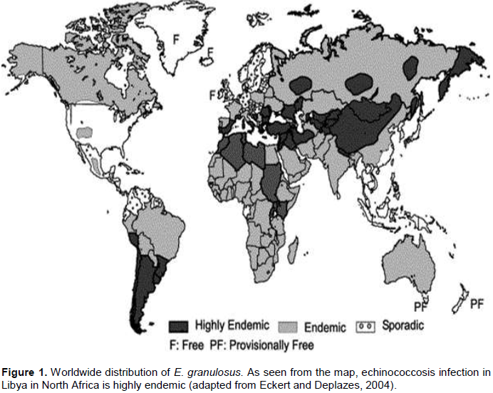
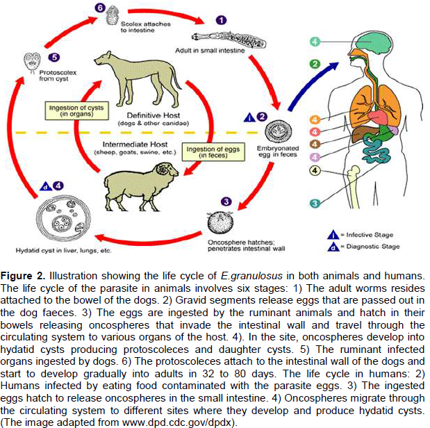
Echinococcus granulosus is a tiny tapeworm that parasitizes the small intestine of canids, mainly dogs, which act as definitive hosts for the parasite. Infected dogs are the main source of infection to humans and livestock which act as intermediate hosts resulting in hydatid disease condition. E. granulosus is widely distributed in many parts of the world, and is very common in North African countries. In Libya, the rate of infection with echinococcosis in dogs was reported to be lower than 7 to 80% in stray dogs, 34.8 to 60% in sheep/guard dogs and 7.7 to 21.6% in farm/house dogs. This data fulfills the world health organization (WHO) criteria and suggests that the incidence of infection with echinococcosis/ hydatidosis in some parts of the country can be reaching the level of hyper endemic. Diagnosis of echinococcosis in infected dogs can be performed by isolating the parasite from their faeces or from the contents of their small intestine after necropsy. Recent developments in immunodiagnostic assays for echinococcosis in dogs have been described. Public health and risk factors as well as ways of hydatid disease treatment and various control strategies, including the use of veterinary vaccines, have also been discussed.


Key words: Dogs, Echinococcus granulosus, prevalence, diagnosis, treatment, risk factors


 INTRODUCTION

Echinococcus granulosus is a tapeworm that causes a condition known as echinococcosis in dogs, and hydatid disease or hydatidosis in humans and other ruminant animals (Chhabra and Singla, 2009). The parasite has been reported to occur in many parts of the world, and is very common in  some  agricultural  regions,  particularly, Northern Africa, Southern South America, Europe, the Middle East, South-Western Asia, and Australia (Figure 1), (Eckert and Deplazes, 2004). In these areas, the infection rate with E. granulosus in dogs was reported to be between endemic and hyper endemic (Dakak, 2010). The    parasite    requires   two    mammalian    hosts   for completion of its life cycle; a definitive host, which is mainly carnivore for the adult stage and an intermediate host, which is mainly ungulates for the larval (hydatid cyst) stage (Figure 2). The life cycle of E. granulosus in Libya has a pastoral or domestic cycle in which dogs acquire the infection by eating the internal organs of infected sheep, goats, camels and cattle.


However, dogs in Libya are considered to be the main source of infection with cystic echinococcosis (CE) to various species of livestock as well as human. This cycle is potentially very important especially in areas where sheep farming is more common (Maureen, 2008). The adult form of the parasite is a minute white tapeworm, few millimeters long (3-7 mm) with three proglottids (segments) and some other features which help in species diagnosis morphologically (Thompson, 1995).
 
Due to the size of the parasite, dogs can carry hundreds or even thousands of them without showing any signs of illness at all. The parasites attach themselves to the wall of the small intestine of the definitive host using their hooks and in this place; they deposit a large number of eggs which are intermittently passed out in the host faeces. When the eggs are in the environment,  they   are   dispersed   by  different  means including wind, water, birds as well as through the fur of the infected dogs which is likely to become contaminated with the parasite eggs.
 
Eggs can also be found on the bodies of other animals sharing the same living environment, making them a source of transmission and distribution of the infection to humans and other ruminant animals. The intermediate hosts become infected with hydatid disease when they ingest the parasite eggs in their contaminated food or water. Once the eggs are ingested, they hatch in the duodenum releasing their embryos (the oncospheres) which subsequently penetrate the intestinal wall of the host entering the mesenteric vessels. They are then carried by the blood to the major filtering organs, mainly liver and lungs, but other sites may become involved, including the abdominal wall, brain, kidneys, bones, muscles and orbits (Polat et al., 2003; Bal et al., 2008).
 
The oncospheres requires about one year to transform and develop into full larval hydatid cysts with numerous tiny protoscoleces which are formed via asexual reproduction. Humans act as an intermediate hosts for E. granulosus and are infected when they accidentally ingest the parasite eggs from the definitive host by any way.
 
The life cycle of the parasite is complete when dogs ingest hydatid cysts containing fully developed protoscoleces, which are subsequently released and attach themselves to the intestinal lining of the host. The protoscoleces start to develop into mature adult tapeworms within 32 - 80 days, depending on the species and the parasite strain. Humans are described as ‘dead-end’ hosts for the parasite, since the life cycle is usually completed when carnivores eat infected herbivores (McManus et al., 2003; Zhang et al., 2003).
 
However, it is mentioned in literature that, infected dead human bodies may contribute to complete the life cycle of the disease in some African villages where burial is not properly practiced and dogs can get access to those dead bodies (Macpherson et al., 1983). The research methodology consists of over 200 published articles on E. granulosus from different sources. The most appropriate publications covering the different sections in this paper were selected. This paper focuses mainly on the prevalence of E. granulosus infections in dogs in Libya since its discovery in 1961 to date and summarises the prospects of diagnosis, risk factors, treatment and the control and prevention strategies of the disease.
 
This study intends to provide base line epidemiological information on the incidence and the status of echinococcosis in dogs in Libya, and can be a source of information for the future studies.
 
Prevalence and the history of Echinococcus granulosus infection in dogs
 
According to scientists, the close relationship between humans and dogs started several thousands of years ago; this relationship facilitates the transmission of so many diseases including cystic hydatid disease.
 
Dogs in Libya, like any other country, can be classified into three groups, based on the type of their relationship to humans and other livestock: stray dogs, herding/sheep dogs and farm/house dogs. Stray dogs are the largest group in all urban and rural areas and they normally wonder freely during the night to scavenge on household waste materials, from which they may become infected with E. granulosus through eating dead animals and discarded offal contaminated with fertile hydatid cysts. These dogs are considered to be the main source of hydatid  disease  to  human  and  livestock   due   to  their ability to spread the parasite eggs over a wide range of areas, especially where the suitable herbivores are grazing.
 
The second group is the herding/sheep dogs which are used for shepherding and guarding sheep. These dogs may become infected with E. granulosus as a result of eating offal contaminated with fertile hydatid cysts provided by their owners or discarded by other people. Herding/sheep dogs are considered to be a source of infection to livestock by contaminating grazing pastures and water pools used for grazing and drinking respectively by the animal flocks. The third group is the farm/house dogs which are used for guarding the farm belongings including animals and the house contents from thieves.These dogs become infected with E. granulosus through contaminated meat or offal provided mostly by their owners.
 
Echinococcosis was discovered in Libya for the first time in 1961, when heavy infections with E. granulosus were detected in 60% of shepherd dogs and 10% of town dogs (Cicogna, 1961). Data available on the prevalence of echinococcosis in dogs is scattered and inadequate. Three different studies have been conducted on the prevalence of echinococcosis in stray dogs between 1986 and 1990, and the obtained results after necropsy were 11.8, 40.3, and 36.8% (Packer and Ali, 1986; Gusbi, 1987a; Awan et al., 1990) respectively. In sheep and house dogs, the infection rate with echinococcosis was reported to be 34.8 and 7.7% respectively (Gusbi, 1987a).
 
The low rate of infection with E. granulosus in house dogs compared to the other two groups of dogs is probably due to the fact that these dogs are kept within premises and under control most of the time and they rarely get access to contaminated offal. Another study on the incidence of echinococcosis in dogs was carried out by Buishi et al. (2005), who reported that 25.8% of stray dogs and 21.6% of farm/house dogs were found to be harbouring the parasite. Using Kato thick smear technique, Ben-Musa and Sadek (2007) investigated 50 samples of faeces from street dogs and found that 58% of the examined specimens were positive for E. granulosus.
 
Moreover, a recent study was conducted by Gusbi (2010) examining 151 stray dogs at post mortem from 14 localities distributed all over the country. The obtained results showed that 27.8% of the examined dogs were infected with E. granulosus. This study also elucidated that, the infection rate was generally greater in the coastal areas of the country which was between 26 and 80%, and was even worse in Zawia, El-Khumes, Misrata, Sirt and Tubruk, where more than 50% of stray dogs were found to be harbouring the parasite.
 
In contrast, a previous study showed that in southern regions such as Sebha and El-Kufra, the infection rate in stray dogs was less than 7% (Gusbi, 1987a). This could simply   be  explained  by  the  fact that huge numbers  of abattoirs exist in the highly populated northern areas and are unfortunately lacking proper disposal of offal. Also moderate temperatures and the relatively high humidity in the northern areas may contribute significantly in prolonging the survival rate of E. granulosus eggs, thus allowing for an increased chance of disease transmission and vice versa in the southern areas (Wachira et al., 1991).
 
Furthermore, the differences in local traditions of slaughtering animals in houses during social occasions and celebrations, which involves the disposal of unwanted offal and remains by feeding it to domestic dogs, may contribute to the indicated variations.
 
Public health risks associated with E. granulosus infection in dogs
 
Despite the establishment of comprehensive and successful control programmes for CE, E. granulosus still continues to have a wide geographical distribution. This may lead to the re-emergence of the disease in many endemic regions worldwide, which would easily spread from endemic to non-endemic areas, causing severe public health problems and considerable economic losses (Craig et al., 2003; McManus et al., 2003).
 
Echinococcosis is a serious zoonosis in certain rural populations where there is close contact with domestic dogs and where human infection with CE is reported to be between > 1 and < 200 cases per 100,000 populations (OIE, 2008). In most endemic areas, where domestic dogs act as a definitive host for E. granulosus, identification of risk factors for canine infection can provide useful information on potential human risk and can be useful for designing and monitoring the parasite control schemes based on treatment of infection in dogs. Agricultural or stock-raising lifestyle, low socio-economic status, climate, bad hygiene, illegal or uncontrolled slaughter, as well as uncontrolled dog populations have all been reported to be risk factors (Cetinkaya et al., 2005; King and Fairley, 2010).
 
Laboratory workers, animal handlers, veterinarians, dog owners are more prone to infection with hydatid disease due to their direct contact with the parasite eggs. On the other hand, Muslim families who have the religious practice of keeping dogs away from homes and avoiding direct contact are reported to be at low risk of being infected with CE (Akalin et al., 2014). Eggs are usually shed to the environment and may therefore, contaminate vegetables, fruits, water, or stick to animals' fur and human hands. Great hygienic care is essential, especially careful hand washing practices which constitute an important preventive measure. In humans, hydatid cysts of E. granulosus are usually developed in organs such as liver and lungs, so the symptoms of infection with the disease will be liver or lungs deficiency,
however,  X-ray,  ultrasound investigation and blood tests should be undertaken regularly for those who are in contact with possible infected dogs. Significant risk factors for copro-positive owned dogs were found to be associated with non-restraint of dogs, in addition, people who do not de-worm their dogs, slaughter animals at home without proper veterinary inspection and have poor knowledge about the parasite transmission were also at high risk of acquiring human CE (Buishi et al., 2005).
 
Diagnosis of E. granulosus in dogs
 
Systematic diagnosis of Echinococcus infection in definitive hosts had always been an important component for establishing epidemiological parameters of echinococcosis and preventing human and livestock infection with CE (Sakai et al., 1995).
 
The problem of diagnosing E. granulosus in dogs has only been partially resolved, even after the introduction of biotechnology. It is more difficult to know when a dog is infected with Echinococcus parasites compared to the other cestods. This is due to the size of the parasite as well as their eggs, which can be easily missed out during faecal examinations and can be hard to differentiate them from Taenia eggs; however, this process still remains the most efficient way to detect the infection and should be performed regularly. Two major diagnostic methods have been used in dogs extensively. These are purgation with arecoline compounds and necropsy (Unruh et al., 1973; Craig et al., 1995; Eckert et al., 2001).
 
The purgation technique has been used in many control programmes all over the world in recent decades. The technique showed 100% specificity, but has certain limitations due to its poor sensitivity, as not all infected dogs respond to the purge and eliminate parasites. In addition to this, the technique is bio-hazardous, time consuming and must be administered by trained personnel (Craig et al., 1995; Eckert et al., 2001). Moreover, most of the epidemiological data and models have been developed from the results of this method (Torgerson et al., 2003). On the contrary, necropsy is the method of choice and is considered to be the more reliable tool for the diagnosis of the disease in dogs, but unfortunately has many limitations (Jenkins et al., 2000; Lopera et al., 2003).
 
Alternatively, immunodiagnostic techniques were used to detect specific antibodies or antigens in dogs. The detection of E. granulosus specific antigens in canine faeces was first reported by Babos and Nemeth (1962). During the last three decades, considerable progress has been achieved in various fields of echinococcosis research when several immunological and serological tests have evolved for the diagnosis of Echinococcus spp. in definitive hosts. Copro-antigen detection enzyme linked immunosorbent assay (cop-Ag-ELISA) test has been developed using polyclonal antibodies to E. granulosus    excretory/secretory   (ES)    antigens,    and appears to be valuable in detecting the infection in dogs with high specificity (96.5%) and sensitivity (87.5%) (Allan et al.,1992). Sandwich ELISA reported to be highly specific and capable in detecting immature and mature stages of Echinococcus spp. and this high specificity was found to be correlated to the worm burden and the duration of the infection (Craig et al., 1995; Ahmad and Nizami, 1998).
 
Some sandwich ELISA systems have been assessed for their ability to detect E. granulosus copro-antigens using monoclonal antibody produced against somatic extract of Echinococcus multilocularis. Although the test was very sensitive (100%) in naturally and experimentally infected animals, there were cases of cross-reactivity with Taenia hydatigena (Sakai et al., 1995; Malgor et al., 1997).
 
Overall, however, the test was the best laboratory-based test for ante mortem diagnosis of canine echinococcosis (Eckert et al., 2001). Parasite copro-antigens have been defined as parasite specific products in the faeces of the host that are amenable to immunological detection and are associated with the parasite metabolism (Allan et al., 2003).
 
Using copro-antigen sandwich ELISA, the sensitivity was between 83.33 and 100%, and the specificity was between 96.94 and 100% (Prathiush et al., 2008; Dalimi et al., 2010). Copro-ELISA test can also detect heat-stable antigens and has been used in a number of studies in the Middle East, Wales, Southern and Eastern Europe, and South America (Deplazes et al., 1992; Sakashita et al., 1995; Eckert et al., 2001).
 
The high sensitivity of monoclonal antibodies (MAb) to parasite specific antigens could increase the reliability of copro-antigen detection. Monoclonal antibodies for E. granulosus copro-antigen detection were produced namely, IgM murine monoclonal antibodies, EgC1 and EgC3, against E/S products of E. granulosus adult worms (Casaravilla et al., 2005).
 
Different studies from many countries suggest that copro-antigen ELISA is a valid test for detecting E. granulosus infection in living dogs. Thus, it is appropriate to apply this test in epidemiological studies (Magnaval et al., 2004; Buishi et al., 2005; Stefania et al., 2006; Kamiya et al., 2007; Zare-Bidaki et al., 2009). It would be useful to develop more specific techniques in cases where the presence of the parasite in the dog population is relatively low (Christofi et al., 2002), as well as for discrimination between dogs with Echinococcus spp. and those with other taeniid infections.
 
Copro-DNA-polymerase chain reaction (Copro-DNA-PCR) technique has been developed, and is only available for a limited number of species or genotypes in particular E. multilocularis and E. granulosus sheep strain (Craig et al., 2003; Mathis and Deplazes, 2006). Bretagna et al. (1993) was the first who developed a species-specific Copro-DNA-PCR for E. multilocularis and  the  technique showed 100% for both specificity and sensitivity, but the later can vary depending on worms quantity and maturity (Mathis and Deplazes, 2006). Copro-DNA-PCR could be improved if the parasite eggs are concentrated by using a process called sequential sieving and zinc chloride flotation (Mathis et al., 1996).
 
A PCR test developed by Cabrera et al. (2002) showed high levels of specificity and sensitivity for the identification of E. granulosus eggs from a contaminated environment. However, it is clear that the test did not cross-react with E. multilocularis but shared similar genetic sequences to other Echinococcus spp. such as Echinococcus oligarthrus and Echinococcus vogeli. A Copro-DNA-PCR assay developed by Stefanic et al. (2004) for detection of E. granulosus sheep strain (G1) showed 100% specificity against other Echinococcus spp. including E. multilocularis and E. vogeli. The PCR test used by Abbasie et al. (2003) gave 100% sensitivity and specificity using DNA samples extracted from 0.3 ml of faeces from 34 infected and 18 non-infected dogs, and the test gave a positive result even with a small number of E. granulosus eggs in the sample. For field application, the cop-Ag-ELISA has the potential for replacing necropsy examinations. The cop-PCR is a valuable method for the confirmation of positive copro-antigen results and the diagnosis of individual animals (Deplazes and Eckert, 1996). It is indeed considered to be the most specific diagnostic technique (Mathis and Deplazes, 2006).
 
Furthermore, when using specific primer along with cop-PCR, the Echinococcus infection can be diagnosed up to species level, with a specificity of 100% (Stefanic et al., 2004; Dinkel et al., 2004).
 
Treatment of echinococcosis in dogs with reference to human cystic hydatidosis
 
Until recently, surgery was the only option for treatment of hydatid disease in humans. However, medication and other surgical techniques (aspiration) are currently widely in use and can replace the need for surgical removal of hydatid cysts (Polat et al., 2002).
 
Nevertheless, in some cases, a surgery may be necessary, along with medication, to prevent the cyst from growing back. It has been agreed that an image-based, stage-specific approach is helpful in determining the choice of human CE treatment, whether it be percutaneous treatment, surgery, and anti-infective drug treatment or watch and wait (Brunetti et al., 2010).
 
Puncture aspiration, re-aspiration, injection and chemotherapy are also available for treatment of CE (PawÅ‚owski et al., 2001). Percutaneous drainage has been widely used as an alternative to surgery in the treatment of hydatid cysts. Unlike surgical procedures, which normally start with the inactivation of the cyst contents followed by removal of all cyst components (Yorganci  and  Sayek,  2002;  Menezes  da Silva, 2003),The percutaneous drainage method does not involve the removal of the cyst membrane which is composed of laminated and germinative layers (Yorganci and Sayek, 2002). Percutaneous drainage may include puncture, aspiration of cyst contents, injection of scolicidal agents and finally re-aspiration of the injected fluid, as described by Ben-Amor et al. (1986), or by catheterization, as described by Akhan and Özmen, (1999).
 
Chemotherapy became a treatment option for hydatid disease decades ago when new anti-helminthic drugs were introduced. Benzimidazole carbamates were shown to kill the entire larval stage of the parasite by inhibiting the formation of microtubules, and thus destructing the uptake of glucose and interfering with the homeostasis of the parasite (Lacey, 1990), whereas praziquantel was found to be effective on protoscoleces (Heath and Chevis, 1974; Schantz et al., 1982).
 
Continuous or irregular treatment with albendazole is recommended for a period of up to 6 months, and to increase the efficacy of the treatment, praziquantel should be used, particularly in the case of cyst spillage (Teggi et al., 1993). It has been reported that the infection caused by adult worms in dogs can be successfully treated with praziquantel and it is advisable to confine dogs and/or use purgatives to facilitate the collection and disposal of infected faeces.
 
Most studies indicate that the effectiveness of albendazole as measured by the disappearance of the cysts, is generally less than 30% under ideal circumstances. However, 40 to 50% of cysts showed some response during the course of therapy such as shrinkage in their size or detachment of their components from the cyst wall. To increase its efficacy, albendazole must be taken daily for 4 to 6 weeks and should be repeated two or three times more. Additionally, using albendazole before and after operation was found to decrease the viability of cysts at the time of surgery, as well as significantly reducing the chances of cyst recurrence (Arif et al., 2008).
 
Oxfendazole is a benzimidazole drug which has been used in veterinary medicine to control nematode infections and was found to be effective against the intestinal stage of E. granulosus, as well as other cestodes in the gastrointestinal tract, thus could be used to treat infections in dogs, the principal reservoir for human infection (Gemmell et al., 1979).
 
Gonzales et al. (1996) examined the effect of the drug on the tissue stage of tapeworm infections and found that a single dose of 30 mg/kg of body weight of oxfendazole in pigs completely eliminated all tissue cysts of Taenia solium, a medically important human tapeworm. Nevertheless, hydatid cysts are much larger than and structurally different from the cysts of cysticercosis, suggesting the possible use of oxfendazole for the treatment of hydatid disease. Further studies carried out by Blanton et al., (1998) and Njoroge et al., (2005) used the same dose of oxfendazole on other animal species.
 
The results obtained after post mortem investigation showed that 97% of cysts from sheep and 93.3% of cysts from goats contained dead or absent protoscoleces, compared to 28 and 27.3% of cysts from untreated control sheep and goats respectively. In addition, 53% of cysts from treated animals were found to be heavily degenerated and even with the potentially viable cysts, there was evidence of severe damage to the wall, severe disorganization of the adventitial layer with invasion of inflammatory cells and in some cases frank necrosis with no apparent adventitial layer (Blanton et al., 1998).
 
More evaluation on oxfendazole was carried out against CE in sheep and the obtained results showed that, the number of fertile cysts decreased, the number of degenerated cysts increased and it was more efficacious against lungs and liver cysts at 49.6 to 61.2% and 91.8 to 100% respectively (Gavidia et al., 2009).
 
Based on the reported results, oxfendazole appears to be a promising alternative drug for the treatment of CE and may potentially become the drug of choice for the treatment of human hydatid disease in the near future. However, with poor response to most chemotherapeutic agents, cystic hydatidosis remains a primarily surgical disease and the importance of using chemotherapy lies in the prophylaxis against spillage during surgery, treatment of none operated cases and for use in areas where adequate surgical facilities are unavailable.
 
Control and prevention of echinococcosis transmission
 
Cystic hydatidosis continues to be a strong cause of morbidity and mortality in many parts of the world, however, complete eradication of CE is difficult to obtain and by using current control options to achieve such a goal will take several years of continuing attempts (Craig et al., 2007).
 
Control of unilocular hydatidosis is based on breaking the cycle of infection, either by preventing dogs from consuming infected organs of intermediate hosts or by preventing intermediate hosts from ingesting eggs present in dogs' faeces and treating infected dogs with effective cestocides, especially in urban environments. Cystic hydatid disease in humans was found to be caused by different genotypes of E. granulosus subspecies. Such genotypes include for example, sensu stricto (G1-G3), equinus (G4), ortleppi (G5) and canadensis (G6-G10) (Thompson, 2008).
 
In Libya, most human cases of CE are caused by sheep strain G1, cattle strain G5 and camel strain G6. These intermediate hosts are the most common reared animals in the country (Abushhiwa et al., 2010). Dogs are the most essential part for hydatid disease transmission to humans and other ruminant animals; however, vaccina-tion of dogs provides a very practical and cost-effective Prevention strategy.A study by Zhang et al. (2006) indicated that vaccination of these animals with soluble native proteins obtained from protoscoleces of E. granulosus promotes significant suppression of worm growth and eggs production. In addition, control strategies need to focus on careful analysis of the local situations such as the cycle, ecology, and ethology of the animal hosts, as well as behavioural characteristics of the population at risk. It is most important to use newly developed tools such as imaging, molecular biology and immunology in both human and animals in any successful control programme. Moreover, anti-parasitic treatment, control of the definitive hosts, control of slaughtering, vaccination of the intermediate hosts, health education are also considered to be essential elements in any control programme (Ito et al., 2003).
 
As it is difficult to completely prevent the exposure to Echinococcus eggs from wild animals, food safety precautions combined with good hygiene can be helpful. All fruits and vegetables, especially those picked up from the wild, should be cleaned thoroughly with water to ensure the removal of the parasite eggs, if any. People who handle pets, or are involved in farming, gardening or preparing food, should wash their hands carefully before eating.
 
Furthermore, fences should be built around vegetable and fruit gardens to keep dogs and other canids away. Untreated water from sources such as lakes may also contain Echinococcus eggs and should therefore, be avoided. Unfortunately, over the past decades, there has been no CE control programme in Libya, but the high incidence of the disease in humans (1.4 to 2%) (Shambesh et al., 1999), and the high prevalence of hydatidosis in livestock, which has been reported to be 1.6 to 40% in sheep, 5.6 to 70% in goats, 2.7 to 56% in cattle, and 2.7 to 48% in camels (Ibrahem et al., 2016), suggests the need for an extensive control programme.
 
A control programme is most effective when implemented on a community or county-wide basis and must include the de-worming of all dogs, especially those with possible access to livestock offal, and this must be repeated at any time after any possible exposure. In endemic areas, where echinococcosis is considered to be a public health concern, dogs should be dosed with praziquantel every 6 weeks. The correct disposal of dead animals or animal viscera, elimination of stray dogs, keeping dogs away from children play grounds, personal hygiene (hand washing) are all essential tools in reducing the chances of the disease endemicity.
 
Despite the available epidemiological information on echinococcosis/hydatidosis in Libya, indicating that the disease is a public health concern, no effective control programme is currently in place. There is evidence suggesting that the incidence of echinococcosis and hydatidosis may have increased in the country during the last few years, due to the major social and political changes   that    affected   veterinary   and   public  health services following the collapse of the country government. Conducting screening surveys, using serological tests, may help in detecting early infections particularly in high risk groups.


 CONCLUSION

Echinococcosis is a prevalent disease in all dog groups in Libya, with variable rates of infection (< 7 to 80% in stray dogs, 34.8 to 60% in sheep or guard dogs and 7.7 to 21.6% in farm or house dogs). Attempts to develop techniques with high specificity and sensitivity for the diagnosis of echinococcosis and hydatidosis are important in understanding the disease's epidemiology, especially in areas where CE is recently discovered. Control of hydatidosis is less effective without the support of dog-owners and this can only be achieved through increasing education and raising community awareness of the disease (Heath et al., 2006). High precautions should be taken in consideration regarding the risk factors influencing the transmission and spread of the disease in areas where the disease is recognised. In areas where home slaughter is practiced, dosing of dogs with a suitable taeniacide will be an important component in the hydatid control programme (Watson-Jones and Macpherson, 1988). In developing countries like Libya, imposing strict measures on offal disposal in abattoirs will certainly reduce disease transmission (Ito et al., 2003).


 CONFLICT OF INTERESTS

The authors have not declared any conflict of interests.


 REFERENCES

Abbasi I, Branzburg A, Campos-Ponce M, Abdel Hafez SK, Raoul F, Craig PS, Hamburger J (2003). Copro-diagnosis of Echinococcus granulosus infection in dogs by amplification of a newly identified repeated DNA sequence. Am. J. Trop. Med. Hyg. 69:324-30.


Abushhiwa MH, Nolan MJ, Jex AR, Campbell BE, Jabbar A, Gasser RB (2010). Genetic classification of E. granulosus cysts from human, cattle and camels in Libya using mutation scanning-based analysis of mitochondrial loci. Mol. Cell Probes 24:346-351.
Crossref


Ahmad G, Nizami WA (1998). Copro-antigens: Early detection and suitability of an immunodiagnostic method for echinococcosis in dogs Vet. Parasitol. 77:237-244.
Crossref


Akalin S, Kutlu SS, Caylak SD, Onal O, Kaya S, Bozkurt AI (2014). Seroprevalence of human cystic echinococcosis and risk factors in animal breeders in rural communities in Denizli, Turkey. J. Infect. Dev. Count. 8: 1188-1194.
Crossref


Akhan O, Özmen MN (1999). Percutaneous treatment of liver hydatid cysts. Eur. J. Radiol. 32:76-85.
Crossref


Allan JC, Craig PS, Garcia Noval J, Mencos F, Liu D, Wang Y, Wen H, Zhou P, Stringer R, Rogan MT, Zeyhle E (1992). Copro-antigen detection for immunodiagnosis of echinococcosis and taeniasis in dogs and humans. Parasitol. 104:347-356.
Crossref


Allan JC, Wilkins PP, Tsang VC, Craig PS (2003). Immunodiagnostic tools for taeniasis. Acta Trop. 87:87-93.
Crossref


Arif SH, Shams-ul-Bari, Wani NA, Zargar SA, Wani MA, Tabassum R, Hussain Z, Baba AA, Lone RA (2008). Albendazole as an adjuvant to the standard surgical management of liver hydatid cyst. Int J. Surg. 6:448-451.
Crossref


Awan MAQ, Gusbi AM, Beesley WN (1990). Echinococcus in Libya. III. Further studies of E. granulosus in dogs. Ann. Trop. Med. Parasitol. 48:473-475.
Crossref


Babos S, Nemeth I (1962). Az echinococcis szerodiagnoztikajanak kerderehez. Mag. Allat. Lap. 17:58-60.


Bal N, Nazim EK, Rabia A, Ali E, Fazilet K (2008). Uncommon locations of hydatid cysts. Saudi Med. J. 29:1004-1008.


Ben-Amor N, Gargouri M, Gharbi HA, Golvan YJ, Ayachi K, Kchouck H (1986). Treatment of hepatic hydatid cyst in sheep by echographic puncture. Tunis. Med. 64:325-331.


Ben-Musa NA, Sadek GS (2007). Prevalence of echinococcosis in street dogs in Tripoli district, Libya. J. Egypt Soc. Parasitol. 37:793-800.


Blanton R, Wachira TM, Zeyhle E, Njoroge EM, Magabo J, Schantz PM (1998). Oxfendazole treatment for cystic hydatid disease in naturally infected animals. Am. Soc. Microbiol. 42:601-605.


Bretagna S., Guilloun JP, Morand M, Houin R (1993). Detection of Echinococcus multilocularis DNA in fox faeces using DNA hybridisation. Parasitol. 106:193-199.
Crossref


Brunetti E, Kern P, Vuitton DA (2010). Expert consensus for the diagnosis and treatment of cystic and alveolar echinococcosis in humans. Acta Trop. 114:1-16.
Crossref


Buishi IE, Njoroge EM, Bouamra O, Craig PS (2005). Canine echinococcosis in northwest Libya: Assessment of coproantigen ELISA, and a survey of infection with analysis of risk-factors. Vet. Parasitol. 130: 223-232.
Crossref


Cabrera M, Canova S, Rosenzvit M, Guarnera E (2002). Identification of Echinococcus granulosus eggs. Diagn. Microbiol. Infect. Dis. 44:29-34.
Crossref


Casaravilla C, Malgor R, Rossi A, Sakai H, Nonaka N, Kamiya M, Carmona C (2005). Production and characterisation of monoclonal antibodies against excretory/secretory products of adult Echinococcus granulosus, and their application to coproantigen detection. Parasitol. Int. 54:43-49.
Crossref


Cetinkaya Z, Ciftci IH, Demirel R, Altindis M, Ayaz E (2005). A seroepidemiologic study on cystic echinococcosis in Midwestern region of Turkey. Saudi Med. J. 26:350.


Chhabra MB and Singla LD (2009). Food-borne parasitic zoonoses in India: Review of recent reports of human infection. J. Vet. Parastol. 23:103-110.


Christofi G, Deplazes P, Christofi N, Tanner I, Economides P, Eckert J (2002). Screening of dogs for E. granulosus coproantigen in a low endemic situation in Cyprus. Vet. Parasitol. 104:299-306.
Crossref


Cicogna D (1961). L,echinociccosi in Tripolitania. Boll. San. Del. Trip. pp. 2:3.


Craig PS, Gasser RB, Parada L, Caprera P, Parietti S, Brgues C, Acuttis A, Agulla J, Snowden K, Paolillo E (1995). Diagnosis of canine echinococcosis: comparison of coproantigen and serum antibody test with arecoline purgation in Uruguay. Vet. Parasitol. 56:293-301.
Crossref


Craig PS, McManus DP, Lightwolers MW, Chabalgoity JA, Garcia HH, Gavidia CM, Gilman RH, Gonzalez AE, Lorca M, Naquira C, NietoA, Schantz PM (2007). Prevention and control of cystic echinococcosis. Latent Infect. Dis. 7:385-394.
Crossref


Craig PS, Rogan MT, Campos-Ponce M (2003). Echinococcosis: disease, detection and transmission. Parasitol. 127:Suppl S5-20.


Dakak A (2010). Echinococcosis/hydatidosis: A severe threat in Mediterranean countries. Vet. Parasitol. 174:2-11.
Crossref


Dalimi A, Motamedi GHR, Syavashi M, Paykari H, Hablolvarid MH (2010). Detection of Echinococcus granulosus infection in dogs using coproantigen enzyme linked immunosorbent assay. Arch. Razi. Inst. 65:9-14.


Deplazes P, Eckert J (1996). Diagnosis of the Echinococcus multilocularis infection in final hosts. Appl. Parasitol. 37:245-252.


Deplazes P, Gottstein B, Eckert J, Jenkins DJ, Ewald D, Jimenez-Palacios S (1992). Detection of Echinococcus coproantigens by enzyme linked immunosorbent assay in dogs, dingoes and foxes. Parasitol. Res. 78:303-308.
Crossref


Dinkel A, Njoroge EM, Zimmermann A, Walz M, Zeyhle E, Elmahdi EI, Mackenstedt U, Romig T (2004). A PCR system for detection of species and genotypes of the Echinococcus granulosus-complex, with reference to the epidemiological situation in eastern Africa, Int. J. Parasitol. 34:645-653.
Crossref


Eckert J, Deplazes P (2004). Biological, epidemiological and clinical aspects of echinococcosis, a zoonosis of increasing concern. Clin. Microbiol. Rev. 17:107-135.
Crossref


Eckert J, Deplazes P, Craig PS, Gemmell MA, Gottstein B, Heath D, Jenkins DJ, Kamiya M, Lightowlers M, Meslin FX, Pawłowski ZS (2001). Echinococcosis in animals: clinical aspects, diagnosis and treatment. WHO/OIE Manual on echinococcosis in humans and animals: a public health problem of global concern. pp. 72-99.


Gavidia CM, Gonzalez AE, Lopera L, Jayashi C, Angelats R, Barron EA, Ninaquispe B, Villarrea L, Garcia HH, Verastegui MR, Gilman RH (2009). Evaluation of nitazoxanide and oxfendazole efficacy against cystic echinococcosis in naturally infected sheep. Am. J. Trop. Med. Hyg. 80: 367-372.


Gemmell MA, Johnstone PD, Oudemans G (1979). The effect of oxfendazole on Echinococcus granulosus and Taenia hydatigena infections in dogs. Res. Vet. Sci. 26:389-390.


Gonzales AE, Garcia HH, Gilman RH, Gavidia CM, Tsang VCW, Bernal T, Falcon N, Romero M, Lopez-Urbina MT (1996). Effective, single-dose treatment of porcine cysticercosis with oxfendazole. Am. J. Trop. Med. Hyg. 54:391-394.


Gusbi AM (1987a). Echinococcosis in Libya. I. Prevalence of Echinococcus granulosus in dogs with particular reference to the role of the dogs in Libyan Society. Ann. Trop. Med. Parasitol. 81:29-34.
Crossref


Gusbi AM (2010). The prevalence of E. granulosus infection in dogs and the parasitic risk in Libya. 4th Sci. Congr. Egypt. Soc. Anim. Manag. 112-119.


Heath D1, Yang W, Li T, Xiao Y, Chen X, Huang Y, Yang Y, Wang Q, Qiu J (2006). Control of hydatidosis. Parasitol. Int. 55:247-252.
Crossref


Heath DD, Chevis RAF (1974). Mebendazole and hydatid cysts. Lanc. 2:218-219.
Crossref


Ibrahem, MM, Ibrahem WM, Abdorrahem MM, Ibrahem KM (2016). Livestock Hydatid Disease (Cystic Hydatidosis) in Libya: A review. Am. J. Anim. Vet. Sci. 11:70-84.
Crossref


Ito A, Urbani C, Jiamin Q, Vuitton DA, Dongchuan Q, Heath DD, Craig PS, Zheng F, Schantz PM (2003). Control of echinococcosis and cysticercosis: A public health challenge to international cooperation in China. Acta Trop. 86:3-17.
Crossref


Jenkins D J, Fraser A, Bradshaw H, Craig PS (2000). Detection of Echinococcus granulosus coproantigens in Australian canids with natural or experimental infection. J. Parasitol. 86:140-145.
Crossref


Kamiya M, Lagapa JT, Ganzorig S, Kobayashi F, Nonaka N, Yuzaburo O (2007). Echinococcosis risk among domestic definitive hosts, Jap. Emerg. Infect. Dis. 13:346-47.
Crossref


King CH, Fairley JK (2010). Cestodes (Tapeworms). In. Mandell GL, Bennett JE, Dolin R (Eds). Principles and Practice of Infectious Diseases. 7th edition. Philadelphia, Churchill Livingstone. pp. 3607-3616.
Crossref


Lacey E (1990). Mode of action of benzimidazole. Parasitol. Tod. 6:112-115.
Crossref


Lopera L, Moro P, Chavez A, Montes G, Gonzales A Gilman R (2003). Field evaluation of a coproantigen enzyme linked immunosorbent assay for diagnosis of canine echinococcosis in a rural Andean village in Peru. Vet. Parasitol. 117:37-42.
Crossref


Macpherson CNL, Karstad L, Stevenson P, Arundel JH (1983). Hydatid disease in the Turkana District of Kenya. III. The Significance of Wild
Crossref


Magnaval JF, Boucher C, Morassin B, Raoul F, Duranton C, Jacquiet P, Giraudoux P, Vuitton DA, Piarroux R (2004). Epidemiology of alveolar echinococcosis in southern Cantal, Auvergne region, France. J. Helminthol. 78:237-242.
Crossref


Malgor R, Nonaka N, Basmadjian I, Sakai H, Carambula B, Oku Y, Carmona C, Kamiya M (1997). Coproantigen detection in dogs experimentally and naturally infected with Echinococcus granulosus by a monoclonal antibody-based enzyme linked immunosorbent assay. Int. J. Parasitol. 27:1605-1612.
Crossref


Mathis A, Deplazes P (2006). Copro-DNA tests for diagnosis of animal taeniid cestodes. Parasitol. Int. 55:87-90.
Crossref


Mathis A, Deplazes P, Eckert J (1996). An improved test system for PCR-based specific detection of Echinococcus multilocularis eggs. J. Helminthol. 70:219-22.
Crossref


Maureen A (2008). More about Echinococcus tape worm in dogs. Worms & Germs Blog, Ont. Vet. Coll., Centre for Public Health and Zoonosis.


McManus DP, Zhang W, Li J, Bartley PB (2003). Echinococcosis. Lanc. 362:1295-1304.
Crossref


Menezes da Silva A (2003). Hydatid cyst of the liver: Criteria for the selection of appropriate treatment. Acta Trop. 85: 237-242.
Crossref


Njoroge E, Mbithi P, Wachira T, Gathuma J, Gathura P, Maitho TE, Magambo J, Zeyhle MSE (2005). Comparative study of albendazole and oxfendazole in the treatment of cystic echinococcosis in sheep and goats. Int. J. Appl. Res. Vet. Med. 3:97-101.


Office International des Epizootics (OIE) (2008). Echinococcosis/ hydatidosis. Terrestrial manual.


Packer DE, Ali TM (1986). Echinococcus granulosus in dogs in Libya. Ann. Trop. Med. Parasitol. 80:137-139.
Crossref


Pawloeski ZS, Eckert J, Vuiton DA, Ammann RW, Kern P, Craig PS, Dar KF, Rosa F, de Filice C, Gottesein B, Grimm F, Macpherson CNL, Sato N, Todorov T, Uchino J, Sinner W, von Wen H (2001). Echinococcosis in humans: clinical aspects, diagnosis and treatment. In. Eckert J, Gemmell MA, Pawloeski ZS, (Eds). Manual on echinococcosis in humans and animals: A public health problem of global concern. WHO/OIE. pp: 20-66.


Polat KY, Balik AA, Oren D (2002). Percutaneous drainage of hydatid cyst of the liver: long-term results. HPB. 4:163-166.
Crossref


Polat P, Mecit K, Fatih F, Selami S, Melike BK, Adnan O (2003). Hydatid Disease from Head to Toe 1. Radiographics 23(2):475-494
Crossref


Prathiush PR, Placid ED, Ananda KJG (2008). Diagnosis of Echinococcus granulosus infection in dogs by a coproantigen sandwich ELISA. Vet. Arch. 78:297-305.


Sakai H, Malgor R, Basmadjian I, Gallardo R, Carmona C, Sato H, Oku Y, Kamiya M (1995). An enzyme linked immunosorbent assay (ELISA) for the detection of Echinococcus granulosus coproantigen in dogs. Japan J. Parasitol. 44:453-461.


Sakashita M, Sakai H, Kohno H, Ooi V, Oku V, Vagì K, Ito M, Kamiya M (1995). Detection of Echinococcus multilocularis coproantigens in experimentally infected dogs using murine monoclonal antibody against adult worms. Jap. J. Parasitol. 44:413-420.


Schantz PM, Van den Bossche H, Eckert J (1982). Chemotherapy for larval echinococcosis in animals and humans: Report of a workshop. Z. Parasit. 53:5-26.
Crossref


Shambesh MK, Craig PS, Macpherson CNL, Rogan MT, Gusbi AM, Eechtuish FE (1999). An extensive ultrasound and serologic study to investigate the prevalence of human cystic echinococcosis in northern Libya. Am. J. Trop. Med. Hyg. 60:462-468.


Stefania S, Avram E, Cozma V (2006). Detection of Echinococcus coproantigens by enzyme linked immunosorbant assay in dogs from the northwest of Romania. Bull. USAMV-CN 63:395-398.


Stefanic S, Shaikenov BS, Deplazes P, Dinkel A, Torgerson PR, Mathis A (2004). Polymerase chain reaction for detection of patent infections of Echinococcus granulosus (sheep strain) in naturally infected dogs. Parasitol. Res. 92: 347-351.
Crossref


Teggi A, Lastilla MG, De Rosa F (1993). Therapy of human hydatid disease with mebendazole and albendazole. Antimicrob. Agents Chemother. 37:1679-1684.
Crossref


Thompson RCA (2008). The taxonomy, phylogeny and transmission of Echinococcus. Exp. Parasitol. 119:439-446.
Crossref


Thompson RCA (1995). Biology and systematics of Echinococcus. In: Thompson RCA, Lymbery AJ (Eds.). The biology of Echinococcus and hydatid disease. Cab International. pp. 1-50.


Torgerson PR, Shaikenov BS, Rysmukhambetova AT, Abdybekova AM, Usenbayev AE, Baitursi nov KK (2003). Modelling the transmission dynamics of Echinococcus granulosus in rural Kazakhstan. Vet. Parasitol. 114:143-153.
Crossref


Unruh DHA, King JE, Eaton RDP, Allen JR (1973). Parasites of dogs from Indian settlements in northwestern Canada: A survey with public health implications. Can. J. Comp. Med. 37:25-32.


Wachira TM, Macpherson CNL, Gathuma JM (1991). Release and survival of Echinococcus eggs in different environments in Turkana, and their possible impact on the incidence of hydatidosis in man and livestock. J. Helminthol. 65:55-61.
Crossref


Watson-Jones DL, Macpherson CNL (1988). Hydatid disease in the Turkana district of Kenya. VI. Man-dog contact and its role in the transmission and control of hydatidosis amongst the Turkana. Ann. Trop. Med. Parasitol. 82:343-356.
Crossref


Yorganci K, Sayek I (2002). Surgical treatment of hydatid cysts of the liver in the era of percutaneous treatment. Am. J. Surg. 184:63-69.
Crossref


Zare-Bidaki M, Mobedi I, Naddaf SR, Kia EB, Mahmoudi M, Piazak N, Nekouie H, Sadeghieh Ah ari S, Habibzadeh Sh, Siavashi M (2009). Prevalence of Echinococcus spp. infection using coproantigen ELISA among canids of Moghan plain, Iran. Iran J. Public Health 38:112-118.


Zhang W, Li J, McManus PD (2003). Concepts in immunology and diagnosis of hydatid disease. Clin. Microbiol. Rev. 16:18-36.
Crossref


Zhang W., Zhang Z., Shi B, Li J, You H, Tulson G, Dang X, Song Y, Yimiti T, Wang J, Jones MK, McManus DP. (2006). Vaccination of dogs against Echinococcus granulosus the cause of cystic hydatid disease in humans. J. Infect. Dis.194:966-974.
Crossref


          */?>