Full Length Research Paper

ABSTRACT
A cross-sectional study was conducted in Tellalak district of Afar region to determine the sero-prevalence of small ruminant brucellosis and its association with major reproductive health problems. Serum samples (272 from goats and 142 from sheep) were collected from three randomly selected peasant associations along with a questionnaire survey. Animals above six months of age with no history of previous vaccination for brucellosis were randomly selected. Modified Rose Bengal Plate Test (mRBT) was used as a screening test while Complement Fixation Test (CFT) was used to confirm reactors. Overall sero-prevalence of 13.7% was observed in both goats and sheep. The prevalence in goats was much higher (15.4%) than in sheep (10.6%). The prevalence among females with a history of retained fetal membrane was significantly higher (P<0.001) than those without. Age, sex and flock size were not associated with the sero-prevalence of brucellosis. However, the presence of pregnancy was a known risk factor in both goats and sheep. Sero-positive animals were likely to abort (OR=5.1) or give rise to a weak offspring (OR=9.4). This study showed brucellosis to be widespread in the study area with a much higher potential for further spread to other sites as well as be a public health risk.
Key words: Brucellosis, reproductive problem, small ruminants, Tellalak, Afar region.
INTRODUCTION
Ethiopia has over 25 million head of sheep and 21 million goats of which 25% of the sheep and 73% of the national goat population inhabit the lowlands. Most goat popu-lations in Ethiopia are raised under pastoral conditions (PFE, 2004; CSA, 2010). Although small ruminants represent a huge resource; production from this important asset does not realize its full potential due to a number of factors including various diseases (Singla, 1995; Ibrahim, 1998). Sheep and goats are two completely different species of animals that tend to be affected by similar bacterial agents that contribute significantly to abortions. Of all the disease problems which can affect flocks of goats or sheep, those causing abortion and reproductive failure are always the most costly (Bruce, 2004). Reproductive proficiency is one of the core profiles of economic consideration in any livestock production enterprise. Loss of a calf, lamb or kid due to abortion and its squeal frequently leads![]() to infertility (Radostitis et al., 2000). Among the most common infectious agents that cause abortions in small ruminants worldwide brucellosis caused by Brucella melitensis.
to infertility (Radostitis et al., 2000). Among the most common infectious agents that cause abortions in small ruminants worldwide brucellosis caused by Brucella melitensis.
In Ethiopia, only few studies have been conducted on small ruminants brucellosis. Reported prevalence include 1.5% in sheep and 1.3% in goats in the central highlands of Ethiopia (Teklye and Kasali, 1999), 15% in sheep and 16.5% in goats in Afar (Teshale et al., 2005), 3.2% in sheep and 5.8% in goats in Afar (Ashenafi et al., 2007), and 3.2% in goats and 1.6% in sheep (Mengistu, 2007) in Konso. Pastoralists usually consume raw milk and meat and there is a high proximity between the pastoralists and their animals which predisposes them to zoonotic diseases. In spite of higher occurrence of abortion in small ruminants, only few studies have tried to study the risk factors, the specific causative agents or it association with brucellosis. Currently, the livelihood of the pastorals is more and more dependent on small ruminants for milk, meat and source of cash. Unfortunately, the status of reproductive performance and the role of brucellosis in causing reproductive problems is not fully known. Therefore, the objectives of this study were to determine the sero-prevalence of brucellosis, to assess the asso-ciation of sero-positivity to reproductive problems, and to identify the most prevalent risk factors in the study area contributing to occurrence, transmission and public health hazard.
MATERIALS AND METHODS
Study area and animals
The study was conducted in Tellalak district of Afar region, North East Ethiopia. Tellalak is a district located in the Southwest part of the region in the lower Awash basin covering a total area of 1418 km2. The topography of the area is mainly plain land with small hills, valleys and riversides. Rainfall is bimodal consisting a main season (karma) and a short rain (Sugum) that together give an average annual rainfall of around 400 mm. In addition, Wata, Tellalak, Gewes and Awash are perennial rivers that serve as a key water source for the area. The vegetation is composed of bushes, consisting most liked forage for camels and goats. There is scattered grassy plain which also serve as good source of pasture for cattle and sheep.
During Gill (October to January) and Hagay season (May to July), cattle and camels migrate from the villages located in the south east part of Tellalak district to Gewane and Awash river side while the villages located in the eastern part migrate to Kewet and Mafud areas of neighboring region. Livestock move back to the area during the rainy season; however, migration of sheep and goats is usually within the same district (APADO, 2007). There are about 162,338 goats, 86,492 sheep, 83,623 cattle, and 46,612 camels in the same order of economic importance (APADO, 2007).
The study animals comprised indigenous Afar goats (also known as the Adal, Denakil or Abyssinian short-eared goat) and sheep (previously known as Adal or Denakil breed of ‘fat tailed sheep’) (ILRI, 2006). Tellalak district has 11 peasant associations; 3 PAs
(Tellalak-Abaro with 32,680 shoats, Adalil-Dewie with 36,320 shoats, and Aware-Ared with 29,900 shoats) were purposively selected for this study based on the size of sheep and goat population and accessibility of the PAs. Goats and sheep of both sexes that are ³6 months of age and with no previous history of vaccination were randomly selected.
Study design
A cross-sectional study was conducted to assess the sero-prevalence of brucellosis. A questionnaire survey was used to investigate risk factors and to evaluate association of brucellosis to selected reproductive disorders such as abortion, retained fetal membrane, stillbirth, and birth of weak offspring. On-spot observational study was also conducted for reproductive health problems.
Sample size determination and sampling method
Two stage cluster sampling method was employed. The primary-sampling unit was a flock defined as a household having at least one sheep or goat. The secondary sampling units were the indivi-dual animals. Since the between-cluster variance of small ruminant brucellosis in the area was unknown, a simple random sampling method was applied to calculate the number of animals to be inclu-ded in the study. Win Episcope 2.0, an improved epidemiological software for veterinary medicine was employed with an infinite population and multiplying the estimated sample size by two to account for potentially large variation (Thrusfield, 2005). A 5% absolute precision, a 95% confidence interval, and an overall prevalence of 16% previously reported for the region were considered in the sample size determination. Accordingly, 414 animals consisting of 272 goats (18 Male and 254 Female) and 142 sheep (17 Male and 125 Female) were included in the study. An ownership of 20 animals per households was assumed to determine the number of clusters, hence 21 (414/20) randomly selected animal owners were included in the study.
Questionnaire survey
Information on potential risk factors of small ruminant brucellosis was collected using pre-tested questionnaire. The individual animal characterstics such as species, sex, age, breeds, pregnancy status and source of replacement stock, movement of sheep and goats in search of feed and water, dry season watering and feeding points, lambing/kidding conditions and its management together with the reproductive disorders such as history of abortion, retention of fetal membrane, stillbirths or births of weak lambs/kids, methods of disposal of fetal membranes and other periparturent reproductive problems were recorded.
Collection of blood samples
Approximately, 6 ml blood sample was collected through jugular venipuncture using sterile plain vacutainer tubes. The samples were properly labeled (date and location of collection, species, sex and age of the animals) and left for 24 h at room temperature to allow clotting and the serum was separated by gently decanting it to sterile cryovials. The serum samples were then transported using an ice box and later stored at -20°C at Kombolcha Regional Veterinary Laboratory until testing for Brucella antibodies.
Serological test
Modified Rose Bengal Test (mRBT)
The mRBT was undertaken at Sebeta National Animal Health Diagnostic and Investigation Institute. Brucella abortus antigen Strain 99 manufactured by Lilliale Diagnostics (Badbury view, UK) was used for the test.
Complement fixation test (CFT)
All the sera that tested positive to mRBT were subjected to CFT for confirmation. CFT was carried out at the National Veterinary Institute (NVI), Department of Immunology. All the reagents required for CFT were evaluated by titration. A 2% sheep red blood cell (SRBC) suspension was prepared before being used in the test proper. The preparation of reagents and CFT procedures were performed according to the protocols of the Federal Institute for Consumer Protection and Veterinary Medicine Service Laboratory, Berlin, Germany (OIE, 2008). Sheep and goats that serially tested positive to both mRBT and CFT tests were considered to be positive for brucellosis.
Data management and analysis
Data obtained from both serological tests and questionnaire surveys were entered into Microsoft excel spreadsheet. A descrip-tive stastical analysis was carried out using SPSS 15.0 (SPSS 2006) to determine the seroprevalence. The individual animal level sero-prevalence was calculated on the basis of mRBT and CFT positive results divided by the number of animals tested (Thrusfield, 2005).
The results obtained from questionnaire survey were compared with those of serological tests. Pearson’s Chi-square test (c2) and Fisher’s exact test were used to study the association of risk factors and reproductive problems with the sero-prevalence of brucellosis. The degree of association between potential risk factors and sero-prevalence were determined using Odds ratio. Statistical significance was held at p<0.05
RESULTS
Overall sero-prevalence
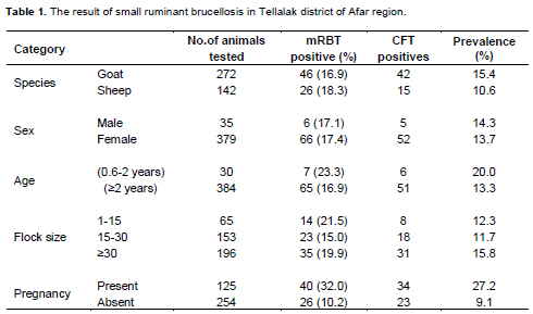
The overall sero-prevalence in both sheep and goats after mRBT was 17.4% (72/414). Out of the mRBT positive sera, 79.2% (57/72) further showed a positive reaction for CFT. The prevalence of brucellosis in both sheep and goats after mRBT for the different study sites was in the order of 20% Tellalak-Abaro, 25% Adalil-Dewi and 23% Aware-Ared. Summary of results of mRBT and CFT categorized by, species, age, sex, flock size and pregnancy status is given as shown in Table 1. Samples positive with both mRBT and CFT were considered as true seropositive and were taken for the subsequent data analyses. The overall true sero-prevalence of brucellosis in small ruminants was 13.7%.
Sero-prevalence among the potential risk factors of small ruminant brucellosis
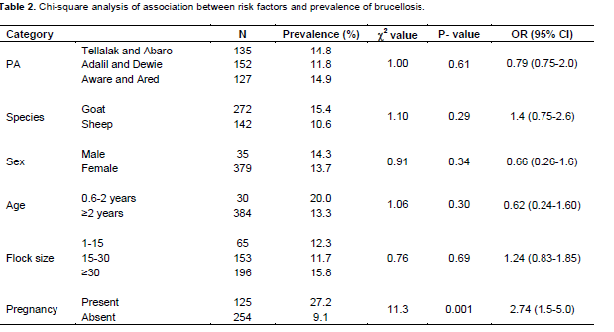
The true sero-prevalence of brucellosis among the study sites is as shown in Table 2. Except for the pregnancy status, there was no stastically significant difference (p>0.05) in the true sero-prevalence between the PAs, between sheep and goats, between males and females, and between the various flock sizes. However, true sero-prevalence was significantly higher (p<0.05) in pregnant animals as compared to non-pregnant ones. The true sero-prevalence of brucellosis among the pregnant goats 82.4% (28/34) was significantly higher (P=0.001) than in sheep 17.6% (6/34).
Reproductive problems of small ruminants in the study area
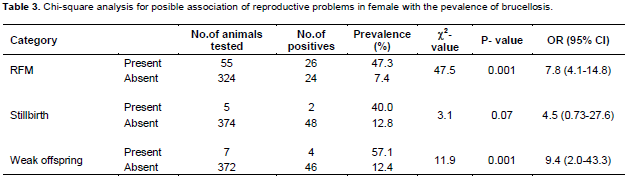
The prevalence of abortion, RFM, still birth and weak offspring were 17.2, 14.5, 1.3 and 1.8%, respectively. History of previous abortion and retained fetal membrane were highly associated (P=0.00) with the highest prevalence of brucellosis (Table 3). Animals with previous history of abortion were 5 times more at risk of being sero-positive as compared to those without history of abortion while animals with RFM were about 8 times more at risk. The odds of animals with history of stillbirth contracting brucellosis was lower (OR=4.5) and not statistically significant as compared to those without stillbirth. However, animals that gave rise to weak offspring also showed positive reaction to brucellosis test and the relationship was statistically significant (P<0.05, Table 3).
Results of questionnaire survey on husbandry practices and associated risk factors
In this study, 21 individuals whose animals were sampled for blood collection were asked about awareness of brucellosis, presence of regular veterinary service, grazing system and disposal of fetal membrane, the practice of migration, method of milk consumption, and handling aborted fetus to find out the presence of associated risk factors with the occurrence of brucellosis. All households used same communal grazing area and do migrate seasonally in search of feed and water for their animals. Most of the respondents (71.2%) commonly leave the afterbirth on the ground while the remaining 28.8% just throw the afterbirth on a tree. Almost all households do not properly take care of aborted fetus. Awareness of small ruminant brucellosis was very low (19.1%) among owners and further, a good proportion (66%) of owners also did not have a regular veterinary service. Milk is commonly consumed raw.
DISCUSSION
The data collected revealed high sero-prevalence (13.7%) of small ruminant brucellosis in the study area. Previous study on brucellosis in Afar region reported a higher prevalence (16%) although the study did not ad-dress specific areas of the region (Teshale et al., 2005).
Subsequent more detailed reports for different districts of the same region indicated lower prevalence of brucellosis over wider areas: 2.3% in Tellalak district, 8.0% in Samurobi, 1.7% in Fursi and 10% in Awash Fentale and neighboring districts (Ashenafi et al., 2007) with an overall prevalence of 4.8% for the whole of Afar region. In the presence of seasonal mobility, lack of veterinary service, and very poor understanding of the disease, higher prevalence probably indicates that the lack of awareness from the owner’s side has contributed to the increase in the prevalence and spread of the disease over time. Additional reason for the difference in the prevalence might also be due to absence of vaccination against brucellosis, coupled with the traditional use of communal grazing that brings the risk population close to one another. The difference in sample size and the variation in degree of sensitivity between CFT and I-ELISA could also be another source of difference in the prevalence reports.
The present study indicates that the prevalence of brucellosis in goats (15.4%) was higher than sheep (10.6%). Although not statistically significant, goats were 1.45 times more at risk of being sero-positive as compared to sheep. This finding is comparable to that of Teshale et al. (2005) and Ashenafi et al. (2007) who also reported higher prevalence in goats than in sheep in Afar region, and Mengistu (2007) in Konso, southern Ethiopia. Similar findings were also reported from Nigeria (Bale et al., 2003; Ojo et al., 2007; Bertu et al., 2010). However, study by Tekelye and Kasali (1999) in the central high lands of Ethiopia, and by Samaha et al. (2008) in Egypt showed a higher prevalence in sheep as compared to goats mainly due to differences in husbandry system and in susceptibility of the sheep and goat breeds in the particular area.
The prevalence in males was higher than the female animals, but this was not statistically significant. It is difficult to draw a strong conclusion, because of the small numbers of male animals sampled in this study. The prevalence of brucellosis was higher in younger (20.0%) animals than in older (13.3%) animals. Unlike the present finding, another study (Ashenafi et al., 2007) reported a prevalence of 5.3% in adult animals and 1.6% in younger sheep and goats. It is known that sexually mature and pregnant animals are more prone to Brucella infection than sexually immature animals of either sex. This may result from the fact that sex hormones and erythritol, which stimulate the growth and multiplication of Brucella species organisms, tend to increase in concentration with age and sexual maturity (Radiostits et al., 2000). However, the fewer number of young animals sampled in the present study has clearly inflated the prevalence as compared to that in adult animals. The prevalence of brucellosis among larger flock size was higher than small flock size; however, the difference was not significant. Literature (Walker, 1999) shows that herd sizes and animal densities are directly related to prevalence of the disease and create difficulty in controlling infection in a population. In the present study, however, it perhaps shows watering and grazing site where all animals have similar chance of exposure to brucella organism to be more important than densities of animals in larger flock.
A total of 379 female animals were included to study association of sero-positivity with the presence of reproductive problems, and with the presence or absence of pregnancy. Male animals of both species were not included in this discussion because by chance no male animal was found with reproductive organ problems at the time of sampling serological test. There was a statistically significant difference (P=0.001) among the pregnant and non pregnant animals. This might be due to susceptibility of the reproductive tracts of pregnant animals. Allantoic factors including erythritol, possibly steroid hormones and other substances stimulate the growth of most of the Brucella spp. (Anonymous, 2007).
The sero-prevalence of brucellosis was also higher (P=0.000) in small ruminants having history of abortion and retained fetal membrane than those without these problems. The higher rate of infection in pregnant sheep and goat might be due to infection within the reproductive tract that provide a potential reservoir site for the orga-nism which eventually propagated and become active as pregnancy advanced. The presence of a statistically significant association (P=0.000) between a positive reaction to brucellosis and the birth of weak offspring is a common sign of brucella infection. Often when not aborted, the offspring is delivered as a weak individual prone to early mortality.
CONFLICT OF INTEREST
The authors declare that there is no conflict of interest
REFERENCES
| Afar Pastoral And Agricultural Development Office (APADO), 2007). Afar National Regional State Agricultural Sector Status and Development Opportunity Record Office. pp. 5-20. | ||||
| Anonymous. (2007): Bovine brucellosis. In: Animal disease cards. Vet. J. 27:145- 151. | ||||
| Ashenafi F, Teshale S, Ejeta G, Fikru R, Laikemariam Y (2007). Distribution of brucellosis among small ruminants in the pastoral region of Africa, Eastern Ethiopia. Rev. Sci. Tech. Off. Inf. Epiz. 26:731-739. | ||||
| Bale J, Nuru S, Addo P (2003). Serological study of sheep and goats Brucellosis in Northern Nigeria. Bull. Anim. Health Prod. Afr. 30:73-79. | ||||
| Bertu, W. Ajogi, J. Bale, J. Kwaga and Ocholi, R. (2010): Sero epidemiology of brucellosis in small ruminants in Plateau State, Nigeria. Afr. J. Micro. Res. 4:1935-1938. | ||||
| CSA (2010). Ethiopia Livestock Estimate, volume I. Bulletin No. 52, Addis Ababa, Ethiopia. | ||||
| Ibrahim H (1998). Small Ruminant Production Techniques. ILRI Training Manual. Nairobi, Kenya. pp. 11-47. | ||||
| International Livestock Research Institute (ILRI) (2006). Domestic animal genetic resources information system (DAGRIS): access to biodiversity data underpins future livestock option for Africa. Addis Ababa. | ||||
| Mengistu M (2007). Sero epidemiology of brucellosis in small ruminants in Southern Ethiopia. Master's thesis, AAU, Debre Zeit. P 45. | ||||
| OIE (2008). Bovine brucellosis, Manual of Standards for Diagnostic Tests and Vaccines. OIE, Paris, France. pp. 624-659. | ||||
| Ojo OE, Oyekunle MA., Omotainse SO, Ocholi RA, Ogunleye AO, Bertu WJ (2007). Serological evidence of brucellosis in a goat flocks with recurrent abortion in Abeokuta. Nigeria. Trop. Vet. 25:26-33. | ||||
| PFE (2004). Background to the Ethiopian livestock industry. In: Proceedings of the 3rd National Conference on Pastoral Development in Ethiopia: pastoralism and sustainable pastoral development, 23-24 December, Addis Ababa. PFE, Addis Ababa. pp. 78-79. | ||||
| Radostitis OM, Gay CC, Blood DC, Hinchcliff KW (2000). Veterinary Medicine: A Text book of the disease of cattle, sheep, goats, pigs and horses, 9th edition. New York W.B. Saunders Company Ltd. pp. 867-882. | ||||
|
Samaha H, Al-Rowaily M, Khoudair RM, Ashour HM (2008). Multicenter Study of Brucellosis in Egypt. Emerg. Infect. Dis. 14:1916-1918. Crossref |
||||
| Singla LD (1995). A note on sub-clinical gastro-intestinal parasitism in sheep and goats in Ludhiana and Faridkot districts of Punjab. Indian Vet. Med. J. 19:61-62. | ||||
| Tekelye B, Kasali OB (1999). Brucellosis in sheep and goats in Central Ethiopia. Bull. Anim. Health Prod. Afr. 38:23-25. | ||||
| Teshale S, Muhie Y, Dagne A, Kidanemariam A (2005). A seroprevalence study of small ruminant brucellosis in selected sites of the Afar and Somali regions, Ethiopia. Rev. Sci. Tech. Off. Int. Epiz. 26 (3) 739. | ||||
| Thrusfield M (2005). Veterinary Epidemiology. 3rd ed. UK. Blackwell science. Ltd, P. 233 | ||||
| Walker RL (1999). Brucella In: Veterinary Microbiological Science, Dwright, C.H. and Chunge, Z.Y. (eds.); Black wells, Massachusetts, Pp. 196-203. | ||||
Copyright © 2024 Author(s) retain the copyright of this article.
This article is published under the terms of the Creative Commons Attribution License 4.0


