Review

ABSTRACT
The use of electrospun fibers in diabetic wound healing assays represents an extremely important topic for investigation. Electrospun fibers have been applied in the immobilization of drugs, allowing sustained and controlled release of active materials. This review aimed to summarize the state-of-art in the application of electrospun fibers on diabetes, hyperglycemic and diabetic ulcers treatment. Regarding the diabetes control and treatment, electrospinning technique contributes to application of wound healing (in vitro and in vivo experiments). The glycemic control is favored due to controlled release which has been sustained and improved. The data suggest that the development of encapsulated drugs in electrospun fibers has a promising application in the treatment of diabetes and related complications.
Key words: Electrospun fibers, nanofibers, electrospinning technique, anti-diabetic drugs, hypoglycemic drugs, wound healing.
INTRODUCTION
Diabetes mellitus is a metabolic disease characterized by insufficiency in insulin secretion, insulin action, or both (ADA, 2014) that results in hyperglycemia. The International Diabetes Federation (IDF) estimates that in 2040, 10% of adult population will be diabetics. Considering the South and Central America, this number increases to 65% (IDF, 2015).
Numerous pathogenic processes are consequence of diabetes, due to the several pathologies involving heart, blood vessels, eyes, kidneys and nerves (ADA, 2014; IDF, 2015; Chen et al., 2015). In addition, diabetic wound healing has been considered one of major complications of diabetes mellitus (Siersma et al, 2014) which results in peripheral neuropathy and/or large vessel disease complicated by trauma (Singh et al., 2005; Boulton, 2008).
The glycemic control is critically important for minimi-zation in the impacts of diabetes and corresponding complications. Recent studies have demonstrated that new therapies can be used in the control of blood glucose and the diabetic complications, such as wound healing (Schneider et al., 2009; Arnolds and Heise, 2007; Moura et al, 2014; Nishimura et al., 2012; ITA, 2015).
Electrospinning technique is an extensively studied technology for nanofiber production (FORMHALS, 1934) recently applied in biomedical issues (Yu et al., 2014; Ding et al., 2014) with focus in the development of methods for diabetes treatment and its complications, such as diabetic ulcers (Xie et al., 2013). The action of electrospun fibers is improved due to the unique properties such as high surface area, high loading and simultaneous delivery for diverse therapies and reasonable cost-effectiveness that provided expansion in the use of fibers for drug delivery (Hu et al., 2014). Natural and synthetic polymers can be used in the electrospinning, including soluble and spinnable polymers in water, biocompatible and biodegradable polymers (Agarwal et al., 2009).
Drug-loaded electrospun fibers have been considered as promising strategies to achieve bioactive wound dressings for chronic non-healing wounds (Schneider et al, 2009) and for controlled release in the gastrointestinal tract. The nanoscale multi-agent delivery system has been applied in the drug loading (Yang et al., 2014); thereby the present systematic review summarizes the current studies about the application of electrospun fibers in the treatment of diabetes, hyperglycemic and diabetic ulcers.
Previous work reveals the potential of application of optimized structures for drug-loaded electrospun fibers production (Araujo et al, 2016; da Costa et al, 2015). Corresponding association with natural products (such as Morus nigra (Almeida et al, 2011)) represent an important topic of research with the aim of developing new nanostructured systems for application as prototype of wound dressings for non-healing wounds.
DIABETES CONTROL
As consequence of preponderance of individuals with elevation in the plasma glucose that remains untreated and/or undiagnosed (Buysschaert et al, 2015), there is a global trend of increasing diabetes cases. Numerous studies have described new therapies related to the control of glucose, insulin levels and other diabetic disorders (Nacht et al., 2015; Yong et al., 2015; Yang et al., 2016; Du et al., 2016; Chen et al., 2016).
This development requires the use of in vivo or in vitro models. The diabetes induction in animal models is typically induced by two mechanisms viz. alloxan and streptozotocin (Lenzen, 2008). However, streptozotocin-induced diabetes is a more stable model for experimental diabetes and it can be used for both shorter and longer experimental studies (Radenković et al., 2016). Kuricová et al. (2016) recently demonstrated that is possible to simulate in vitro culture conditions mimicking human glycemic variability. The nanotechnology in diabetes research has potential to treat the diabetics complications and to improve the quality of life of the patients (Disanto et al., 2015). Studies have demonstrated that electrospun nanofibers can be used as glucose sensors due to the different mechanisms (Senthamizhan et al., 2016; Ramasamy et al., 2015). In addition, the incorporation of nanostructures promotes the enhanced insulin production in response to glucose stimulation, which enables the development of new diagnostic test for drug development (Blackstone et al, 2014).
PROCESS PARAMETERS FOR ELECTROSPINNING
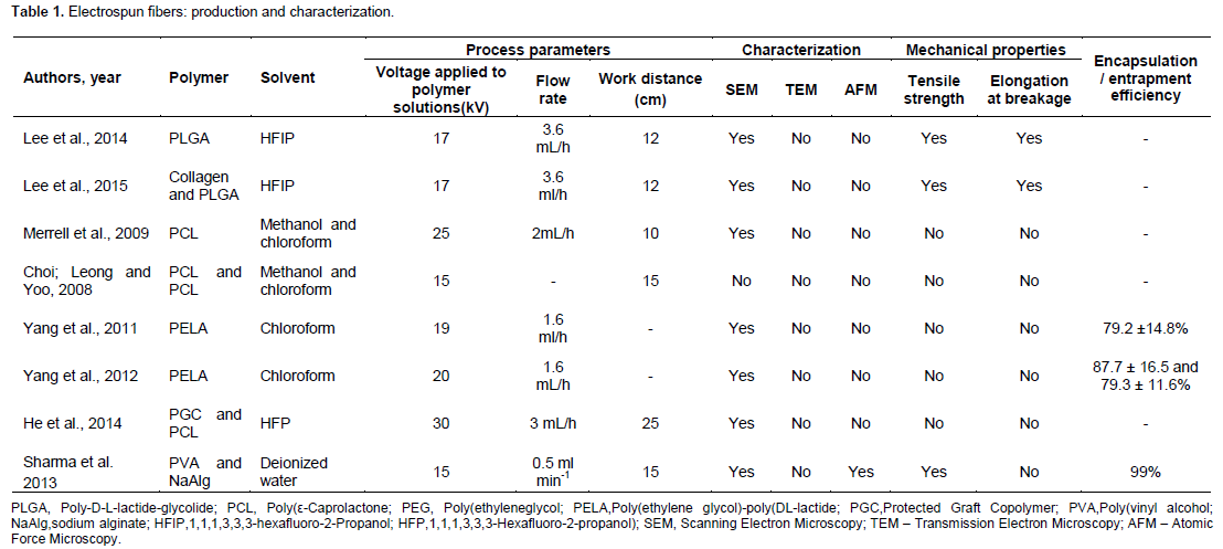
Relative to electrospun fibers production, the control in the process parameters is a critical parameter in order to obtaining nanofibers with desired morphologies and diameters. The polymer molecular weight, solution concentration and solvent type also affect the diameters and morphologies of the fibers (Cui et al, 2007). According to Jang et al. (2015) pivotal parameters such as polymer concentration, molecular weight, input voltage and gap distance affects the production of nanofibers.
Therefore, there was the broad range in the characteristic fiber diameters and fiber morphologies in different studies due to the use of different process parameters, polymers and solvents.
The definition of appropriate polymer represents a key component for development of sustained release electrospun fibers (Chou et al., 2015). Polymers used in these studies are all biocompatible and may be employed for applications involving the development of control drug delivery systems (Li et al., 2000; Zong et al., 2003, Yao et al., 2003; Li et al., 2003; Zhou et al., 2003) (Table 1).
LOADED AND CONTROLLED RELEASE OF DRUGS
The encapsulation efficiency contributes to improve the absorption and prolonged release of components (corresponding data is summarized on Table 1). In all reported data, there were enhancements on resulting release. Sustaining drug release from electrospun nanofibers shows regular release kinetics that depend upon material surface chemistry, drug loading, and processing parameters (Chou et al., 2015).
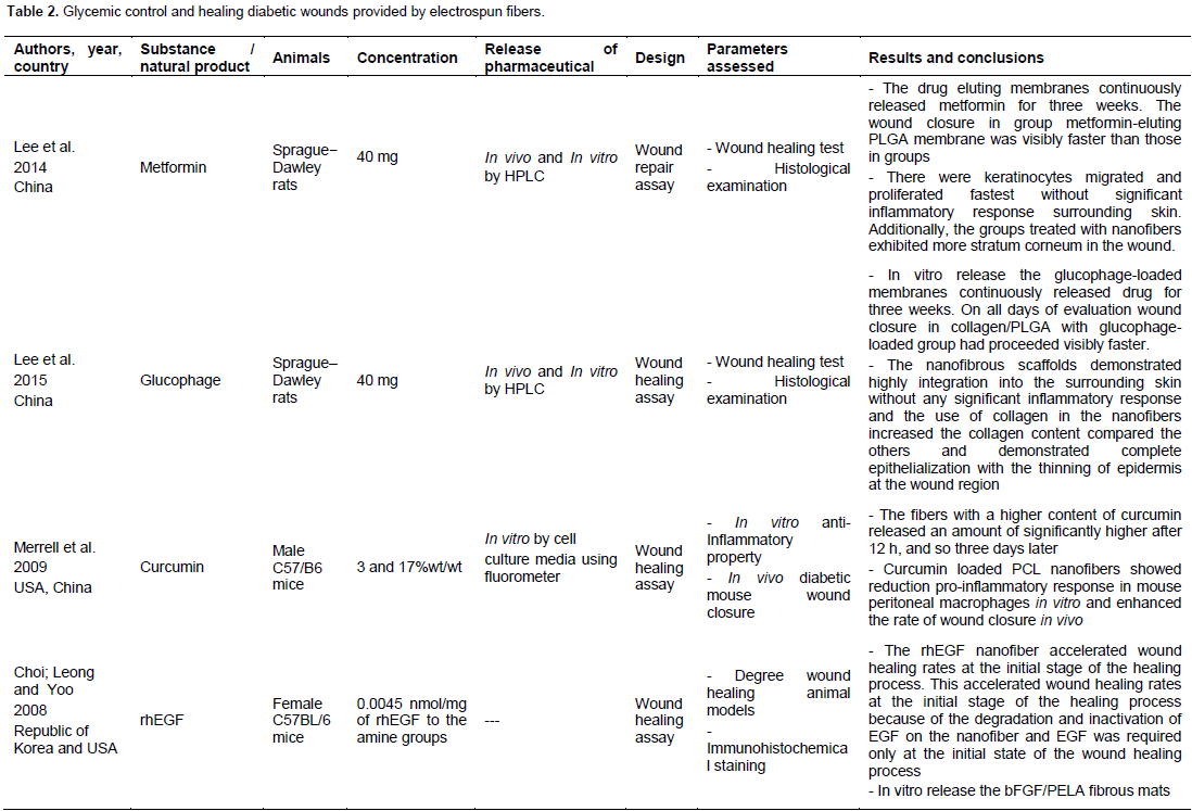
In these studies, the electrospun fibers show improved activity of the substances followed by controlled release of drugs and/or substances. The profile of in vitro assays shows sustained and controlled release of drugs, substances and natural product (Lee et al., 2015, 2014; He et al., 2014; Sharma et al., 2013; Merrell et al., 2009; Yang et al., 2011, 2012; Choi et al., 2008), while for different systems the drug release is order of hours (Sharma et al., 2013), days (He et al., 2014; Merrell et al., 2009; Choi et al., 2008) or weeks (Lee et al., 2015, 2014; Yang et al., 2011, 2012).
WOUND HEALING ACTIVITY
The absence of reepithelialization requires the development of acute or chronic non-healing wounds, in which the use of electrospun nano-fibers accelerate wound healing rates in diabetics rats (Lee et al., 2015, 2014; Merrell et al., 2009; Yang et al., 2011, 2012; Choi et al., 2008) from sustained release of substances that increase the re-epithelialization and regeneration of the skin by various mechanisms which depend on the substance/drug, the polymer and the process parameters applied in the production of the fiber (Table 2).
On the other side, the metformin is used in the treatment of type 2 diabetes. It represents the first-line pharmacological therapy associated with low weight gain and a reduced amount of hypoglycemic occurrence (UKPDS, 1998). In spite of this application, no trials about the effect of metformin on wound healing in diabetic patients (Salazar et al., 2015) were performed. Studies have demonstrated that metformin acts in wound healing around oral implants (Inouye et al., 2014) and promotes nerve regeneration after sciatic nerve injuries in diabetes mellitus, providing its therapeutic values for peripheral nerve injury repair in diabetes mellitus (Ma et al., 2015).
The use of metformin-loaded electrospun fibers was active in the treatment of diabetic wounds and accelerated for healing wound (Lee et al., 2014, 2015). Furthermore, the use of collagen associated with metformin in membranes fibrous increases the content of collagen I, suggesting that nanofibrous containing metformin and collagen can be effective tissue-engineering scaffold for regenerating skin around diabetic wounds (Lee et al., 2015).
Curcumin possesses modulating effects on wound healing (AKBIK et al., 2014). The effect of its incorporation in the nanofibers returned wound closure in order of 80% along 10 days demonstrating the potential of curcumin-loaded nanofibers in the elevation of wound closure rate applied in a diabetic mouse (Merrell et al., 2009). Studies performed with curcumin in its raw state returned no significant response of material (Kant et al, 2015, 2014).
These studies show that the curcumin can be a new agent for accelerating diabetic wound healing in humans and animals since they show potential in the treatment of cutaneous wounds with corresponding improvement in cutaneous wound healing in acute phase diabetic rats (Kant et al., 2015) and still the topical application in the acceleration of cutaneous wound healing, with corresponding decrease in the persistence of the inflammatory state in acute diabetic rats (Kant et al., 2014).
Recent study reported that encapsulation of curcumin circumvents conventional difficulties related with curcumin administration, enabling delivery of this therapeutic substance and allowing its use as a novel topical agent for burn wound infection and cutaneous injuries (Krausz et al., 2015). Thus, studies with the encapsulation of curcumin in fibers have a promising future for the reason that electrospun fibers can improve the delivery of the therapeutic substance.
The inflammatory response, formation of granu-lation tissue, reepithelialization, matrix formation and wound closure are provided and regulated by an equally complex signaling network involving numerous growth factors, cytokines and chemokines (Barrientos et al., 2008). Growth factors are important substances that have the ability to modulate cell behavior and guide tissue repair and renewal (Mitchell et al, 2016). Currently, the release of growth factors has great potential for development of new therapies which favors the healing and skin regeneration (Gainza et al., 2015). Articles suggest that electrospinning process can produce materials containing growth factors that allows the creation of multi-functionalized bioactive nanomaterials to chronic non-healing wounds and that can serve as skin tissue engineering scaffolds for wound healing (Schneider et al, 2009; Bertoncelj et al, 2014; Wang et al, 2016).
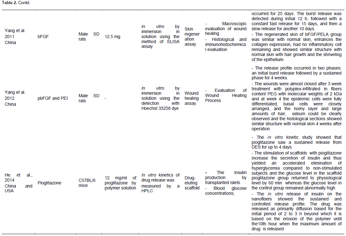
The growth factors, such as basic fibroblast growth factor and recombinant human epidermal growth factor, immobilized or incorporated have healing activity in diabetic animals (Choi et al., 2008; Yang et al., 2011, 2012, Sun et al., 2014). Recombinant human epidermal growth factor (rhEGF) immobilized on the electrospun nanofibers for treating diabetic ulcers in female mice demonstrated enhanced keratinocytic expression of human primary keratinocytes in comparison to rhEGF in solution, accelerating wound healing rates at the initial stage of the healing process (Choi et al., 2008). Basic fibroblast growth factor (bFGF) incorporated into electrospun ultrafine fibers indicated that bFGF-loaded fibrous mats (in vitro investigations) enhances cell adhesion, proliferation, and secretion of extracellular matrix and enhanced collagen deposition and ECM remodeling. The arrangement and components of collagen fibers were similar to normal tissues (Yang et al. 2011).
Polyplexes of basic fibroblast growth factor-encoding plasmid (pbFGF) with poly(ethylene imine) incorporates into electrospun fibers showed a sustained release of 4 weeks and demonstrated significant higher wound recovery rate with collagen deposition and maturation, complete reepithelialization and skin appendage regene-ration (Yang et al. 2012).
Hyperglycemia control
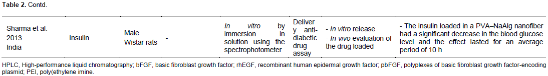
The hyperglycemic is other problem related with the diabetes and all of its complications. The use of electrospun nanofibers allows a sustained and controlled release profile of drugs that can reduce blood glucose. Insulin is a hormone synthesized in the pancreas which plays a critical role in intermediary metabolism. It is the key hormone for regulation of blood glucose and, generally, normoglycaemia is maintained by the balanced interplay between insulin action and insulin secretion (Stumvoll et al., 2005). Recent trials have demonstrated progress in technologies for insulin delivery in vivo and/or in vitro (Zhang et al., 2011; Nishimura et al., 2012; Ling and Chen, 2013; Balducci et al., 2014). The poor oral bioavailability and invasive drug delivery methods represent important barriers to be overcome in this topic.
To our knowledge, there are no studies about the encapsulation of insulin using electrospun fibers. A unique study reports that it is possible to encapsulate insulin on nanofibers. The sublingual route based on composite nanofiber diffusion along transmucosal patch can delivers the insulin in a sustained and controlled manner by controlling the morphology of the composite nanofiber patch (Sharma et al., 2013). This study also shows that the insulin cannot be easily released from the composite nanofiber patch due to the crosslinking between the drug molecule and polymeric chains unless polymeric nanofibers are degraded.
He et al. (2014) reported that scaffold with pioglitazone was able to locally deliver the drug to transplanted islets; hence, there was increased insulin secretion and a lower blood glucose level in diabetic mice. Furthermore, the decrease of hyperglycemic also aided in the recovery of renal functions compromised by the diabetes.
Thus, the encapsulation by electrospun fibers allows for better bioavailability of drugs, facilitates its sustained action and act as an adjuvant for recovery of complications caused by diabetes. In this direction, it is clear that studies are required in order to apply this technology and ensure its applicability in vivo.
The development of long-acting drug formulations has been explored in many applications in order to overcome challenges with adherence and emerging drug resistance (Chou et al., 2015) with the use of electrospun fibers, which has been considered a promising source for the treatment of diabetes and its complications, such as diabetic wounds.
CONCLUSIONS AND FUTURE PERSPECTIVES
The recent studies summarized in this article have showed advances in the treatment of diabetes and opens a discussion to expand the understanding of the use of electrospun fibers for treatment of chronic diseases associated with high cost to public health and quality of life of patients. We have highlighted in this review the future trends for treatment of hyperglycemic and diabetic wound healing diabetics relates with use of electrospun fibers. The electrospinning is a promising technique with increasing interest in health area. The electrospun nanofiber has ability to load or immobilized drugs/ substances, promotes a controlled and sustained release of the drug or substance. Besides that, the nanofibers can be explored for less vulnerable drugs to the attack by enzymes, providing improved stability. In diabetes, electrospinning technique has shown excellent results in relation to wound healing in in vitro and in vivo experiments, due to fast decrease in the area of the wound associated with better healing. In spite of few trials about the action of electrospun fibers in glycemic control, there are strong indications that in vitro and in vivo release has been improved and sustained. Further studies are needed to prove the effectiveness and efficiency of drug encapsulated and the associated risks and side effects of this encapsulation.
CONFLICT OF INTERESTS
The authors have not declared any conflict of interests.
ACKNOWLEDGMENT
This work was supported by Brazilian Funding Agency FACEPE.
REFERENCES
|
Agarwal S, Greiner A, Wendorff JH (2009). Electrospinning of Manmade and Biopolymer Nanofibers-Progress in Techniques, Materials, and Applications. Adv. Funct. Mater. 19:2863-2879. |
|
|
Akbik D, Ghadiri M, Chrzanowski W, Rohanizadeh R (2014). Curcumin as a wound healing agent. Life Sci. 116(1):1-7. |
|
|
Almeida JRGS, Guimarães AL, Oliveira AP, Araujo, ECC, Silva FS, Neves LF, Oliveira RA, Sa PGS, Quintans-Junior LJ (2011). Evaluation of hypoglycemic potential and pre-clinical toxicology of Morus nigra L. (Moraceae). Acta Farmaceutica Bonaerense, 30:96-100. |
|
|
ADA (American Diabetes Association) (2014). Diagnosis and classification of diabetes mellitus. Diabetes Care 37(1):81-90. |
|
|
Araujo ES, Nascimento MLF, de Oliveira HP (2016) Electrospinning of Polymeric Fibres: An Unconventional View on the Influence of Surface Tension on Fibre Diameter. Fib. Text. East. Eur. 24: 22-29. |
|
|
Arnolds S, Heise T (2007). Inhaled insulin. Best Pract. Res. Clin. Endocrinol. Metab. 21(4):555-571. |
|
|
Balducci AG, Cagnani S, Sonvico F, Rossi A, Barata P, Colombo G, Colombo P, Buttini F (2014). Pure insulin highly respirable powders for inhalation. Eur. J. Pharm. Sci. 51:110-117. |
|
|
Barrientos S, Stojadinovic O, Golinko MS, Brem H, Tomic-Canic M (2008). Growth factors and cytokines in wound healing. Wound Repair Regen. 16(5):585-601. |
|
|
Blackstone BN, Palmer AF, Rilo HR, Powell HM (2014). Scaffold architecture controls insulinoma clustering, viability, and insulin production. Tissue Eng. Part A. 20(13-14):1784-1793. |
|
|
Bertoncelj V, Pelipenko J, Kristl J, Jeras M, Cukjati M, Kocbek P (2014). Development and bioevaluation of nanofibers with blood- derived growth factors for dermal wound healing. Euro. J. Pharm. Biopharm. 88(1):64-74. |
|
|
Boulton AJ (2008). The diabetic foot: grand overview, epidemiology and pathogenesis. Diabetes Metab. Res. Rev. 24(1):3-6. |
|
|
Buysschaert M, Medina JL, Bergman M, Shah A, Lonier J (2015). Prediabetes and associated disorders. Endocrine 48:371-393. |
|
|
Chen H, Jia P, Kang H, Zhang H, Liu Y, Yang P, Yan Y, Zuo G, Guo L, Jiang M, Qi J, Liu Y, Cui W, Santos HA, Deng L (2016). Upregulating Hif-1α by Hydrogel Nanofibrous Scaffolds for Rapidly Recruiting Angiogenesis Relative Cells in Diabetic Wound. Adv. Healthc. Mater. 5(8):907-918. |
|
|
Chen Y, Sloan FA, Yashkin AP (2015). Adherence to diabetes guidelines for screening, physical activity and medication and onset of complications and death. J. Diabetes Complications 29(8):1228-1233. |
|
|
Choi JS, Leong KW, Yoo HS (2008). In vivo wound healing of diabetic ulcers using electrospun nanofibers immobilized with human epidermal growth factor (EGF). Biomaterials. 29(5):587-596. |
|
|
Chou SF, Carson D, Woodrow KA (2015). Current strategies for sustaining drug release from electrospun nanofibers. J. Control. Release 220:584-591. |
|
|
Cui W, Li X, Zhou S, Weng J (2007). Investigation on Process Parameters of Electrospinning System through Orthogonal Experimental Design. J. Appl. Polym. Sci. 103(1):3105-3112. |
|
|
da Costa FPC, Araujo ES, NASCIMENTO MLF, de Oliveira HP (2015) Electrospun Fibers of Enteric Polymer for Controlled Drug Delivery. Int. J. Polym. Sci. Article ID 902365:1-8. |
|
|
Ding F, Deng H, Du Y, Shi X, Wang Q (2014). Emerging chitin and chitosan nanofibrous materials for biomedical applications. Nanoscale 6(16):9477-9493. |
|
|
DiSanto RM, Subramanian V, Gu Z (2015). Recent advances in nanotechnology for diabetes treatment. Wiley Interdiscip. Rev. Nanomed Nanobiotechnol. 7(4):548-564. |
|
|
Du Y, Zhang W, Wang ML (2016). Sensing of salivary glucose using nano-structured biosensors. Biosensors 6(1): 1-14. Formhals A. US patent 1,975,504, 1934. |
|
|
Gainza G, Villullas S, Pedraz JL, Hernandez RM, Igartua M (2015). Advances in drug delivery systems (DDSs) to release growth factors for wound healing and skin regeneration. Nanomedicine. 11(6):1551-1573. |
|
|
He Q, Zhang X, Han B, Xu J, Tang K, Fu Z, Yin H (2014). A synergistic therapeutic scheme for hyperglycemia and nephrotic disorders in diabetes. Theranostics. 4(5):556-564. |
|
|
Hu X, Liu S, Zhou G, Huang Y, Xie Z, Jing X (2014). Electrospinning of polymeric nanofibers for drug delivery applications. J. Control. Release 185:12-21. |
|
|
Inouye KA, Bisch FC, Elsalanty ME, Zakhary I, Khashaba RM, Borke JL (2014). Effect of metformin on periimplant wound healing in a rat model of type 2 diabetes. Implant Dent. 23(3):319-327. |
|
|
International Diabetes Federation IDF (2015). IDF Atlas, 7thed. Brussels, Belgium: International Diabetes Federation. |
|
|
Ita K (2015). Transdermal delivery of drugs with microneedles: Strategies and Outcomes. J. Drug Deliv. Sci. Technol. 29:16-23. |
|
|
Jang S, Kim Y, Oh JH (2015). Influence of Processing Conditions and Material Properties on Electrohydrodynamic Direct Patterning of a Polymer Solution. J. Electron. Mater. 45(4):1-8. |
|
|
Kant V, Gopal A, Kumar D, Pathak NN, Ram M, Jangir BL, Tandan SK, Kumar D (2015). Curcumin-induced angiogenesis hastens wound healing in diabetic rats. J. Surg. Res. 193(2): 978-988. |
|
|
Kant V, Gopal A, Pathak NN, Kumar P, Tandan SK, Kumar D (2014). Antioxidant and anti-inflammatory potential of curcumin accelerated the cutaneous wound healing in streptozotocin-induced diabetic rats. Int. Immunopharmacol. 20(2):322-330. |
|
|
Krausz AE, Adler BL, Cabral V, Navati M, Doerner J, Charafeddine RA, Chandra D, Liang H, Gunther L, Clendaniel A, Friedman JM, Nosanchuk JD, Friedman AJ (2015). Curcumin-encapsulated nanoparticles as innovative antimicrobial and wound healing agente. Nanomedicine. 11(1):195-206. |
|
|
Kuricová K, Pácal L, Šoupal S, Prázný M, Kaňková K (2016). Effect of glucose variability on pathways associated with glucotoxicity in diabetes: Evaluation of a novel in vitro experimental approach. Diabetes Res. Clin. Pract. 114:1-8. |
|
|
Lee CH, Chang SH, Chen WJ, Hung KC, Lin YH, Liu SJ, Hsieh MJ, Pang JH, Juang JH (2015). Augmentation of diabetic wound healing and enhancement of collagen content using nanofibrous glucophage-loaded collagen/PLGA scaffold membranes. J. Colloid. Interface Sci. 439:88-97. |
|
|
Lee CH, Hsieh MJ, Chang SH, Lin YH, Liu SJ, Lin TY, Hung KC, Pang JH, Juang JH (2014). Enhancement of diabetic wound repair using biodegradable nanofibrous metformin-eluting membranes: in vitro and in vivo. ACS Appl. Mater. Interfaces 6(6):3979-3986. |
|
|
Lenzen S (2008). The mechanisms of alloxan- and streptozotocin-induced diabetes. Diabetologia. 51(2):216-226. |
|
|
Li WJ, Danielson KG, Alexander PG, Tuan RS (2003). Biological response of chondrocytes cultured in three-dimensional nanofibrous poly(epsilon-caprolactone) scaffolds. J. Biomed. Mater. Res. A. 67(4):1105-1114. |
|
|
Li X, Deng X, Yuan M, Xiong C, Huang Z, Zhang Y, Jia W (2000). In Vitro Degradation and Release Profiles of Poly-DLLactide-Poly(ethylene glycol) Microspheres with Entrapped Proteins. J. Appl. Polym. Sci. 78:140-148. |
|
|
Ling MH, Chen MC (2013). Dissolving polymer microneedle patches for rapid and efficient transdermal delivery of insulin to diabetic rats. Acta Biomater. 9(11):8952-8961. |
|
|
Ma J, Liu J, Yu H, Chen Y, Wang Q, Xiang L (2015). Beneficial Effect of Metformin on Nerve Regeneration and Functional Recovery After Sciatic Nerve Crush Injury in Diabetic Rats. Neurochem. Res. 41(5):1130-1137. |
|
|
Merrell JG, McLaughlin SW, Tie L, Laurencin CT, Chen AF, Nair LS (2009). Curcumin Loaded Poly(ε-Caprolactone) Nanofibers: Diabetic Wound Dressing with Antioxidant and Anti-inflammatory Properties. Clin. Exp. Pharmacol. Physiol. 36(12):1149-1156. |
|
|
Mitchell AC, Briquez PS, Hubbell JA, Cochran JR (2016). Engineering growth factors for regenerative medicine applications. |
|
|
Moura LI, Dias AM, Suesca E, Casadiegos S, Leal EC, Fontanilla MR, Carvalho L, de Sousa HC, Carvalho E (2014). Neurotensin-loaded collagen dressings reduce inflammation and improve wound healing in diabetic mice. Biochim. Biophys. Acta. 1842(1):32-43. |
|
|
Nacht B, Larndorfer C, Sax S, Borisov SM, Hajnsek M, Sinner F, List-Kratochvil EJW, Klimant I (2015). Integrated catheter system for continuous glucose measurement and simultaneous insulin infusion. Biosen. Bioelectron. 64:102-110. |
|
|
Nishimura A, Hayakawa T, Yamamoto Y, Hamori M, Tabata K, Seto K, Shibata N (2012). Controlled release of insulin from self-assembling nanofiber hydrogel, PuraMatrix™: Application for the subcutaneous injection in rats. Eur. J. Pharm. Sci. 45(1-2):1-7. |
|
|
Radenković M, Stojanović M, Prostran M (2016). Experimental diabetes induced by alloxan and streptozotocin: The current state of the art. J. Pharmacol. Toxicol. Methods 78:13-31. |
|
|
Ramasamy R, Ramachandran K, Philip GG, Ramachandran R, Therese HA, Kumar GG (2015). Design and development of Co3O4/NiO composite nanofibers for the application of highly sensitive and selective non-enzymatic glucose sensors. RSC ADVANCES 5(93): 76538-76547 |
|
|
Salazar JJ, Ennis WJ, Koh TJ (2015). Diabetes Medications: Impact on Inflammation and Wound Healing. J. Diabetes Complications (15): 1056-8727. |
|
|
Schneider A, Wang XY, Kaplan DL, Garlick JA, Egles C (2009). Biofunctionalized electrospun silk mats as a topical bioactive dressing for accelerated wound healing. Acta Biomater. 5(7):2570-2578. |
|
|
Sharma A, Gupta A, Rath G, Goyal A, Mathura RB, Dhakate SR (2013). Electrospun composite nanofiber-based transmucosal patch for anti-diabetic drug delivery. J. Mater. Chem. B 1:3410-3418. |
|
|
Siersma V, Thorsen H, Holstein PE, Kars M, Apelqvist J, Jude EB, Piaggesi A, Bakker K, Edmonds M, Jirkovská A, Mauricio D, Ragnarson Tennvall G, Reike H, Spraul M, Uccioli L, Urbancic V, van Acker K, van Baal J, Schaper NC (2014). Health-Related Quality of Life Predicts Major Amputation and Death, but Not Healing, in People With Diabetes Presenting With Foot Ulcers: The EurodialeStudy. Diabetes Care 37(3):694-700. |
|
|
Singh N, Armstrong DG, Lipsky BA (2005). Preventing Foot Ulcers in Patients With Diabetes. J. Am. Med. Assoc. 293(2):217-28. |
|
|
Senthamizhan A, Balusamy B, Uyar T (2016). Glucose sensors based on electrospun nanofibers: a review. Anal. Bioanal. Chem. 408:1285-1306. |
|
|
Stumvoll M, Goldstein BJ, van Haeften TW (2005). Type 2 diabetes: principles of pathogenesis and therapy. Lancet. 365(9467):1333-1346. |
|
|
Sun X, Cheng L, Zhao J, Jin R, Sun B, Shi Y, Zhang Lu, Zhang Y, Cui W (2014). bFGF-grafted electrospun fibrous scaffolds via poly(dopamine) for skin wound healing. J. Mater. Chem. B. 2:3636-3645. |
|
|
UK Prospective Diabetes Study UKPDS. (1998). Effect of intensive blood-glucose control with metformin on complications in overweight patients with type 2 diabetes (UKPDS 34). Lancet. 352(9131):854-865. |
|
|
Wang Z, Qian Y, Li L, Pan L, Njunge LW, Dong L, Yang L (2016). Evaluation of emulsion electrospun polycaprolactone/hyaluronan/ epidermal growth factor nanofibrous scaffolds for wound healing. J. Biomater. Appl. 30(6):686-698. |
|
|
Xie Z, Paras CB, Weng H, Punnakitikashem P, Su LC, Vu K, Tang L, Yang J, Nguyen KT (2013). Dual growth factor releasing multi-functional nanofibers for wound Healing. Acta biomater. 9(12):9351-9359. |
|
|
Yang G, Wang J, Li L, Ding S, Zhou S (2014). Electrospun Micelles/Drug-Loaded Nanofibers for Time-Programmed Multi-Agent Release. Macromol. Biosci. 14(7):965-976. |
|
|
Yang Y, Xia T, Chen F, Wei W, Liu C, He S, Li X (2012). Electrospun Fibers with Plasmid bFGF Polyplex Loadings Promote Skin Wound Healing in Diabetic Rats. Mol. Pharm. 9(1):48-58. |
|
|
Yang Y, Xia T, Zhi W, Wei L, Weng J, Zhang C, Li X (2011). Promotion of skin regeneration in diabetic rats by electrospun core-sheath fibers loaded with basic fibroblast growth factor. Biomaterials. 32(18):4243-4254. |
|
|
Yang J, Tang S, Cheke RA (2016). The regulatory system for diabetes mellitus: Modeling rates of glucose infusions and insulin injections Commun Nonlinear Sci Numer Simul. 37:305-325. |
|
|
Yao L, Haas TW, Guiseppi-Elie A, Bowlin GL, Simpson DG, Wnek GE (2003). Electrospinning and Stabilization of Fully Hydrolyzed Poly(Vinyl Alcohol) Fibers. Chem. Mater. 15(9):860-1864. |
|
|
Yong C, Wang Z, Zhang X, Shi X, Ni Z, Fu H, Ding G, Fu Z, Yin H (2014). The therapeutic effect of monocyte chemoattractant protein-1 delivered by an electrospun scaffold for hyperglycemia and nephrotic disorders. Int J Nanomedicine. 17(9):985-993. |
|
|
Yu H, Jia Y, Yao C, Lu Y (2014). PCL/PEG core/sheath fibers with controlled drug release rate fabricated on the basis of a novel combined technique. Int. J. Pharm. 469(1):17-22. |
|
|
Zhang Y, Wei W, Lv P, Wang L, Ma G (2011). Preparation and evaluation of alginate-chitosan microspheres for oral delivery of insulin. Eur. J. Pharm. Biopharm. 77(1):11-19. |
|
|
Zhou S, Deng X, Yang H (2003). Biodegradable poly(É›-caprolactone)-poly(ethylene glycol) block copolymers: characterization and their use as drug carriers for a controlled delivery system. Biomaterials 24(20):3563-3570. |
|
|
Zong X, Ran S, Kim KS, Fang D, Hsiao BS, Chu B (2003). Structure and Morphology Changes during in Vitro Degradation of Electrospun Poly(glycolide-co-lactide) Nanofiber Membrane. Biomacromolecules 4(2):416-423. |
|
Copyright © 2024 Author(s) retain the copyright of this article.
This article is published under the terms of the Creative Commons Attribution License 4.0


