African Journal of
Biochemistry Research

  • Abbreviation: Afr. J. Biochem. Res.
  • Language: English
  • ISSN: 1996-0778
  • DOI: 10.5897/AJBR
  • Start Year: 2007
  • Published Articles: 425

Full Length Research Paper

Evaluation of the effects of Azadirachta indica leaf on haematology, lipid profile, body weight and organ-system functions of streptozotocin-induced diabetic male rats

Ezeigwe Obiajulu Christian
  • Ezeigwe Obiajulu Christian
  • Department of Applied Biochemistry, Faculty of Biosciences, Nnamdi Azikiwe University, Awka, Anambra State, Nigeria.
  • Google Scholar
Okani Chukwudi Onyeaghana
  • Okani Chukwudi Onyeaghana
  • Department of Histopathology, Chukwuemeka Odumegwu Ojukwu University (Formerly: Anambra State University), Awka Campus, Anambra State, Nigeria.
  • Google Scholar
Nnadi Naomi Ngozi
  • Nnadi Naomi Ngozi
  • Department of Applied Biochemistry, Faculty of Biosciences, Nnamdi Azikiwe University, Awka, Anambra State, Nigeria.
  • Google Scholar
Obiukwu Onyinye Olivia
  • Obiukwu Onyinye Olivia
  • Department of Medicine and Surgery, Faculty of Medicine, College of Health Sciences, Nnamdi Azikiwe University, Nnewi Campus, Anambra State, Nigeria.
  • Google Scholar
Ekwunoh Peter Okwukwe
  • Ekwunoh Peter Okwukwe
  • Department of Biochemistry, Chukwuemeka Odumegwu Ojukwu University Uli, Anambra State, Nigeria.
  • Google Scholar
Obayuwana Erhunmwense Ann
  • Obayuwana Erhunmwense Ann
  • Department of Applied Biochemistry, Faculty of Biosciences, Nnamdi Azikiwe University, Awka, Anambra State, Nigeria.
  • Google Scholar
Okibedi Frances Uchenna
  • Okibedi Frances Uchenna
  • Department of Applied Biochemistry, Faculty of Biosciences, Nnamdi Azikiwe University, Awka, Anambra State, Nigeria.
  • Google Scholar
Obi Chioma Henrietta
  • Obi Chioma Henrietta
  • Department of Applied Biochemistry, Faculty of Biosciences, Nnamdi Azikiwe University, Awka, Anambra State, Nigeria.
  • Google Scholar


  •  Received: 11 May 2020
  •  Accepted: 01 July 2020
  •  Published: 31 July 2020

 ABSTRACT

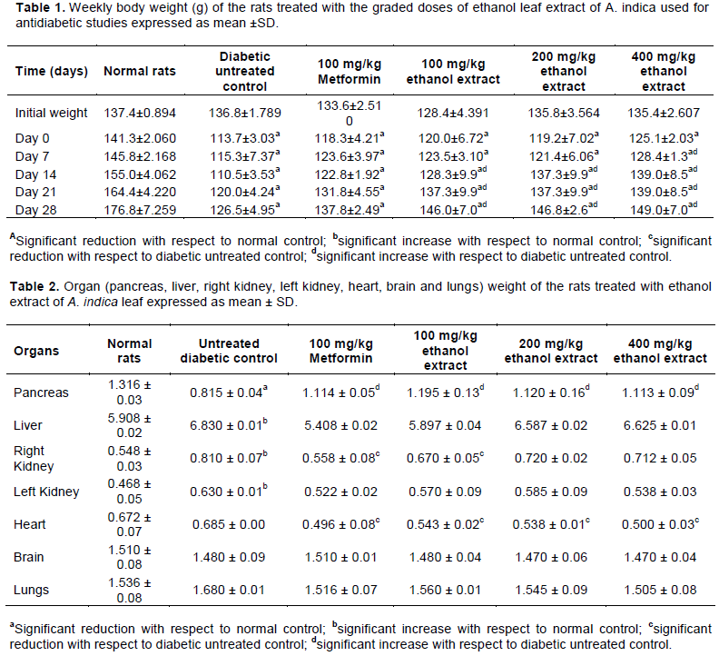
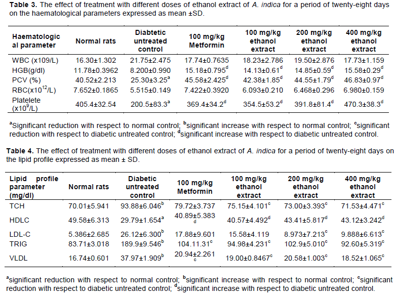
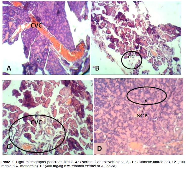
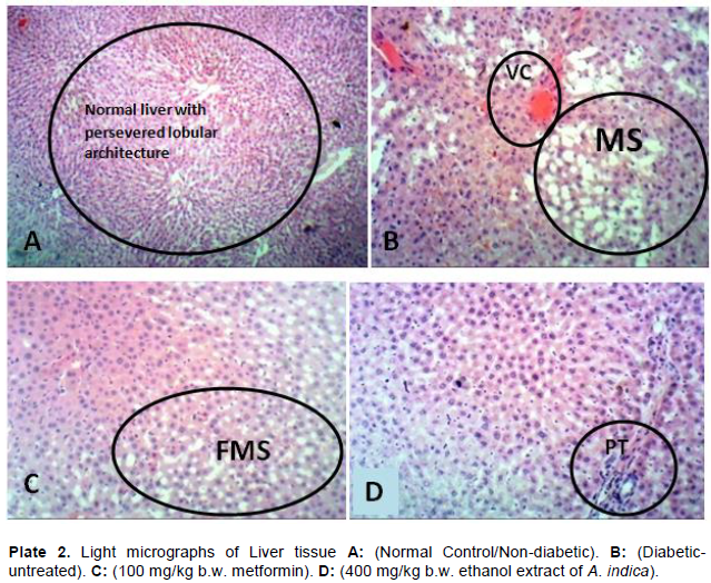
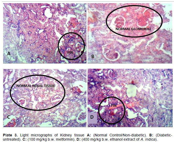
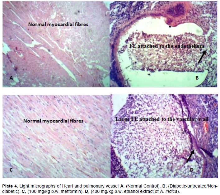
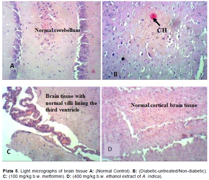
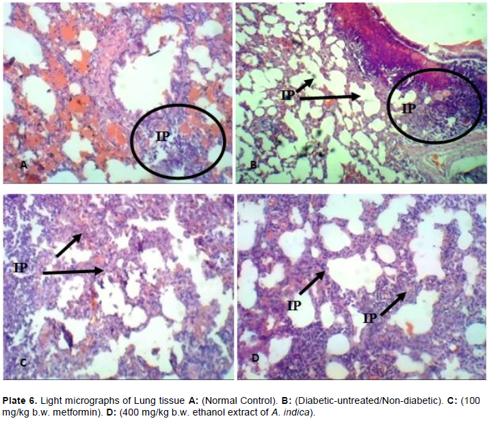
This study was carried out to evaluate the effects of ethanol extract of Azadirachta indica leaf on haematological parameters, lipid profile, body weight, organ weight and histopathological functions of streptozotocin-induced diabetic rats. Diabetes was induced by a single intraperitoneal administration of streptozotocin (50 mg/kg bw.). The haematological parameters, lipid profile and histopathological investigations were performed using standard methods. Continuous administration of ethanol extract of A. indica leaf for a period of four weeks significantly (p<0.05) increased the bodyweight of the streptozotocin-induced diabetic rats compared with the diabetic-untreated control. There was a significant (p<0.05) increase in the haemoglobin concentration, packed cell volume, red blood cells, platelet count and a significant (p<0.05) reduction in the total serum cholesterol, low-density lipoprotein, triglycerides and very-low-density lipoprotein of the groups treated with ethanol extract of A. indica compared with the diabetic-untreated control. The result of the histopathological studies showed regeneration of the organs for the groups treated with 400 mg/kg bw of the extract compared with the diabetic-untreated control. These results suggest that the ethanol extract of A. indica can be considered as an excellent remedy for diabetes and an alternative to antidiabetic drugs in reducing the complications associated with type II diabetes mellitus.


Key words: Diabetes, Azadirachta indica, haematological parameters, Lipid profile, Bodyweight, Histopathological functions.


 INTRODUCTION

Diabetes mellitus is characterized by the presence of hyperglycaemia. This is as a result of the  body's  inability to produce required amounts of insulin (the hormone that regulates  blood  sugar)  known  as  type 1  diabetes or to efficiently use the insulin it produces; in other words, reduced sensitivity of the cells of the body to insulin. In this case, it is known as type 2 diabetes (WHO, 2019). It is known to be the fifth leading cause of death (Kazi, 2014). Diabetes mellitus (DM) is a silent killer that is not only assuming pandemic proportions worldwide but also poses threats to the economies of low-income countries of the world much more than their high-income counterparts. It is one of the leading causes of death worldwide. Diabetes mellitus has been known for many centuries as far back as the fifth century (Karamanou et al., 2016). It was derived from the Greek word "Diabetes" meaning "a siphon" while the "Mellitus" mean "sweet" (Piero, 2015). 
 
Type 1 diabetes mellitus often times occur in childhood, and its onset can happen in adults. It has been estimated that about 84% of people living with type 1 diabetes mellitus are adults. Type 1 diabetes mellitus is caused by autoimmune destruction of pancreatic β cells in genetically predisposed individuals and results in severe insulin deficiency. It is usually regarded as a disease of childhood and adolescence, but its onset can happen at any age (Ziegler and Neu, 2018). Accurate diagnosis of type 1 diabetes in young individuals (less than 20 years) responsible for about 85% of diabetes mellitus cases in that population and it is responsible for less than 5% of all diabetes cases (Diaz-Valencia et al., 2015). This type of diabetes requires treatment with insulin (DeWitt and Hirsch, 2003).
 
Type 2 diabetes mellitus is a progressive condition in which the cells of the body become resistant to insulin action and/or gradually the pancreas loses its capacity to produce adequate insulin (American Diabetes Association, 2019). It is a disease of adulthood but affects both old and young people with females and patients aged 61-65 years mostly affected (Debrah et al., 2020). The driving factors for the production of type 2 diabetes include obesity, sedentary lifestyle, increased consumption of energy-dense diets, sugar-sweetened beverages (Yan et al., 2018). Type 2 diabetes mellitus accounts for over 90% of diabetes mellitus cases (Holman et al., 2015). Research has shown that type 2 diabetes could be prevented and managed by maintaining healthy body weight, engaging in a healthy diet, exercising daily for at least 30 min, avoiding smoking and consuming alcohol in moderation (Schellenberg et al., 2013). Type 2 diabetes mellitus (T2DM) is sometimes undiagnosed at an early stage because hyperglycemia gradually develops a year before its symptoms could be noticed (American Diabetes Association, 2019). 
 
Apart from the two classifications of diabetes, there is gestational diabetes mellitus, which is characterized by glucose intolerance  during  preg­nancy  (Coustan,  2013).
 
Women with gestational diabetes mellitus have an increased risk of developing T2DM when compared to normoglycaemic pregnancy (Bellamy et al., 2009). Pregnant women with gestational diabetes mellitus are always at risk of birth complications for both the mother and the baby because they can have babies that are large. Diabetes mellitus causes complications like diabetic peripheral neuropathy (nerve damage) (Said, 2007), diabetic retinopathy, most common blindness among working-age individuals (Klein et al., 2006), diabetic nephropathy (kidney disease) (Jain, 2012). Diabetes mellitus also increases the morbidity and mortality associated with cardiovascular disease (Chiha et al., 2012; Lee et al., 2000). Diabetic patients are more likely to die after myocardial infarction (Donahoe et al., 2007).
 
Globally, 463 million people were estimated to be living with diabetes, and the number is predicted to be 578 million by the year 2030. If the trend is continued, by the year 2045 700 million people will be living with diabetes mellitus. Over 4 million people between the ages of 20-79 years were estimated to die of diabetes-related complications (IDF Diabetes Atlas, 2019). In 2019 over one million children and adolescent have type 1 diabetes and 231.9 million of the 463 million adults living with type 2 diabetes with women most affected. About 20.4 million live births are estimated to be affected by high blood glucose in pregnancy (IDF Diabetes Atlas, 2019). Research conducted shows that diabetes prevalence is increasing in sub-Saharan Africa, with a regional prevalence of 2–3% in the mid-1990s rising to about 4.6% in 2010 (Mbanya et al., 2010).
 
According to the IDF Diabetes Atlas 9th Edition, 19.4 million adults (20-79 years) are living with diabetes representing a regional prevalence of 3.9%. Africa has the highest number of undiagnosed diabetes cases, with 60% of adults living with diabetes. In Nigeria, the South-South region has the highest pooled prevalence of T2DM at 8.5% followed by the North–East and South-East regions, at 4.6 and 3.7% respectively. The North-Central had the lowest pooled prevalence at 2.0%. The highest prevalence of T2DM was observed in the period 2000-2009 and 2010-2015 at 6.9 and 4.0% respectively (Adeloye et al., 2017).
 
Diabetes mellitus does not only affect an individual, or the society but also, the economy of a country. It has caused a regional economic loss of about 25.5 billion US$ (about $3633 per diabetic case) in Africa as of the year 2000. Insulin and other medications were responsible for the bulk amount of money spent on diabetes (Kirigia et al., 2009).
 
Globally, the yearly health expenditure on diabetes is estimated   to   be  US$  760  billion.  It  is  predicted  that
 
spending will reach US$ 825 billion by 2030 and US$ 845 billion by 2045 (IDF Diabetes Atlas, 2019). The global increase of diabetes mellitus and its adverse effect on the individual, economy and the world at large has attracted the need for more research on treatment options for the disease and its complications, especially as the cost of insulin continues to rise (Robert, 2019). Medicinal Plants are vastly used in the treatment of various diseases as they exhibit essential phytochemicals that are therapeutic with lesser or no side effect and are cost-effective. Azadirachta indica is a member of the Meliaceae family prevalent in India, Bangladesh and Nepal. It possesses a therapeutic property in the treatment and prevention of diseases due to its rich source of antioxidant and phytochemicals. A. indica has both antidiabetic and antidiabetogenic effects and could be of great use in the treatment and management of diabetes mellitus, controlling blood sugar level as well as in preventing or delaying the onset of diabetes mellitus. Pre-treatment with the aqueous extract of A. indica at a dose of 100 mg/kg bw for fourteen days showed significant protection from alloxan-induced diabetogenic effect in rats resulting in a 39.5% reduction in blood glucose level when diabetes was induced (Ezeigwe et al., 2015).
 
The various components in A. indica, including Nimbin, Nimbidin, Nimbolide, and limonoids aid in disease treatment through modulation of different genetic pathways and other activities. Nimbolide displays anticancer activity by selective modulation of multiple cell signaling pathways that are related to inflammation, survival, growth, invasion, angiogenesis and metastasis (Bodduluru and Sistla, 2014). It is a chemotherapeutic agent for bladder cancer as it inhibits the proliferation of bladder cancer cells via Chk2-mediated (antibodies against checkpoint kinase) G2/M phase cell cycle arrest (Shin et al., 2019). A team of scientists reported that Nimbolide isolated from A. indica can stop pancreatic cancer from growing and spreading without harming normal, healthy cells. In their, report Nimbolide induces the excessive generation of reactive oxygen species (ROS), thereby regulating both apoptosis and autophagy in pancreatic cancer cells (Subramani et al., 2016). Quercetin and ß-sitosterol were first polyphenolic flavonoids extracted from the leave of A. indica, and they have antifungal and antibacterial activities (Govindachari et al., 1998). Nathan et al. (2005) reported that azadirachtin and other limonoids components of A. indica extracts are active on malaria vectors. Seed kernel of unripe A. indica reduces about 30% proportion of red blood cell infected with the malaria parasite in C57BL/6 mice. There was a high level of TNF (Tumour necrosis factor) and MMP-9 (Matrix metalloproteinase-9 Mmp9), establishing a pro-inflammatory effect of the plant (Habluetzel et al., 2019).
 
A. indica inhibits the growth of Aspergillus flavus and Alternaria solani (Shrivastava and Swarnkar, 2014). In a study conducted by Anjali et  al.  (2013),  it  was  reported that aqueous extracts of neem inhibit the spore germination of fungi such as C. lunataH. pennisetti, and C. gloeosporioidesA. indica also exhibit antiulcer activity. In a clinical study, the lyophilized powder of A. indica extract controls gastric hypersecretion to about 77%. The bark extract almost completely healed a duodenal ulcer at the dose of 30-60 mg twice daily for 10 weeks. One case of oesophagal ulcer and the gastric ulcer was healed completely when administrated at the dose of 30 mg twice daily for 6 weeks (Bandyopadhyay et al., 2004). The antidiabetic property and antioxidant potentials of ethanol extract of A. indica leaf in streptozotocin-induced diabetic rats have earlier been reported (Ezeigwe et al., 2020). This study was carried out to evaluate the effects of A. indica Leaf on haematology, lipid profile, bodyweight, organ weight and organ-system functions of streptozotocin-induced diabetic male rats. This study aims to produce a more reliable alternative treatment to type II diabetes mellitus.


 MATERIALS AND METHODS

Collection and Identification of plant materials
 
The leaves of A. indica were collected from Nnamdi Azikiwe University, Awka, Anambra State. The sample was identified by a botanist in the Department of Botany, Nnamdi Azikiwe University, Awka. The voucher number as deposited in the herbarium of Nnamdi Azikiwe University, Awka is 14.
 
Preparation of ethanol extract of A. indica leaf
 
The leaves were washed and air-dried at room temperature. The dried leaves were pulverized into powder using Corona manual grinding machine. Then 1 kg of the ground leaves powder of A. indica was soaked in 5 L of 80% ethanol for 24 h for complete extraction. The ethanol extraction was sieved using a muslin cloth and filtered using Whatman number 1(125 mm) filter paper. The filtrate was evaporated to dryness using a rotary evaporator. The extract was stoppered in a universal bottle and preserved in the refrigerator for use. The extract was solubilized with distilled water on a daily basis and administered to the experimental animals (extract-treated groups) for a period of 28 days.
 
Chemicals
 
Streptozotocin was manufactured by Sigma, Germany. All other chemicals used in this study were analytical grade.
 
Experimental animals
 
A total of 30 male albino rats of Wistar strains were bred within the animal house of Chris Experimental Animals Farm, Awka, Anambra State, Nigeria. They were maintained and housed in aluminum cages in the Department of Applied Biochemistry Laboratory, Nnamdi Azikiwe University, Awka with optimum condition and were allowed to acclimatize with the environment freely for one week before use. The animals were allowed free access to guinea growers mash pellets (Vital feed, Agro products) and water ad libitum. The floors of the cage were filled with saw specks of dust and cleaned daily.


Animal grouping and extract administration
 
Thirty Albino rats of Wistar strains weighing between 120 and 150 g were randomly grouped into six (Groups A-F). Group A was not induced. In groups B to F diabetes was induced by giving an intraperitoneal injection using 50 mg/kg bodyweight of streptozotocin. Group B was diabetic but didn’t receive treatment, Group C was treated with 100 mg/kg bodyweight metformin (a standard antidiabetic drug used for the treatment of diabetes), Groups D to F were treated with 100, 200 and 400 mg/kg bodyweight of the ethanol extract respectively. The treatment was carried out by oral gavage daily for a period of 28 days. At the end of the treatment period, the animals were anesthetized and blood was collected by cardiac puncture before the organs were harvested.
 
Determination of body and organ weight
 
The weight of the rats and their organs (liver, kidney, heart, lungs, pancreas and brain) were determined using a compact electronic scale (Alpha-SRS 130).
 
Hematological analysis
 
Hematological parameters that were analyzed include Red Blood Cells (RBC), White Blood Cells (WBC), Haemoglobin (Hb), Packed Cell Volume (PCV) and Platelets. They were determined using automated hematology analyzer (Mindray-BC-28000).
 
Lipid profile
 
The lipid profile (Total Cholesterol, Triglycerides, HDL, LDL and VLDL) were determined using Randox test kits (Trinder, 1969; Tietze et al., 1990). Low-density Lipoprotein-Cholesterol (LDL-C) was calculated using a standard formula (Friedewald et al., 1972). The procedure used was according to the manufacturer's instructions.
 
Histopathological studies
 
Immediately the animals were sacrificed, the organs were eviscerated and fixed in 10% buffered formalin. The tissues were grossed and processed after 48 h of fixation. Tissue procession involved: Dehydration using graded alcohol concentration (starting with 70% alcohol, to 80, to 90 and 95% alcohol, and finally absolute alcohol). The clearing was done with xylene. Molten paraffin wax was subsequently used for  infiltration  and  embedding.  Microtomy
was done, and the slides were stained using Haematoxylin and Eosin method (Titford, 2009). The slides were interpreted by a Histopathologist.
 
Statistical analysis
 
Data obtained from the experiments were analyzed using the Statistical Package for Social Sciences (SPSS) software for Windows version 21 (SPSS Inc., Chicago, Illinois, USA). All the data were expressed as Mean ± SD. Statistical analysis of the results obtained was performed by using one-way analysis of variance test to determine if significant difference exists between the mean of the test and control groups. The limit of significance was set at p<0.05.


 RESULTS

Body weight
 
The result of the body weights of the animals shows a significant (p<0.05) decrease in all the weights of the animals after the induction of diabetes with the exception of the normal control group which was not induced (Table 1). The weight of the normal rats increased gradually but consistently for the period of twenty-eight days. The weight of the groups treated with graded doses of A. indica leaf extract slightly increased. However, the weight of the extract-treated groups and the group treated with standard antidiabetic drug increased in the second, third and fourth week of treatment although the increase was not statistically significant (p>0.05) when compared with the weights before the induction of diabetes. The weight of the diabetic-untreated rats remained low in the course of treatment, although there was a slight increase on day 21 and 28. The weight of the treated groups was observed to be more than their initial weights while the weight of the diabetic-untreated rats was lower than the initial weight before the induction of diabetes (Table 1).
 
Organ weight
 
Induction of diabetes caused a significant (p<0.05) decrease in the weight of  the  pancreas.  Treatment  with the graded doses of A. indica extracts significantly (p<0.05) increased the weight of the pancreas compared to the diabetic-untreated control (Table 2). Diabetes caused a significant (p<0.05) decrease in the weight of the liver when compared to that of the normal control group, which was not induced. Treatment with the different doses of ethanol extract of A. indica leaf and metformin restored the weight of the liver close to normal when compared with the nondiabetic control group. The results revealed that the right kidney weigh more than the left kidney (Table 2). Induction of diabetes significantly (p<0.05) increased the weight of the right kidney compared to that of nondiabetic control. Continuous treatment for twenty-eight days significantly (p<0.05) reduced the weight of the kidney for the groups treated with Metformin (100 mg/kg b.w.) and A. indica leaf extract (100 mg/kg b.w.). Induction of diabetes did not cause a marked difference in the weight of the heart. However, continuous treatment for twenty-eight days significantly, (p<0.05) reduced the weight of the heart in all the treatment groups. The brain and the lungs did not show a significant difference in their weight after the induction of diabetes and in the cause of treatment (Table 2).
 
 
Haematological analysis
 
The result of the twenty-eight day's treatment with ethanol leaf extract of A. indica on the haematological parameters is reported in Table 3. The ethanol extract of A. indica leaf triggered significant (p<0.05) increases in haemoglobin concentration, packed cell volume, red blood cells and platelets compared with the diabetic untreated and non-diabetic groups. This effect is dose-dependent. The white blood cells significantly (p<0.05) increased in the diabetic-untreated group compared with the extract-treated groups (Table 3). There was a significant (p<0.05) increase in haemoglobin concentration and the packed cell volume of the groups that  were  administered  the  graded  doses   of   ethanol extract of A. indica leaf compared with the group that was treated with 100 mg/kg bw metformin. 
 
There was an increase (p<0.05) in haemoglobin (HGB) concentration and packed cell volume (PCV) levels of rats administered A. indica leaf extracts compared with both normal non-diabetic and diabetic untreated rats. While diabetes appears to increase WBC, administrations of A. indica leaf extract appear to have normalized the WBC count in tested groups. RBC also appears to be normalized in diabetic rats treated with A. indica leaf extracts. Appreciable recovery in platelet counts in tested animals was observed (Table 3).
 
Result of the lipid profile test
 
The effect of treatments with ethanol leaf extract of A. indica on the lipid profile (total cholesterol, high-density lipoprotein cholesterol, low-density lipoprotein cholesterol, triglycerides and very-low-density lipoprotein) are shown in Table 4. It is apparent from table 4 that diabetes upsets the lipid profile of the experimental animals, increasing the TCH, LDL, TRIG and VLDL while reducing HDL. The administration of A. indica extract modulated these profiles to differing degrees. The diabetic-untreated group showed a significant (p<0.05) increase in the total cholesterol level compared to the normal non-diabetic group. There was a significant (p<0.05) decrease in serum total cholesterol level in all instances of treatment compared with the untreated diabetic rats. The modulatory effect of A. indica extract on total cholesterol is dose-dependent, declining with increasing concentration of extract and approximating the concentration in normal non-diabetic groups at 400 mg/kg bw. Results show a dose-dependent increase in serum HDL-Cholesterol for groups treated with ethanol extract of A. indica leaf. This marked increase shows a significant (p<0.05) difference when compared with the diabetic-untreated group. The diabetic-untreated group showed    a    significant    (p<0.05)    decrease    in     the HDL-cholesterol compared to the normal non-diabetic group. The HDL cholesterol concentration of the extract-treated groups maintained a close level with that of the normal non-diabetic rats. A dose-dependent decrease in serum LDL-Cholesterol was observed for groups treated with ethanol extract of A. indica leaf. The observed decrease in the treatment groups is significant (p<0.05) compared with the diabetic-untreated group. The LDL-cholesterol of the diabetic-untreated group significantly (p<0.05) increased compared to the extract administered groups and the normal non-diabetic group. There was a significant (p<0.05) decrease in the serum triglyceride level of the group of rats treated with ethanol extracts of A. indica leaf compared with the diabetic-untreated rats. The diabetic-untreated group showed a significant (p<0.05) increase in the triglyceride level compared with the groups treated with the ethanol extract of A. indica leaf and normal non-diabetic group. There was a significant (p<0.05) decrease in the serum VLDL level of the group of rats treated with ethanol extract of A. indica leaf compared with the diabetic-untreated rats though the values remain significantly higher than the VLDL values observed in the normal non-diabetic (Table 4).
 
 
Histopathological analysis
 
Macroscopy of the pancreas
 
Normal non-diabetic rat: A lobulated yellowish tissue weighing 1.34 g and measuring 0.5 cm × 0.4 cm x 0.2 cm. Cut sections show normal lobulated appearance.
 
Diabetic-untreated rat: A lobulated yellowish tissue weighing 0.85 g and measuring 0.5 cm × 0.4 cm x 0.2 cm. Cut sections show normal lobulated appearance.
 
Diabetic-treated rat: A lobulated yellowish tissue weighing 1.12 g and measuring 0.4 cm × 0.3 cm × 0.1 cm. Cut sections show normal lobulated appearance.
 
Macroscopy of the Liver
 
Normal non-diabetic rat: Mahogany colored liver weighing 5.32 g and measuring 3 cm × 2 cm × 1.5 cm.
 
Diabetic-untreated rat: Enlarged yellowish liver tissue weighing 6.96 g and measuring 3.3 cm x 2.1 cm x 1.6 cm.
 
Cut sections show acute congestion with focal yellowish discolouration suggesting fatty change.
 
Diabetic-treated rat: Moderately enlarged yellowish liver tissue weighing 6.67 g and measuring 3 cm x 2 cm x 1.5 cm. Cut sections show mild congestion.
 
Macroscopy of the kidney
 
Normal non-diabetic rat: Brownish right and left kidneys weighing 0.56 and 0.45 g respectively. The kidneys measure 0.9 × 0.7 × 0.4 cm and 0.8 × 0.6 × 0.3 cm respectively. Cut sections show normal corticomedullary differentiation.
 
Diabetic-untreated rat: Brownish right and left kidneys weighing 0.86g and 0.64g respectively. The kidneys measure 0.9 × 0.7 × 0.4 cm and 0.8 × 0.6 × 0.3 cm respectively. Cut sections show normal corticomedullary differentiation.
 
Diabetic-treated rat: Brownish right and left kidneys weighing 0.72 and 0.55 g respectively. The kidneys measure 0.9 × 0.7× 0.4 cm and 0.8 × 0.6 × 0.3 cm respectively. Cut sections show normal corticomedullary differentiation.
 
Macroscopy of the heart
 
Normal non-diabetic rat: A heart tissue weighing 0.76 g and measuring 1 × 1 × 0.8 cm. Cut sections show no focal lesion, and the left and right ventricles appear normal. Wall thickness is normal.
 
Diabetic-untreated rat: A heart tissue weighing 0.67 g and measuring 1 × 1 × 0.8 cm. Cut sections show no focal lesion, and the left and right ventricles appear normal. Wall thickness is normal.
 
Diabetic-treated rat: A heart tissue weighing 0.49 g and measuring 1 × 1 × 0.8 cm. Cut sections show no focal lesion, and the left and right ventricles appear normal. Wall thickness is normal.
 
Macroscopy of the brain
 
Normal non-diabetic rat: A brain weighing 1.41 g, measuring 1 × 0.7 × 0.2 cm. The cerebral hemispheres are symmetrical. Coronal sections through the cerebral hemispheres appear normal. Radical sections through the cerebellum also appear normal. Transverse sections through the brain are also normal.
 
Diabetic-untreated rat: A brain weighing 1.34 g, measuring 1 × 0.7 × 0.2 cm. The cerebral hemispheres are symmetrical. Coronal sections through the cerebral hemispheres appear normal. Radical sections through the cerebellum also appear normal. Transverse sections through the brain are also normal.
 
Diabetic-treated rat: A brain weighing 1.44 g, measuring 1 × 0.7 × 0.2 cm. The cerebral hemispheres are symmetrical. Coronal sections through the cerebral hemispheres appear normal. Radical sections through the cerebellum also appear normal. Transverse sections through the brain are also normal.
 
Macroscopy of the lungs
 
Normal non-diabetic rat: Left and right lungs weighing 1.32 g and measuring 2.5 × 2 × 1.8 cm. Cut sections show no focal lesion. Floatation tests are negative on the lobes.
 
Diabetic-untreated rat: A moderately heavy lung is weighing 1.80 g and measuring 2.6 x 2 x 1.8 cm. Cut sections show patchy areas of consolidation with positive floatation test.
 
Diabetic-treated rat:  1.82 g and measuring 2.5 × 2 × 1.8 cm. Floatation tests are negative on the lobes.
 
Microscopy of the pancreas, liver, kidney, heart, brain and lung
 
The normal non-diabetic control shows a normal pancreas. The diabetic-untreated control shows features of insulitis, as evidenced by a focal aggregate of Chronic Inflammatory Cells (CIC). This can cause inflammatory infiltration of the islets of Langerhans. The infiltrate classically consists of cytotoxic T cells, macrophages, T helper cells and B cells. The standard drug (100 mg/kg b.w. metformin) treated group shows centroacinar cells which are inconspicuous small cells with minimal cytoplasm and oval nuclei. A large Congested Vascular Channel (CVC) is seen running through the lobular formation of the acini. The group treated with 400 mg/kg b.w. The extract shows nuclei with a Stippled Chromatin Pattern (SCP), and there is moderate amphophilic cytoplasm (Plate 1).
 
The normal non-diabetic control shows normal liver tissue. The diabetic-untreated control shows hypoxic injury evidenced by foci of micro and Macrovesicular Steatosis (MS). The central vein shows Vascular Congestion (VC). This is the primary cause of liver injury during transplantation. The standard drug (100 mg/kg b.w. metformin)  treated  group  shows  liver  tissues  with Focal Mild Steatohepatosis (FMS). The implication of this is a severe fatty liver disease if not diagnosed early and treated. The group managed with 400 mg/kg b.w. The extract shows liver tissue with mild Microvesicular Steatosis (FMS) portal triaditis (PT). It is often associated with characteristics of the metabolic syndrome and is considered to be the hepatic manifestation of the metabolic syndrome (Plate 2).
 
 
The normal non-diabetic control is showing normal kidney tissue. The diabetic-untreated control shows Focal Interstitial Nephritis (FIN) with normal glomeruli, tubules and vascular channels. Interstitial nephritis is a kidney condition characterized by swelling in between the kidney tubules. Swelling of these tubules can cause some kidney symptoms that range from mild to severe conditions.     The    standard    drug    (100   mg/kg   b.w. metformin) treated group shows normal glomerular on light microscopy. The group treated with 400 mg/kg b.w. The extract shows normal kidney tissue (Plate 3).
 
The normal non-diabetic control shows a normal heart. The diabetic-untreated control shows pulmonary trunk displaying Fat Embolism (FE) that is partly attached to the vascular wall. Fat embolism obstructs the pulmonary blood flow completely. It can also cause endothelial damage and respiratory failure. The standard drug (100 mg/kg b.w. metformin) treated group shows the myocardium of the heart, which appears normal. The group treated with 400 mg/kg b.w. The
extract shows a myocardium of the heart, which appears like normal issue (Plate 4).


The normal non-diabetic control shows a normal cerebellum. The diabetic-untreated control shows evidence  of Cerebral Hypoxia (CH) and acute ischaemic injury, which causes increased eosinophilia of neuron with evidence of shrinkage, creating vacuoles around the neurons. The standard drug (100 mg/kg b.w. metformin)
 
treated group shows normal brain with normal villi lining the third ventricle. The group treated with 400 mg/kg b.w. the extract shows a normal brain (Plate 5).
 
The normal non-diabetic control shows a normal lung. The diabetic-untreated control shows lung having features of Interstitial Pneumonia (IP) with intense infiltration of the stroma by lymphoid cells, forming follicles with germinal centres. The standard drug (100 mg/kg b.w. metformin) treated group shows lung with evidence of a diffuse Interstitial Pneumonia (IP), causing damage to the interstitium. The interstitium provides support to the lungs microscopic air sacs (alveoli). The group treated with 400 mg/kg b.w. extract shows a normal lung (Plate 6).


 DISCUSSION

The result of the body weights of the animals shows a significant (p<0.05) decrease in the body weights after the induction of diabetes except for the normal control group, which was not induced. The study is in  agreement with Alese et al. (2013), Daye et al. (2013) and Zafar and Naqvi,  2010) that observed reduction in body weight of rats after administration of streptozotocin (STZ). STZ-induced Diabetes goes along with weight loss (Akbarzadeh et al., 2007). STZ-induced loss of body weight is as a result of its alkylation of DNA (Zafar and Naqvi, 2010). The weight of the groups treated with graded doses of A. indica leaf extract slightly increased. This is consistent with the study of Das et al. (2010), which observed an increase in the weight of the diabetic rat at 250 mg/kg body weight of A. indica extract. Gupta et al. (2017) stated that A. indica extract prevents weight loss. The maintenance of high blood glucose level requires a regular breakdown of structural proteins of the body which affect the body weight, resulting in weight loss in the diabetic rat (Irfan et al., 2016).
 
The white blood cells significantly (p<0.05) increased in the diabetic-untreated group compared with the extract- treated groups and the non-diabetic control. It is apparent from the results that the ethanol extract of A. indica leaf triggered significant (p<0.05) increases in haemoglobin concentration, packed cell  volume,  red  blood  cells  and
 
 platelets compared with the diabetic untreated and non-diabetic groups. This result is in agreement with Iyare and Obaji (2014), stating that A. indica is a hematopoietic agent with the potential of improving anaemia during pregnancy. The rise in the blood parameters could be traced to its components (flavonoids and quercetin) that have hematopoietic properties (Raja et al., 2011). It has also been reported to increase the body's macrophage response, which stimulates the lymphatic system and also increase the production of WBCs (Ray et al., 1996; Sen et al.1992).
 
Diabetes upsets the lipid profile of the experimental animals, increasing the TCH, LDL, TRIG and VLDL while reducing HDL. The reports of Ebaid et al. (2019) and Erukainure et al. (2013) were in agreement with the present result. Insulin resistance and insulin deficiency observed in type 2 diabetic patients are likely to contribute to these lipid changes, as insulin functions in regulating lipid metabolism, a significant factor for the risk of cardiovascular diseases (Bruno, 2015). Several studies have established the fact that increased hyperlipidaemia increases lipid peroxidation and reduces the hepatic antioxidant  defence  mechanism  in  rats  fed with high cholesterol diet for 30 days (Oh et al., 2006; Kumar et al., 2007).
 
There was a significant (p<0.05) decrease in the serum triglyceride, LDL, and VLDL level of the group of rats treated with ethanol extracts of A. indica leaf compared with the diabetic-untreated rats. In a research study conducted by Adekunle et al. (2016) in other to determine the hypoglycemic, antihyperglycemic, antihyperlipidemic and antioxidative properties of 2 different doses of A. indica (100 and 200 mg/kg) in comparison with glibenclamide (a reference drug), the researchers reported a significantly (p<0.05) reduction in total cholesterol, triglyceride and LDL-cholesterol concentrations when compared with corresponding values in the untreated diabetic groups after 21 days. The HDL cholesterol concentration of the extract-treated groups maintained a close level with that of the normal non-diabetic rats. The increased HDL-cholesterol facilitates the transport of triglyceride cholesterol from serum to liver through reverse cholesterol transport, where it is catabolized and excreted. HDL-cholesterol transports cholesterol from peripheral tissues to the liver for catabolism, causing a significant reduction in  TCHOL,
 
TRIG, and VLDL-cholesterol, (Srinivasan-Rao and Saileela, 2013). It also exacts antioxidative and anti-inflammatory capacities (Femlak et al., 2017).
 
Induction of diabetes caused a significant (p<0.05) decrease in the weight of the pancreas. The diabetogenic action of STZ led to the irreversible destruction of the pancreatic beta cells resulting in degranulation and loss of its ability to secrete insulin leading to the loss of weight of pancreases (Kim et al., 2006; Heidari et al.; 2008). The untreated diabetic group revealed a breakdown of micro-anatomical features including necrotic changes, β-cell degranulation and severe vacuolation in the islet when viewed under the microscope. The islet cells show an irregular shape. This is in line with the observations of Alese et al. (2013). STZ-induced diabetic mice show severe damage to the pancreas, liver and kidney as well as glomerular proliferation, cell necrosis and hypochromatosis (Zhang et al., 2017).
 
Treatment with the graded doses of A. indica extracts significantly (p<0.05) increased the weight of the pancreas compared with the diabetic-untreated control. The increase in the weight of the pancreatic tissue could be as a result of the antioxidative properties of the extract. Oxidative stress occurs when free radicals act on biological molecules causing damage to the cell by pulling an electron from the molecule destabilizing the molecule and turning it into a free radical. An excessive amount of free radicals causes oxidative damage on proteins, lipids and nucleic acid. It is the primary source of inflammation and is associated with diseases like cancer, atherosclerosis, myocardial infarction and many others (Katerji et al., 2019). According to Biney et al. (2020), A. indica contains phenol a principal phytochemical constituent that exhibits a significant role in reducing free radicals found in the body. This phytochemical is responsible for the increase in antioxidant enzyme activity in experimental animals (Ezeigwe et al., 2020). This explains the increase in weight of the pancreas. A. indica reduces the free radicals in the body thereby reducing the rate at which proteins and lipids are broken down, which in turn causes an increase in weight of the pancreas.
 
 
Diabetes caused a significant (p<0.05) decrease in the weights of the liver and kidney when compared with the normal control group, which was not induced. Treatment with 400 mg/kg bodyweight of the ethanol extract of A. indica leaf and standard antidiabetic drug restored the weight of the liver close to normal when compared to the normal control group.  This is in line with the researches carried out by Shailey and  Basir  (2012). In the report, it stated that both A. indica leaf extract and A. indica bark extract increased the weight of the kidney of diabetic rats.
 
Histological investigation of the organs of the normal non-diabetic rats, untreated diabetic rats, rats treated with 100 mg/kg bw. metformin and the rats treated with 400 mg/kgb.w ethanol extracts of A. indica leaf respectively revealed  important  changes  in  the  photomicrograph of the organs. The pancreas of untreated diabetic rats showed evidence of insulitis as evidenced by focal aggregate of chronic inflammatory cells. This can cause inflammatory infiltration of the islets of Langerhans if not treated. The groups treated with the extract did not show any sign of insulitis revealing that the extract may be responsible for restoring the pancreas to normal. The liver of the untreated diabetic rats showed evidence of hypoxic injury with focal micro and macro-vascular steatosis with associated mild steato hepatitis. The implication of this is serious fatty liver disease if not diagnosed early and treated. The liver of the extract treated groups appeared normal with mild microvesicular steatosis. The kidney of the untreated diabetic rats showed focal interstitial nephritis. Interstitial nephritis is characterized by swelling in between the kidney tubules. Swelling of these tubules can cause a number of kidney symptoms that range from mild to severe. The photomicrographs of the diabetic rats treated with the extract showed normal kidney tissue.
 
The heart  of  the  untreated  diabetic  rats  showed  fat embolism that is partly attached to the vascular wall. Fat embolism causes obstruction of the capillary blood flow completely with associated endothelial damage and respiratory failure. The photomicrographs of the diabetic rats treated with the extracts did not show any evidence of fat embolism. The brain of the untreated diabetic rats had cerebral hypoxic, acute ischaemic injury, increased eosinophilia of neurone with evidence of shrinkage, creating vacuoles around the neurons. The brain of the diabetic rats treated with the extracts appeared normal. The lung of the untreated diabetic rats had evidence of interstitial pneumonia with intense infiltration of the stroma by lymphoid cells, forming follicles with germinal centers. These abnormalities were observed to be mild in the photomicrographs of the lung of diabetic rats treated with the extracts. These observations made from the histological studies of the organs of the diabetic untreated rats could be responsible for the high mortality rate of the rats in the diabetic  untreated  group  compared  with  the group of rats that were treated with extracts and a reference drug in the course of the experiment. The ethanol extract of A. indica leaf ameliorated some of these observations made in the organs of the diabetic untreated rats. The rats treated with the A. indica leaf extract showed evidence of recovery from the abnormalities created by the induction of diabetes.


 CONCLUSION

Some of the major complications of diabetes mellitus are perturbations in haematological parameters, aberrant lipid profile, weight loss, and organ damage. The result of this study shows that A. indica in addition to having hypoglycemic effects; it also protects against the adverse effects of diabetes mellitus and thus can be used as a remedy for the treatment and management of diabetes mellitus and its complications.


 CONFLICT OF INTERESTS

The authors have not declared any conflict of interests.


 ACKNOWLEDGEMENTS

The authors appreciate the management of Chris Experimental Animal Farms and Research Laboratory, Awka for providing the experimental animals used for this study. They also appreciate Dr. (Mrs.) B. O. Aziagba for assisting in the plant identification. their unreserved gratitude goes to the Chief Laboratory Technologist, Mr C. O. Anagonye of the Department of Applied Biochemistry, Faculty of Biosciences, Nnamdi Azikiwe University Awka, Anambra State, Nigeria, for his technical assistance.


 ETHICAL APPROVAL

The study was carried out in strict compliance with the recommendations in the guide for the Institutional Animal Care and Use Committee (IACUC) of Nnamdi Azikiwe University, Awka, Nigeria in line with the detailed protocols of Animal Care and Use in Research, Education and Testing (ACURET).


 REFERENCES

Adekunle AS, Adelusi TI, Kamdem J, Ishmael A, Akintade BB (2016). Insulinomimetic, Antihyperlipidemic and Antioxidative Properties of Azadirachta indica. Possible Mechanism of Action. British Journal of Medicine and Medical Research 17(5):1-11.
Crossref


Adeloye D, Ige JO, Aderemi AV, Adeleye N, Amoo E,Auta A, Oni G (2017). Estimating the prevalence, hospitalisation and mortality from type 2 diabetes mellitus in Nigeria: a systematic review and meta-analysis. BMJ Open 7(5).
Crossref


Akbarzadeh A, Norouzian D, Mehrabi MR., Jamshidi SH., Farhangi A, Allah VA, Mofidian SMA, Lame RB (2007). Induction of diabetes by streptozotocin in rats. Indian Journal of Clinical Biochemistry. 22(2):60-64.
Crossref


Alese MO, Adewole SO, Ijomone MO, Ajayi SA, Omonisi A (2013). Histological studies of pancreatic β-cells of streptozotocin-induced diabetic wistar rats treated with methanolic extract of Sphenocentrum jollyanum. Journal of Pharmaceutical Science and Innovation 2(2):8-12.
Crossref


American Diabetes Association (ADA) (2019) "Classification and diagnosis of diabetes: standards of medical care in diabetes." Diabetes Care 42(5):13-28.
Crossref


Anjali K, Ritesh K, Sudarshan M, Jaipal SC, Kumar S (2013). Antifungal efficacy of aqueous extracts of neem cake, karanj cake and vermicompost against some phytopathogenic fungi. The Bioscan 8:671-674.


Bandyopadhyay U, Biswas K, Sengupta A (2004). Clinical studies on the effect of Neem (Azadirachta indica) bark extract on gastric secretion and gastroduodenal ulcer. Life Sciences 75(24):2867-2878.
Crossref


Bellamy L, Casas JP, Hingorani AD, Williams D (2009). Type 2 diabetes mellitus after gestational diabetes: A systematic review and meta-analysis. Lancet 373:1773-1779.
Crossref


Biney EE, Nkoom M, Darkwah WK, Puplampu JB (2020). High-performance liquid chromatography analysis and antioxidant activities of extract of Azadirachta indica (neem) leaves. Pharmacognosy Research 12(1):29-34.


Bodduluru LN, Sistla R (2014). Chemopreventive and therapeutic effects of nimbolide in cancer: The underlying mechanisms. Toxicology In Vitro 28(5):1026-1035.
Crossref


Bruno V (2015). Pathophysiology of diabetic dyslipidaemia: Where are we? Diabetologia 58(5):886-899.
Crossref


Chiha M, Njeim M, Chedrawy EG (2012). Diabetes and coronary heart disease: A risk factor for the global epidemic. International Journal of Hypertension. Article ID 697240:7. 
Crossref


Coustan DR (2013). Gestational diabetes mellitus. Clinical Chemistry 59:1310-1321.
Crossref


Das AR, Mostofa M, Hoque ME, Das S, Sarkar AK (2010). Comparative efficacy of neem (Azadirachta indica) and metformin hydrochloride (comet®) in streptozotocin induced diabetes melitus in rats. Bangladesh Journal of Veterinary Medicine 8(1):75-80.
Crossref


Daye C, Bin L, Yunhui L (2013). Antihyperglycemic Effect of Ginkgo biloba Extract in Streptozotocin-Induced Diabetes in Rats. BioMed Research International. Article ID 162724:7.
Crossref


DeWitt DE, Hirsch IB (2003). Outpatient insulin therapy in type 1 and type 2 diabetes mellitus: scientific review. Journal of the American Medical Association 289(17):2254-2264.
Crossref


Debrah A, Godfrey OM, Ritah K (2020). Prevalence and Risk Factors Associated with Type 2 Diabetes in Elderly Patients Aged 45-80 Years at Kanungu District Hindawi Journal of Diabetes Research Volume 2020, Article ID 5152146:5.
Crossref


Diaz-Valencia PA, Bougneres P, Valleron AJ (2015). Global epidemiology of type 1 diabetes in young adults and adults: A systematic review. BMC Public Health 15:255.
Crossref


Donahoe SM, Stewart GC, McCabe CH, Mohanavelu S, Murphy SA, Cannon CP, Antman EM (2007). Diabetes and mortality following acute coronary syndromes. Journal of the American Medical Association 298:765-775.
Crossref


Ebaid H, Bashandy SAE, Alhazza IM, Hassan I, Al-Tamimi J (2019). Efficacy of a Methanolic Extract of Adansonia digitata Leaf in Alleviating Hyperglycemia, Hyperlipidemia, and Oxidative Stress of Diabetic Rats. Biomedical Research International. Article ID 2835152:10. 
Crossref


Erukainure OL, Ebuehi OAT, Adeboyejo FO, Aliyu M, Elemo GN (2013). Haematological and biochemical changes in diabetic rats fed with fiber-enriched cake. Journal of Acute Medicine 3(2):39-44.
Crossref


Ezeigwe OC, Ononamadu CJ, Enemchukwu BN, Umeoguaju UF, Okoro JC (2015). Antidiabetic and antidiabetogenic properties of the aqueous extracts of Azadirachta indica leaves on alloxan induced diabetic wistar rats. International Journal of Biosciences 7:116-126.
Crossref


Ezeigwe OC, Ezeonu FC, Igwilo IO (2020). Antidiabetic property and antioxidant potentials of ethanol extract of Azadirachta indica leaves in streptozotocin-induced diabetic rats. The Bioscientist 8(1):1-11.


Femlak M, Gluba A, Cialkowska-Rysz A, Rysz J (2017). The role and function of HDL in patients with diabetes mellitus and the related cardiovascular risk. Lipid in Health and Disease. 16.10.1186/s12944-017-0594-3.
Crossref


Friedewald WT, Levy RI, Fredrickson DS (1972). "Estimation of the concentration of low-density lipoprotein cholesterol in plasma, without use of preparative ultracentrifuge. Clinical Chemistry 18:499-02.
Crossref


Govindachari TR, Suresh G, Gopalakrishnan G, Banumathy B, Masilamani S (1998). Identification of antifungal compounds from the seed oil of Azadirachta indica. Phytoparasitical 26(2):109-116.
Crossref


Gupta NK, Srivastva N, Bubber P, Garg S, Mohammad O (2017). Protective potential of Azadirachta Indica leaf extract in diabetic rat liver. International Journal of Pharmacognosy and Phytochemical Research 9(2):174-180.
Crossref


Habluetzel A, Pinto B, Tapanelli S, Saviozzi M, Nkouangang J, Chianese G, Lopatriello A, Tenoh AR, Yerbanga RS, Taglialatela-Scafati O, Esposito F, Bruschi F (2019). Effects of Azadirachta indica seed kernel extracts on early erythrocytic schizogony of Plasmodium berghei and pro-inflammatory response in inbred mice. Malaria Journal 18(35).
Crossref


Heidari Z, Mahmoudzadeh-Sagheb H, Moudi BA (2008). Quantitative study of sodium tungstate protective effect on pancreatic beta cells in streptozotocin-induced diabetic rats. Micronutrients 39(8):1300-1305.
Crossref


Holman N, Young B, Gadsby R (2015). Current prevalence of type 1 and type 2 diabetes in adults and children in the UK. Diabetes Medicine 32:1119-1120.
Crossref


International Diabetes Federation (IDF) Diabetes Atlas (2019). ninth edition. 


Irfan HM, Asmawi MZ, Khan NA, Sadikun A (2016). Effect of ethanolic extract of Moringa oleifera lam. leaves on body weight and hyperglycemia of diabetic rats. Pakistan Journal of Nutrition 15(2):112.
Crossref


Iyare E, Obaji NN (2014). Effect of aqueous leaf extract of Azadirachindica on some haematological parameters and blood glucose level in female rats. Nigerian Journal of Experimental and Clinical Biosciences 2:54.
Crossref


Jain M (2012). Histopathological changes in diabetic kidney disease. Clinical Queries and Nephrology 102:127-133.
Crossref


Karamanou M, Protogerou A, Tsoucalas G, Androutsos G and Poulakou-Rebelakou E (2016). Milestones in the history of diabetes mellitus: the main contributors. World Journal of Diabetes 7:1-7.
Crossref


Katerji M, Filippova M, Duerksen-Hughes (2019). Approaches and method to measure oxidative stree in clinical samples: Research application in the cancer field. Oxidative Medicine and Cellular Longevity. Article ID127950 page 29. 
Crossref


Kazi S (2014). Use of traditional plants in diabetes mellitus. International Journal of Pharmaceutics 4(4):283-9.


Kim JD, Kang SM, Seo BI, Choi HY, Choi HS, Ku SK (2006). Anti-diabetic activity of SMK001, a poly herbal formula in streptozotocin-induced diabetic rats: therapeutic study. Biological and Pharmaceutical Bulletin 29(3):477-82.
Crossref


Kirigia JM, Sambo HB, Sambo LG (2009). Economic burden of diabetes mellitus in the WHO African region. BMC Int Health Hum Rights 9:6.
Crossref


Klein R, Klein BE, Moss SE, Wong TY (2006). The relationship of retinopathy in persons without diabetes to the 15-year incidence of diabetes and hypertension: Beaver dam eye study. Transactions of the American Ophthalmological Society 104:98-107.


Kumar BR, Praveen TK, Nanjan MJ, Karvekar MD, Suresh B (2007). Serum glucose and triglyceride lowering activity of some novel glitazones against dexamethasone-induced hyperlipidemia and insulin resistanance. Indian Journal of Pharmacology 39:299-302.
Crossref


Lee WL, Cheung AM, Cape D, Zinman B (2000). Impact of diabetes on coronary artery disease in women and men: A meta-analysis of prospective studies. Diabetes Care 23:962-968.
Crossref


Mbanya JC, Motala AA, Sobngwi E (2010). Diabetes in sub-Saharan Africa. Lancet 375:2254-2266.
Crossref


Nathan SS, Kalaivani K, Murugan K (2005). Effects of neem limonoids on the malaria vector Anopheles stephensi Liston (Diptera: Culicidae). Acta Tropica 96(1):47-55.
Crossref


Oh PS, Lee SJ, Lim KT (2006). Hypolipidemic and antioxidative effects of the plant glycoprotein (36 kDa) from Rhus verniciflua stokes fruit in Triton WR-1339-induced hyperlipidemic mice. Biosciences Biotechnology and Biochemistry 70:447-56.
Crossref


Piero MN, Nzaro GM, Njagi JM (2015). Diabetes mellitus - A devastating metabolic disorder. Asian Journal of Biomedical and Pharmaceutical Sciences 5:1.
Crossref


Raja SB, Murali MR, Kumar NK and Devaraj SN (2011). Isolation and partial characterisation of a novel lectin from Aegle marmelos fruit and its effect on adherence and invasion of Shigellae to HT29 cells. PloS One 6:e16231.
Crossref


Ray A, Banerjee BD, Sen P (1996). Modulation of humoral and cell-mediated immune responses by Azadirachta indica (Neem) in mice. Indian Journal of Experimental Biology 34:698-701.


Robert DK (2019) the deadly cost of insulin. American Journal of Managed Care Volume 25.


Said G (2007). Diabetic neuropathy-A review. Nature Clinical Practice Neurology 3:331-340.
Crossref


Schellenberg ES, Dryden DM, Vandermeer B, Ha C, Korownyk C (2013). Lifestyle interventions for patients with and at risk for type 2 diabetes: A systematic review and meta-analysis. Annals of Internal Medicine 159:543-551.
Crossref


Sen P, Medira PK, Ray A (1992). Effects of Azadirachta indica A Juss on some biochemical, immunological and viscera parameters in normal and stressed rats. Indian Journal of Experimental Biology 30:1170-5.


Shailey S, Basir SF (2012). Strengthening of antioxidant defense by Azadirachta indica in alloxan-diabetic rat tissues. Journal of Ayurveda Integrated Medicine 3(3):130-135.
Crossref


Shin S, Hwang B, Muhammad K, Gho Y, Song J, Kim W, Kim G, Moon S (2019). Nimbolide Represses the Proliferation, Migration, and Invasion of Bladder Carcinoma Cells via Chk2-Mediated G2/M Phase Cell Cycle Arrest, Altered Signaling Pathways, and Reduced Transcription Factors-Associated MMP-9 Expression. Evidence-Based Complementary and Alternative Medicine. ID 3753587: 12 pages.
Crossref


Shrivastava DK, Swarnkar K (2014). Antifungal activity of leaf extract of neem (Azadirachta indica Linn) International Journal of Current Microbiology and Applied Sciences 3(5):305-308.


Srinivasan-Rao BD, Saileela CH (2013). Anti-hyperlipidemic activity of methanolic extract of Rhinacanthus nasutus. International Journal of Research in Pharmacy and Chemistry 3:708-11.


Subramani R, Gonzalez E, Arumugam A, Narayan M, Nandy S, Gonzalez V, Medel J, Camacho F, Ortega A, Bonkoungou S, Dwivedi AK, Lakshmanaswamy R (2016). Nimbolide inhibits pancreatic cancer growth and metastasis through ROS-mediated apoptosis and inhibition of epithelial-to-mesenchymal transition. Science and Reproduction 6:19819.
Crossref


Tietze NW, Finley PR, Pruden EL (1990). Clinical Guide to Laboratory Tests. 2nd Edition, WB. Saunders, Philadelphia, 304-306.


Titford M (2009). Progress in the development of microscopical techniques for diagnostic pathology. Journal of Histotechnology 32:9-19.
Crossref


Trinder P (1969). Determination of glucose in blood using glucose oxidase with an alternative oxygen acceptor. Annals of Clinical Biochemistry 6:24-27.
Crossref


World Health Organization (WHO) (2019). Classification of Diabetes mellitus, pp. 13-14. ISBN 978-92-4-151570-2.


Yan Z, Sylvia HL, Frank BH (2018). Global aetiology and epidemiology of type 2 diabetes mellitus and its complications. Europe PMC 14:88-98.
Crossref


Zafar M, Naqvi SN (2010). Effects of STZ-Induced diabetes on the relative weights of kidney, liver and pancreas in albino rats: A comparative study. International Journal of Morphology 28(1):135-142.
Crossref


Zhang C, Li J, Hu C, Wang J, Zhang J, Ren Z, Song X, Jia L (2017). Antihyperglycaemic and organic protective effects on pancreas, liver and kidney by polysaccharides from Hericium erinaceus SG-02 in streptozotocin-induced diabetic mice. Scientific Reports 7:10847.
Crossref


Ziegler R, Neu A (2018). Diabetes in childhood and adolescence. Deutsches Arzteblatt International 115(9):146-156.
Crossref


          */?>