ABSTRACT
The present study aims to evaluate acute oral toxicity of leaf extract of Moringa oleifera and determine the effect of the leaf’s meals of this plant on hematological and biochemical parameters of the rabbit. Ten rats weighing 150 ± 200 g were used for the oral acute toxicity study. Five rats received orally a single dose of 2000 mg/kg of weight of aqueous extract and 5 rats used as control. Forty-eight New Zealand rabbits, weighing 968±100 g were randomly spread into 4 treatments groups containing 12 growing rabbits each. Rabbits are fed rations T0, T1, T2 and T3 containing respectively 0, 5, 10 and 15% of the leaf powder M. oleifera for 56 days. The results show that M. oleifera is not toxic at 2000 mg/kg. The results of the biochemical and hematological parameters obtained in the rabbits do not show any significant difference (p > 0.05) between the treatments. These results suggest that feeding M. oleifera up to 15% inclusion in rabbit diet will not have a deleterious effect on the health of rabbits. However, histological studies of the liver and kidney would be necessary to confirm the innocuousness of the plant.
Key words: Moringa oleifera, rats, rabbits, oral toxicity, hematological-biochemical parameters.
Rabbit breeding is one of the most accessible breeding for most of the rural and peri-urban population because of the many benefits it offers (Akouango et al., 2014). Indeed, the domestic rabbit (Oryctolagus cuniculus) is an important source of good quality meat production for humans (Ahemen et al., 2013). Its consumption allows, among other things, a significant intake of proteins and essential amino acids such as lysine, leucine and arginine (Combes, 2004). The rabbit has a good lipid profile. Its tender and tasty meat is indeed an appreciable source of vitamins B3, B12, phosphorus and selenium (Ahemen et al., 2013; Combes, 2004). A white meat is recommended for individuals with diabetes, hypertension, etc.
In the light of all qualities that abound in this meat, it has an undeniable place as part of a healthy diet. As a result, the acceptability of this animal's meat is not a problem in the countries of the world.
In Benin, 64% of the population consumes farm rabbit meat at least once, and almost all (95% of consumers) appreciated it (Kpodekon et al., 2015). As a result, rabbit breeding is becoming a growing activity in Benin. This activity allows breeders to have a subsidiary income and provide animal protein of good quality for families. However, one of the factors hindering this breeding is feeding, which causes cases of various nutritional pathologies, the elongation of reproduction cycles, very long unproductive periods (Akouango et al., 2014), as well as mortality (Kpodekon et al., 2015). These pathologies of digestive origin have a detrimental effect causing rapid decline in production of meat of rabbit. Food-related expenses account for 70% of production costs. To overcome these problems, the use of unconventional local foods in the diet of these animals appears as a reasonable alternative (Aboh et al., 2002). Recently, the use of Moringa oleifera leaves as a source of cheaper protein in livestock feeding is becoming common place (Sarwatt et al., 2002). Indeed, this plant is rich in carotene, ascorbic acid, iron, methionine and cysteine. In addition, the leaves of this plant are energizing, rich in vitamins, and have the ability to strengthen the immune system (Ologhobo et al., 2014). They also cure diseases related to malnutrition, on the one hand, and on the other hand, diseases related to body mass and improve some blood parameters in rabbits (Osman et al., 2012). Indeed, the analysis of blood parameters is a means of establishing the state of health of an animal and thus determining the effect of the ingestion of food components on the blood composition (Church et al., 1984). Blood testing can therefore be considered as an appropriate measure of long-term nutritional status.
This study therefore aims to study the effect of the gradual incorporation of M. oleifera leaf meal on the blood parameters of domestic rabbits (O. cuniculus) in southern Benin; and also, to determine toxicological effects in rats.
Plant source and processing
M. oleifera leaves were collected from Abomey-Calavi (Benin) city starting from month October to November 2016, and were certified at the National Herbarium of Benin under the reference AA66/1645/HNB. The plant material was air dried at room temperature in the laboratory for 10 days at a temperature of 24°C. The leaves were then milled with a grinder.
Extraction of plant
Cold extraction was done with water for 72 h at room temperature with intermittent shaking. A rotary evaporator set at 50°C was used to concentrate the extract. The dry extract obtained was kept in a refrigerator for later use. The plant was used in its powdered or meal form to determine its biochemical and hematological parameters on rabbits.
Experimental animals
Mice
Ten healthy female Wistar strain albino rats aged between 8 and 12 weeks, weighing 150 and 200 g were used for the toxicological study. The animals used were nulliparous and non-pregnant.
Rabbits
Forty-eight rabbits of New Zealand breeds weighing 968±100 g were used in this study. The animals were between 49 and 56 days old. The rabbits were randomly allocated to four (4) treatment groups with twelve (12) rabbits per treatment. The experimental groups were arranged as follows: group 1 (T0) feeds with a ration containing 0% of M. oleifera leaf meal (control ration); group 2 (T5) feeds with a ration containing 5% of M. oleifera leaf meal; group 3 (T10) feeds with a ration containing 10% of M. oleifera leaf meal and group 4 (T15): feeds with a ration containing 15% of M. oleifera leaf meal. Each group was then replicated thrice with 4 rabbits per replicate. The experimental feed and water were supplied ad-libitum twice daily at 7.00 and 16.00 h and the experiment lasted for 8 weeks.
Acute toxicity studies
This assay was done in accordance with the Organization for Economic Cooperation and Development (OECD, 2001)guideline. Five female and non-gravid Wistar rats weighing 150 to 200 g and aged between 8 and 12 weeks received by gavage, 2000 mg/kg aqueous extract of M. oleifera after being kept on fasting the previous night, while another four female rats (control) received water. The animals are observed closely during the first 4 h and daily for 14 days in order to monitor the weight changes, tremors, convulsions, salivation, diarrhea, lethargy, sleep, coma, death, and also changes in the skin, fur, eyes and behavioural pattern such as ingestive and physical (grooming, locomotion, inactivity). At the end of 14 days, hematological and biochemical parameters were measured.
Collection of blood and serum samples
Blood was taken at the end of 14 days. To sample blood, the rats were briefly anesthetized with isoflurane/oxygen and blood collected from the retro-orbital plexus according to Lenarczyk et al. (2013) in the heparinezed and dry tubes.
The blood in the heparinized tubes was used for haematological examination and the blood in the dry tubes was used for biochemical analysis on the same day.
Hematological and biochemical analyses of blood of rat
The hematological examinations were made using blood samples collected from retro-orbital of the experimental rats and conserved in capillary tubes (EDTA). These bloods were used to determine the Red Blood Cell (RBC), White Blood Cell (WBC), haemoglobin (Hb), and Packed Cell Volume (PCV) according to Duncan et al. (1994)methods. Blood constants such as Mean Corpuscular Haemoglobin Concentration (MCH), Mean Corpuscular Volume (MCV), and Mean Corpuscular Haemoglobin Concentration (MCHC) are calculated according to Ewuola and Egbunike (2008)methods. The serum used for biochemistry analyses such as Aspartate Aminotransferase (AST), Alanine Aminotransferase (ALT), Creatinine and Urea, were also measured.
Effect of M. oleifera leaf meal on hematological and biochemical parameters of rabbits
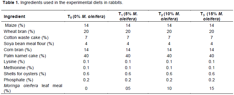
The meal that was obtained by grinding M. oleifera leaf was stored in airtight container until when needed for compounding or rather manufacturing. Four experimental diets comprising M. oleifera leaf meal has been used as a dietary supplement (Table 1).
T0: received 0% of M. oleifera leaf meal
T5: received 5% of M. oleifera leaf meal
T10: received 10% of M. oleifera leaf meal
T15: received 15% of M. oleifera leaf meal
Blood collection and evaluation of blood parameters on rabbits
At the end of the feeding period, blood samples were collected from the ear vein of each rabbit in the various groups, using a sterilized disposable syringe and needle. 2 ml blood was collected into labelled sterile vacuum tube containing ethylene-diamine-tetra-acetic acid (EDTA) as anticoagulant, of which another 3 ml of blood was collected into labelled sterile sample bottles without anticoagulant according to Ewuola et al. (2012).
The blood samples collected in EDTA was used for the determination of haematological parameters such as the RBC, WBC, Hb, and PCV as describe in Ewuola and Egbunike (2008). Blood constants such as MCH, MCV, MCHC and White blood differential counts (that is, Lymphocytes, Monocytes and Granulocytes) were determined to use appropriate formulae as described by Jain (1983). Biochemistry parameters such as glucose, urea, creatinine, cholesterol, total serum protein, serum albumin, Aspartate Aminotransferase (AST), Alanine Aminotransferase (ALT) and Alkaline phosphatase (ALP) were determined.
Statistical analysis
Analysis of variance (ANOVA) was done on the data collected. Duncan Multiple Range Test (Duncan, 1955)at a significant level of 0.05 was carried out in comparisons among dietary means. All computation was performed using statistical package SPSS 17.0 (SPSS Inc., Chicago, IL, USA).
Toxicological study
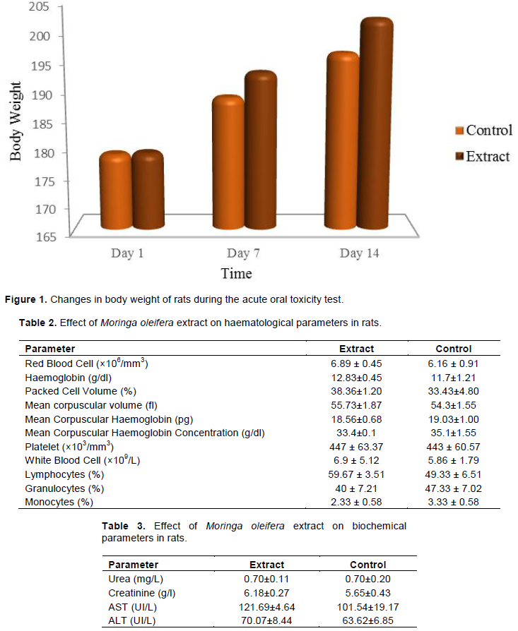
At the end of the two weeks of the experimental period (Day 14), no adverse effects were observed such as seizures, agitation, diarrhea, tremors, breathing difficulties and weight loss. Similarly, no mortality was recorded in animals throughout the experimental period as a consequent effect of administration of the M. oleifera leaf meal aqueous extract at a single dose of 2000 mg/kg body weight. This result shows that the dose of 2000 mg of the aqueous extract of M. oleifera leaves is lower than the LD50.
Figure 1 shows the weight variation of the rats tested as the controls throughout this test. There was an increase in the weight of the rats that received aqueous extract of M. oleifera leaf meal at a dose of 2000 mg/kg body weight.
Determination of hematological and biochemical parameters of rats
After 14 days of follow-up, blood samples were taken from the rats that received the extract as well as from the control rats for blood tests (Table 2). From this table it appears that no significant difference was noted on the two batches of rats, on both the globular constants and the blood cells.
Table 3 shows that the mean values of urea and creatinine in the test rats are not significantly different (p > 0.05) from the control rats. Similarly, the mean value of ALT and AST in test rats was not significantly influenced (p > 0.05) by the aqueous extract compared to control rats.
Physiological responses of rabbits fed graded levels of M. oleifera leaf meal on haematological and biochemical parameters
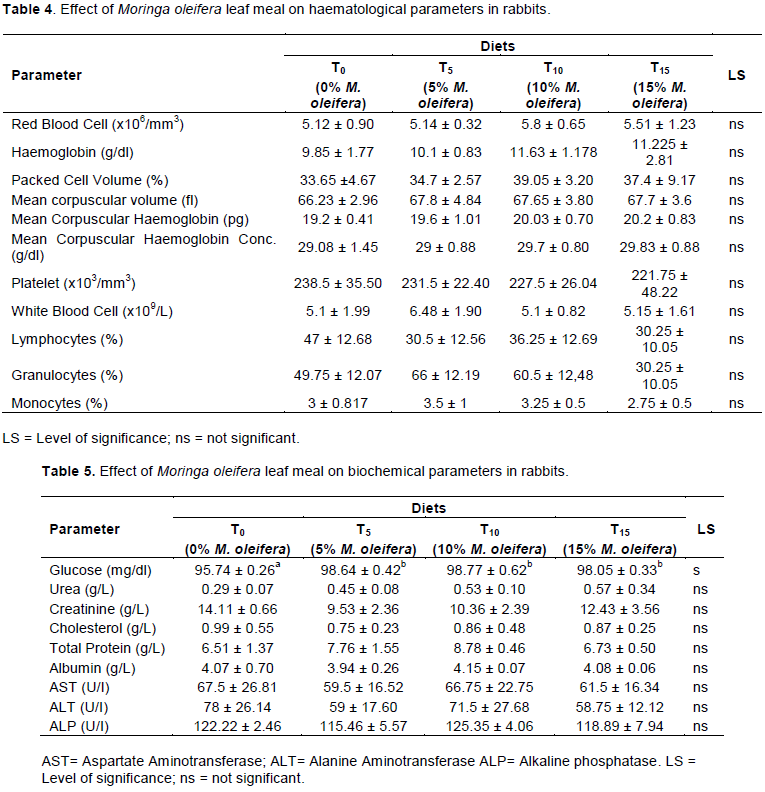
The results of the haematological parameters of rabbits fed different diets are shown in Table 4. RBC, WBC, Blood Platelets, Haemoglobin, MCV, MCHC, and MCH level showed no significant difference (P> 0.05) in different treatments compared to the control group.
The results of the effect of the M. oleifera leaf meal incorporated into the granulated feed of rabbits on the biochemical parameters are presented in Table 5. In fact, alanine aminotransferase, aspartate amino transferase, cholesterol, rabbit creatinine and urea in the different treatments showed no significant difference (p> 0.05). However, there is a slight increase of urea of subjects treated compared to the control. Similarly, a non- significant decrease in cholesterol levels is noted in the subjects treated relative to the control.
The acute oral toxicity of M. oleifera leaves meal was evaluated in this study to determine the tolerance limits of this plant just as used by traditional healers. Indeed, two weeks after the administration of the aqueous extract of M. oleifera leaf meal at the single dose of 2000 mg/kg body weight to the rats, no mortality within the batches was recorded. In addition, no adverse effects such as seizures, agitation, diarrhea, tremors, breathing difficulties and hair loss were observed within 14 days post-treatment. A significant difference was observed in body weight of animals in both the control and test groups. This weight gain observed in the animals of the test batch may be due to a beneficial action of the plant extract inducing an increase in appetite of rats causing them to increase their daily ration. In addition, it has been reported that, in addition to their therapeutic value, medicinal plants can also positively affect the nutritional status of animals (OECD, 2001).
According to Dougnon et al. (2013), any plant whose toxicity does not exceed 1000 mg/kg is said to be nontoxic and in this present study, the extract was administered at a dose of 2000 mg/kg. The aqueous extract of M. oleifera leaf meal administered at a single dose of 2000 mg/kg body weight had no significant effect on the hematological parameters of the rats. Therefore, the aqueous extract from the plant could have an effect against anemia, enhance the immune system and facilitate blood clotting for increased blood platelet levels. Moreover, no deformation of the appearance of Red Blood Cells and White Blood Cells has been observed and this further reveals the non-toxic nature of plant extracts (Oduola et al., 2007). Aminotransferases (AST and ALT) are biomarkers of liver malfunction and can be used to evaluate liver cytolysis with ALT as being a more sensitive biomarker of hepatotoxicity than AST (Pramyothin et al., 2006). In our study, transaminase levels (AST and ALT) that reflect liver function status did not significantly change the test batch compared to the control group. This is an indication that the extract did not affect normal liver function. In addition, a significant increase or decrease in transaminase activity, especially ALT, is often associated with evidence of hepatocellular damage (Wannang et al., 2007). M. oleifera leaf meal are non-toxic in rats at a dose of 1000 mg/kg (Jonathan et al., 2014). In addition, no significant difference, of plasma glucose, cholesterol and creatinine levels were noted in test rats compared to control rats. These results converge with those of Akouango et al. (2014)who evaluated the acute toxicity of M. oleifera in mice orally at doses ranging from 100 to 5000 mg/kg orally (PO) and from 10 to 2000 mg/kg intraperitoneal (PI). The results obtained for the acute oral toxicity test on the aqueous extract of M. oleifera leaf meal made it possible to affirm that the oral use of the leaves of this plant seems relatively safe.
The analysis of blood parameters is a means of establishing the state of health of an animal and thus determining the effect of the ingestion of food components on the blood composition (Church et al., 1984). Blood testing can therefore be considered an appropriate measure of long-term nutritional status.
Iheukwuemere et al. (2006) have shown that changes in hematological and biochemical parameters in animals indicate their physiological state. Thus, the hematological components are useful in the monitoring of food toxicity especially with feeding components, which may affect the formation of blood. PCV is a parameter for measuring relative blood mass. PCV levels obtained in this study are not significantly (p>0.05) influenced by different dietary treatments. Their values ​​were in the 33 to 50% reference range considered normal Packed Cell Volume of a healthy rabbit reported by Burns and De Lannoy (1966). The normal value of hematocrit shows the proper nutritional status of rabbits (Church et al., 1984). The result of our study is in agreement with the conclusion of Jiwuba et al. (2016)who observed no significant difference (p>0.05) in the PCV of rabbits fed M. oleifera leaf meal. There is no significant difference in the number of red blood cells (specialized blood cells in oxygen transport) in this study which is in the reference range (5.46 × 10¹² to 7.94 × 10¹² L-1) according to Mitruka and Rawnsley (1977). This result corroborates that of Jiwuba et al. (2016), who also found no significant influence of feeding in the red blood cells of rabbits fed M. oleifera leaf meal. The hemoglobin levels obtained showed no significant difference (p>0.05) with respect to the control. However, the hemoglobin levels of the treatments are slightly higher than that of the control. Overall, it should be noted that these levels are within the reference range (9.40 to 17.90 g/dL) according to Campbell (2015). MCV, MCHC, and MCH values ​​obtained in this study are not affected by the different dietary treatments and are included in the normal values ​​of healthy rabbits. These parameters being important morphological characters of anemia (Campbell, 2015), show that rabbits are not anaemic. The White Blood Cell was not significantly (p>0.05) influenced by dietary treatments. The values ​​obtained in this study are within the reference values ​​recommended by Campbell (2015). These values ​​show that rabbits fed with the gradual concentrations of M. oleifera leaf meal in the diet are in good health. Indeed, leukopenia is an indicator of allergies, anaphylactic shock, and certain parasitism, while the high number of white blood cells indicates the existence of a recent infection (Ahamefule et al., 2008). Granulocytes, lymphocytes and monocytes do not have a significant difference (p> 0.05). This indicates a probably normal physiological state in these rabbits subjected to these different treatments. The presence of monocytes in rabbit blood with the different treatments contradicts the results of the works of Bitto et al. (2006)that recorded the complete absence of monocytes in rabbits.
The biochemical results reveal no significant difference (p> 0.05) with respect to the control. The values of urea, creatinine, alanine aminotransferase, aspartate aminotransferase and cholesterol are included in the reference values. The absence of a significant difference in transaminases shows the protective hepatotoxic effect of M. oleifera leaves on the health of rabbits. The absence of significant differences in creatinine and urea also shows the nephroprotective property of M. oleifera leaves on the health of rabbits.
These results show that M. oleifera leaves can be incorporated into the granulated feed of rabbits up to 15% without any deleterious effect on the health of the rabbits if the good hygienic practices are maintained.
This study indicated that oral administration of aqueous extract of M. oleifera leaf at 2000 mg/kg body weight showed no changes in clinical signs and blood parameters. Therefore, M. oleifera have no toxicity at 2000 mg/kg body weight. From this study, it was concluded that M. oleifera leaf meal did not affect the biochemical and haematological parameters of rabbits. M. oleifera leaf meal may be incorporated into the rabbit's feed formulation at 15% rate. M. oleifera leaf meal can be used for increase in growth and health performance of rabbits and also subsidiary income of breeders.
ETHICAL COMMITTEE APPROVAL
The study was carried out in strict compliance with the recommendations of the guide of the Research Ethics Committee of the National University of Agriculture (UNA), Porto Novo, Republic of Benin and in line with detailed protocols of Animal Care and Use in Research, Education and Testing: N° 062- 2016/ P-Ethic Committee/SA.
The authors have not declared any conflict of interests.
The authors acknowledged the laboratory of Ethnopharmacology and Animal Health.
REFERENCES
|
Aboh A, Olaafa M, Dossou-Gbété G, Dossa A, Djagoun N (2002). Ingestion volontaire et digestibilité apparente d'une ration à base de la farine de graines de Mucuna pruriens var. utilis complétée de fourrages chez les lapins. Tropicultura 20(4):165-169.
|
|
|
|
Ahamefule F, Obua B, Ukweni I, Oguike M, Amaka R (2008). Haematological and biochemical profile of weaner rabbits fed raw or processed pigeon pea seed meal based diets. African Journal of Agricultural Research 3(4):315-319.
|
|
|
|
|
Ahemen T, Abu AH, Iorgilim LK (2013). Physiological responses of rabbits fed graded levels of Moringa oleifera leaf meal (MOLM): Some aspects of haematology and serum biochemistry. Archives of Applied Science Research 5(2):172-176.
|
|
|
|
|
Akouango P, Opoye I, Ngokaka C, Akouango F (2014). Contribution à la réduction des périodes improductives du cycle de reproduction des lapines (Oryctolagus cuniculus) dans un élev age fermier. Afrique Science: Revue Internationale des Sciences et Technologie 10(2): 356-364.
|
|
|
|
|
Bitto I, Arubi J, Gumel A (2006). Reproductive tract morphometry and some haematological characteristics of female rabbits fed pawpaw peel meal based diets. African Journal of Biomedical Research 9(3).
Crossref
|
|
|
|
|
Burns KF, De Lannoy CW (1966). Compendium of normal blood values of laboratory animals, with indication of variations: I. Random-sexed populations of small animals. Toxicology and Applied Pharmacology 8(3):429-437.
Crossref
|
|
|
|
|
Campbell T (2015). Exotic animal hematology and cytology: John Wiley & Sons, Inc. 402 p.
Crossref
|
|
|
|
|
Church JP, Judd JT, Young CW, Kelsay JL, Kim WW (1984). Relationships among dietary constituents and specific serum clinical components of subjects eating self-selected diets. The American Journal of Clinical Nutrition 40(6):1338-1344.
Crossref
|
|
|
|
|
Combes S (2004). Valeur nutritionnelle de la viande de lapin. INRAE Productions Animales 17(5):373-383.
Crossref
|
|
|
|
|
Dougnon V, Bankolé H, Edorh P, Klotoé JR, Dougnon J, Fah L, Loko F, Boko M (2013). Acute toxicity of Solanum macrocarpon Linn (Solanaceae) on Wistar rats: study about leaves and fruits. American Journal of Biochemistry 3:84-88.
|
|
|
|
|
Duncan D (1955). Multiple range and multiple F tests. Biometrics 11(1):1-42.
Crossref
|
|
|
|
|
Duncan GJ, Brooksâ€Gunn J, Klebanov PK (1994). Economic deprivation and early childhood development. Child Development 65(2):296-318.
Crossref
|
|
|
|
|
Ewuola E, Egbunike G (2008). Haematological and serum biochemical response of growing rabbit bucks fed dietary fumonisin B1. African Journal of Biotechnology 7(23):4304-4309.
|
|
|
|
|
Ewuola E, Jimoh O, Atuma O, Soipe O (2012). Haematological and serum biochemical response of growing rabbits fed graded levels of Moringa oleifera leaf meal. World Rabbit Science Association Proceedings 3(6):683-679.
|
|
|
|
|
Iheukwuemere F, Abu A, Ameh M (2006). Effect of human menopausal gonadotropin on haematological and serum biochemical parameters of the Nigerian Indigenous chickens. International Journal of poultry Science 5(7):632-634.
Crossref
|
|
|
|
|
Jain N (1983). Scanning electron micrograph of blood cells. Schalm's Veterinary Haematology 4:63-70.
|
|
|
|
|
Jiwuba P, Ikwunze K, Dauda E, Ugwu D (2016). Haematological and serum biochemical indices of growing rabbits fed diets containing varying levels of Moringa oleifera leaf meal. British Biotechnology Journal 15(2):1-7.
Crossref
|
|
|
|
|
Jonathan AE, Etuk EU, Bello SO, Gwarzo MS, Egua MO, Nkwoka IJ (2014). In vitro Antibacterial Activities of Aqueous and Ethanolic Stem Bark Extracts of Bridelia ferruginea Benth. International Journal of Current Research in Biosciences and Plant Biology 1(5):28-31.
|
|
|
|
|
Kpodekon M, Toleba S, Boko C, Dagnibo M, Djago Y, Dossa F, Farougou S (2015). Fréquence des Escherichia coli entéropathogènes chez les lapins (Oryctolagus cuniculus) dans la commune d'Abomey-Calavi en zone sub-équatoriale du Bénin. Revue de Médecine Vétérinaire 166(3-4):84-89.
|
|
|
|
|
Lenarczyk M, Lam V, Jensen E, Fish B, Su J, Koprowski S, Komorowski R, Harmann L, Migrino R, Li X, Hopewell J, Moulder J, Baker J (2013). Cardiac Injury after 10 Gy Total Body Irradiation: Indirect Role of Effects on Abdominal Organs. Radiation Research 180(3):247.
Crossref
|
|
|
|
|
Mitruka BM, Rawnsley HM (1977). Clinical biochemical and hematological reference values in normal experimental animals: Masson Pub. USA pp. 1981-413.
|
|
|
|
|
Oduola T, Adeniyi F, Ogunyemi E, Bello I, Idowu T, Subair H (2007). Toxicity studies on an unripe Carica papaya aqueous extract: biochemical and haematological effects in wistar albino rats. Journal of Medicinal Plants Research 1(1):001-004.
|
|
|
|
|
OECD (2001). Guidelines 420: Acute oral toxicity-fixed dose procedure.
View
|
|
|
|
|
Ologhobo A, Adejumo I, Akangbe E (2014). Comparison effect of Moringa oleifera leaf meal and oxytetracycline on haematology and serum biochemical profile of broiler finishers. International Blood Research Reviews 2(1):29-36.
Crossref
|
|
|
|
|
Osman HM, Shayoub ME, Babiker EM (2012). The effect of Moringa oleifera leaves on blood parameters and body weights of albino rats and rabbits. Jordan Journal of Biological Sciences 5(3):47-150.
|
|
|
|
|
Pramyothin P, Samosorn P, Poungshompoo S, Chaichantipyuth C (2006). The protective effects of Phyllanthus emblica Linn. extract on ethanol induced rat hepatic injury. Journal of Ethnopharmacology 107(3):361-364.
Crossref
|
|
|
|
|
Sarwatt S, Kapange S, Kakengi A (2002). Substituting sunflower seed-cake with Moringa oleifera leaves as a supplemental goat feed in Tanzania. Agroforestry Systems 56(3):241-247.
Crossref
|
|
|
|
|
Wannang NN, Jimam NS, Omale S, Dapar ML, Gyang SS, Aguiyi JC (2007). Effects of Cucumis metuliferus (Cucurbitaceae) fruits on enzymes and haematological parameters in albino rats. African Journal of Biotechnology 6(22).
|
|