African Journal of
Biochemistry Research

  • Abbreviation: Afr. J. Biochem. Res.
  • Language: English
  • ISSN: 1996-0778
  • DOI: 10.5897/AJBR
  • Start Year: 2007
  • Published Articles: 425

Full Length Research Paper

Evaluation of the effect of vitamin C on caspase 9 and oxidative stress in rheumatoid arthritis patients

Amira M. Elshamy
  • Amira M. Elshamy
  • Department of Medical Biochemistry, Faculty of Medicine, Tanta University, Egypt.
  • Google Scholar
Nagah K. Gaafar
  • Nagah K. Gaafar
  • Department of Medical Biochemistry, Faculty of Medicine, Tanta University, Egypt.
  • Google Scholar
Nadia E. ElAshwah
  • Nadia E. ElAshwah
  • Department of Medical Biochemistry, Faculty of Medicine, Tanta University, Egypt.
  • Google Scholar
Ayman A. Wagih
  • Ayman A. Wagih
  • Department of Medical Biochemistry, Faculty of Medicine, Tanta University, Egypt.
  • Google Scholar
Abeer A. Shahba
  • Abeer A. Shahba
  • Department of Internal Medicine, Faculty of Medicine, Tanta University, Egypt.
  • Google Scholar


  •  Received: 08 May 2018
  •  Accepted: 03 September 2018
  •  Published: 30 November 2018

 ABSTRACT

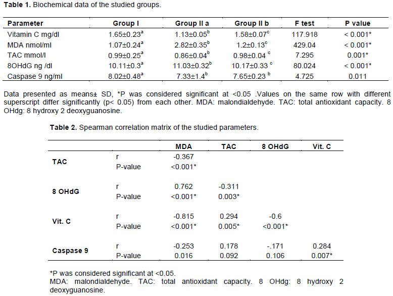
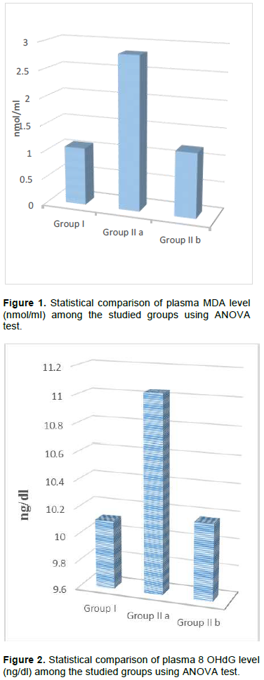
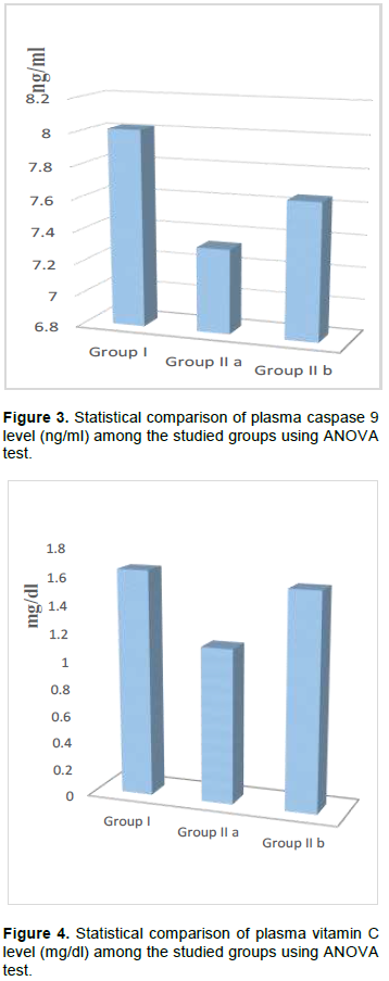
Rheumatoid arthritis (RA) is a chronic and an autoimmune disease of the joints and is widely distributed worldwide. It is characterized by alterations of the antioxidant defense system and increased free radical formation and pro-inflammatory cytokine. The aim of the present study was to evaluate the effect of vitamin C supplementation on oxidative stress biomarkers and caspase 9 level in rheumatoid arthritis patients. This study included 30 RA patients and 30 healthy subjects. Plasma levels of malondialdehyde (MDA), total antioxidant capacity (TAC), caspase 9 and 8-hydroxy-2′-deoxyguanosine (8 OHdG) were assayed as well as blood vitamin C level. These parameters were re-evaluated in RA patients after vitamin C supplementation for one month. Increased MDA and 8 OHdG levels and reduced TAC, caspase 9 and vitamin C. Levels were demonstrated in RA patients. After vitamin C supplementation, RA patients showed significant increase in TAC and vitamin C level and significant decrease in MDA and 8 OHdG levels, plasma caspase 9 level was not significantly affected after vitamin C supplementation. Increased oxidative stress and decreased apoptosis may have an important role in the pathogenesis of RA. The administration of vitamin C supplementation may help to relieve oxidative stress and enhance the antioxidant defense in these patients.


Key words: Rheumatoid arthritis, oxidative stress, apoptosis, caspase 9, vitamin C.


 INTRODUCTION

Rheumatoid arthritis (RA) is a chronic and an autoimmune disease of the joints. It affects 0.5-1% of adults worldwide. About 30% of RA patients would become disabled in the first 2-3 years without sufficient treatment. RA is characterized by alterations of the antioxidant defense system, increased free radical formation and pro-inflammatory cytokine expression at the site of inflammation (Kundu et al., 2012).         
 
Reactive oxygen species (ROS) are produced during oxidative phosphorylation. When the production of ROS exceeds the physiological level, it induces oxidative stress and damages tissue and cellular protein, lipid, and nucleic acids (Hitchon and El-Gabalawy, 2004). Large amounts of ROS have been detected in the synovial fluid and peripheral blood of RA patient .ROS production is induced by TNF-α stimulation. ROS can directly degrade
 
joint cartilage, attacking its proteoglycan and inhibiting its synthesis. ROS have also been linked to mutation of p53 in RA-derived fibroblast-like synoviocytes (RA-FLS) (Sakon et al., 2003). Among the DNA constituents, guanine is highly susceptible to the oxidative damage induced by free radicals. The oxidation product, 8-hydroxy-2′-deoxyguanosine (8-OHdG) is one of the most common biomarkers used to detect oxidative DNA damage. 8-OHdG is eliminated in body fluids and can be measured (Halliwell, 2000).
 
The damaging effect of ROS is antagonized by antioxidants. An antioxidant is any substance that is able to scavenge free radicals or inhibit the oxidation process inside the cell. Enzymatic antioxidants include superoxide dismutase (SOD), catalase (CAT), and glutathione (GSH) related enzymes (glutathione peroxidase [GPx], glutathione reductase [GR], and thioredoxin reductase). Furthermore, the non-enzymatic antioxidants includes vitamins (A, C, and E), β-carotene, antioxidant minerals (copper, ferritin, zinc, manganese, and selenium) and reduced glutathione (GSH) (Kalpakcioglu and Senel, 2008).
 
Vitamin C (ascorbate) is an essential water-soluble vitamin in humans which must be taken in diet. It acts as a cofactor for many biosynthetic enzymes required for the synthesis of amino acid-derived macromolecules, neurotransmitters and neuropeptide hormones. Also, it acts as a cofactor for many hydroxylases enzymes such as those involved in collagen biosynthesis. Moreover, vitamin C acts as an antioxidant and prevents oxidative stress-induced damage (Monfort and Wutz, 2013). RA-FLS proliferate abnormally under the stimulatory effect of many inflammatory cytokines, such as IL-1, IL-6, and TNF- α. RA-FLS  are  resistant  to  apoptosis,  yielding expansion  capabilities  similar  to  tumors. This transforms FLSs from innocent mesenchymal cells to destructive aggressors. This leads to local cartilage destruction and chronic synovial inflammation. Apoptosis induction of RA-FLS is therefore suggested as a potential therapeutic approach for RA (Noss and Brenner, 2008). Apoptosis, programmed cell death, is the physiologically preferred pathway of cell death. Apoptosis is involved in a many physiological processes such as growth, development, differentiation and immune response. Any deregulation of the apoptotic pathway may lead to the development of diseases. Apoptosis occurs through two signaling pathways: the extrinsic pathway and the intrinsic pathway. The extrinsic pathway is induced by extracellular triggers. The intrinsic pathway involves a mitochondrial pathway (Pileczki et al., 2016).
 
Caspases are the central executioners of apoptosis and are responsible for the morphological features of apoptosis. The extrinsic pathway is mediated by caspase-8 while the intrinsic pathway can be initiated through caspase-9. Both pathways trigger apoptosis through the cleavage of the downstream executioner proteins (caspase-3 and -7). Although caspase-9 is an intracellular protein, it was suggested that serum caspases may be noninvasive biomarkers for detection of apoptosis (Kuida, 2000).
 
Disease-modifying anti-rheumatic drugs (DMARDs) are the mainstay of RA therapy. Methotrexate is recommended as the first-line treatment in patients with active RA (Saag et al., 2008). However, MTX can be cytotoxic per se and further increase the degree of cell damage in RA. MTX which is used for a long time exerts cytotoxic effects on ‘S phase’ of the cell cycle, and inhibits cell division. MTX exerts its effects on cells by decreasing cellular antioxidant activity, and exposing cells to the unfavorable effects of ROS, eventually inducing detrimental alterations (Yulug et al., 2013). MTX markedly increases sensitivity of cells to apoptosis mediated via either death receptor or mitochondrial pathways, in part, by increasing expression of genes whose protein products play key roles in induction of apoptosis (Spurlock et al., 2011). The effects of antioxidant materials in relieving toxic effects of MTX have been intensively investigated. Vitamin C is an important agent in enhancing the antioxidant capacity of cells (Vijayprasad et al., 2014). This study therefore should be viewed as a step in assessing potential beneficial effects of the vitamin C supplementation tested on rheumatoid arthritis. We aim to use vitamin C supplementation alongside drug treatment to reduce drug dose and thus side effects of treatment.


 PATIENTS AND METHODS

Patients
 
This study included 30 healthy subjects aged (25-55) years old as a control group (Group I) and  30 RA patients aged (25-55 years old) (Group IIa), these patients were given vitamin C supplementation at a dose of 500 mg twice daily for one month (vitacid 0.5 g tablets) then patients were re-evaluated again (Group IIb). RA patients were diagnosed according to the revised criteria of the American College of Rheumatology (ACR). The disease duration was 8+ 4.6 years. Patients were collected the outpatient clinics of rheumatology unit of internal medicine department, Tanta University Hospitals, Egypt. Ethical approval of this study was obtained from the Ethical Committee of Faculty of Medicine, Tanta University (approval code 30326/05/15) and written informed consents were obtained from all patients. All RA patients were under disease-modifying anti-rheumatic drugs (DMARDs) either as mono-therapy or in combination. The vast majority of RA patients were consuming non-steroidal anti-inflammatory drugs (NSAIDs) on irregular basis.
 
Justification of sample size
 
If the sample size is too small, the study may fail to answer its research question. If the sample size is too large, the study will be more difficult and costly than necessary. So, I adjusted this sample size by the help of previous researches (the antioxidant vitamins A, C, E and selenium in the treatment of arthritis: A systematic review of randomized clinical trials) (Canter, 2007).
 
 
Exclusion criteria
 
Patients suffering from chronic disorders such as diabetes mellitus, thyroid dysfunction, liver or kidney diseases were excluded.
 
Blood sampling
 
Blood samples were collected from patients and controls under aseptic precautions by venipuncture; 5.0 ml in a heparinized vial for vitamin C, TAC, MDA, 8 OHdG and caspase 9 levels assessment. 1 ml of heparinized blood samples was used to measure vitamin C level (as soon as possible within 2 h), and the remaining samples were centrifuged as soon as possible at 2000 g for 10 min at 4ο C. plasma samples were stored at 70°C until the time of analysis of MDA , TAC , 8 OHdG and caspase 9  levels.
 
Assay of apoptotic and DNA damage biomarkers
 
Plasma caspase 9 and 8 OHdG levels were measured by commercial supplied ELISA kit supplied Chongqing Biospes, Co. (Catalog No. BYEK2175, No. BYEK1218 respectively). Assay was carried out according to the manufacturers’ instructions. Using the mean absorbance value for each sample, the corresponding concentration of caspase 9 and 8 OHdG were determined from the standard curve.
 
Assay of oxidative stress biomarkers
 
Plasma levels of both MDA and TAC were spectrophotometric assayed using commercial kit supplied by Bio-diagnostic, Egypt. The absorbance of sample and standard were measured against blank at 534 and 510 nm for MDA and TAC respectively using semiautomatic BTS-350 Biosystems spectrophotometer.
 
Assay of vitamin C level
   
Blood vitamin C level was assayed according to the method of Jagota and Dani using Folin-Ciocalteu reagent which reacts specifically with ascorbic acid in a broad pH range. Vitamin C standard was prepared by dissolving 10 mg of vitamin C powder in 10 ml distilled water. The sample concentrations were calculated by interplotting from the standard curve (Jagota and Dani, 1982).
 
Statistics
 
The results for continuous variables were expressed as means± SD and one-way analysis of variance (ANOVA) were used for their analysis. The statistical significances of differences in frequencies of variants between the groups were tested using the F test. A difference was considered significant at P values less than 0.05.  All statistical calculations were performed using the computer program SPSS (Statistical Package for the Social Science) version 17 for Microsoft Windows. Correlation between the variables was examined using the Pearson’s correlation coefficient.


 RESULTS

Biochemical data of the studied groups are illustrated in Table 1. Plasma MDA level and 8OHdG were significantly increased in RA patients as compared to the healthy control subjects (p< 0.0001) as depicted in Figures 1 and 2 respectively .On the other hand, caspase 9 level, vitamin C level and total antioxidant capacity were significantly decreased in RA patients as compared to control subjects (p< 0.0001) as depicted in Figures 3, 4 and 5 respectively. After vitamin C supplementation, MDA level and 8OHdG level were significantly decreased as depicted in Figures 1 and 2 respectively while vitamin C level and total antioxidant capacity were significantly increased as depicted in Figures 4 and 5 respectively. On the other hand, there was no statistically significant improvement in caspase 9 level after vitamin C supplementation in RA patients (as depicted in Figure 3 and Table 2).


 DISCUSSION

Rheumatoid arthritis (RA) is an autoimmune disease characterized by cartilage and bone destruction and systemic complications. ROS have been implicated to play an important role in RA pathogenesis (Filippin 2008). The present study showed increased level of MDA in RA patients. MDA is one of the most potential biomarkers of lipid peroxidation. The rise in lipid peroxidation is due to increased ROS which tends to increase abundantly during chronic inflammation. ROS lead to cascade stimulation of the activity of mitogen activated protein kinase (MAPK) and nuclear factor-kappa B (NF-Κb) pathways and increase the inflammatory cytokines’ gene expression which finally creates immune responses and causes inflammation (Srivastava and Shrivastava, 2016). In agreement with these findings, El-barbary et al. (2011); Alver  et  al. (2011); Datta et al.  (2014); Karaman   et   al. (2011); Shah et al. (2011); Mishra et al. (2012) and Hassan et al. (2011) found a remarkable elevation in MDA levels in RA patients.


On the other hand, Jacobson et al. (2012) and Kajanachumpol et al. (2000) reported no significant difference in MDA concentration between RA patients and control group. The present study showed significant elevation of plasma 8-OHdG in rheumatoid arthritis patients. 8-OHdG is the most common stable product of oxidative DNA damage. 8-OHdG is currently used as a recognized biomarkers of oxidative DNA damage mainly because of its abundance in DNA and also because of its reliable detectability (Hakem, 2008). Increased 8-OHdG level in RA patients have also been found by Kageyama et al., 2008; and Ishibashi et al., 2014;  Khan et al., 2011. In contrast to the results of the present study, low level of 8-OHdG in spite of elevation of other oxidative stress biomarkers in several diseases has been reported. Sova et al. (2010) found that Serum 8-OHdG level was significantly lower in women with polycystic ovary syndrome. Inactive DNA repair enzymes may lie behind lower serum 8-OHdG levels in these patients. The main repair enzyme connected with  8-OHdG  is  8-oxoguanine  DNA glycosylase 1 (OGG1).With impaired OGG1 function and/or expression, cells are not able to cleave damaged guanosine from DNA, resulting in decreased 8- OHdG serum levels (Hirano, 2008).
 
The present study revealed significant reduction in vitamin C level and total antioxidant capacity level in RA patients. The continuous production of free radicals in inflamed joint gives rise to failure of antioxidant system and further tissue damages. Also the intake of food antioxidants is lower than the recommended amounts in RA patients (Silva et al., 2014). Reduced vitamin C level in RA patients was also reported by Jaswal et al. (2003); Mateen et al. (2016); and Vijayakumar et al. (2006). Also, Hadi et al. (2014) found significant reduction in TAC in RA. This is attributed to excess antioxidants utilization by the inflamed   tissues to scavenge the excessive ROS generated at inflammatory sites. The present study found significant improvement in vitamin C level and TAC level with significant reduction of MDA and 8-OHdG levels after intake of vitamin C supplementation. Vitamin C is a chain-breaking antioxidant that halts the propagation of per-oxidative processes and reacts with membrane bound oxidized vitamin E, thus reducing it back to its native form. These findings suggest that vitamin C behaves as an ROS scavenger and may be effective in combating oxidative damage.
 
In agreement with these results, Meki et al. (2009) and Kuiper et al. (2011) found significant reduction in lipid peroxidation in response to vitamin C administration. Also, Al-Jassabi and Khalil (2006) found that vitamin C markedly inhibited oxidative DNA damage (as indicated by measurement of 8 OHdG). Moreover, Gęgotek et al. (2017) showed that vitamin C supplementation to fibroblasts increased GSH and SOD level and reduced MDA and total ROS levels. On the other hand, Nath et al. (2010) proved that vitamin C has no effect on 8-OHdG formation in a chemical model.
 
Apoptosis is a mechanism by which cells undergo programmed death. Serum markers of apoptosis may be noninvasive biomarkers in patients. Moreover, measurement of caspases in serum could be useful in monitoring different diseases (Babas et al., 2010). Owing to its crucial function of converting the death signal to the first proteolytic event and activating executioner protease, we aimed to evaluate plasma caspase 9 level in RA patients and its response to vitamin C supplementation. The present study showed significant reduction in plasma caspase 9 level in rheumatoid arthritis patients compared to control group.
 
Serum caspases were also evaluated in rheumatoid arthritis patients. Alzaidy et al. (2016) detected significant reduction in serum caspase3 level in rheumatoid arthritis patients. RA-FLS play an important role in both the initiation and the perpetuation of RA. RA-FLS are resistant to apoptosis. At the molecular level, synovial fibroblasts are characterized by the activation of signaling cascades that result in the inhibition  of  apoptosis  (Khan et al., 2011). In agreement with these results, Lattuada et al. (2016) proved that the percentage of apoptotic cells (RA-FLSs) cultured in synovial fluid samples aspirated from the joints of RA was lower than in cells cultured in tissue medium alone, probably because of the presence in the SF of anti-apoptotic factors. Resistance to apoptosis in RA-FLSs depends on up regulation of IAPs which are expressed at high levels in RA-FLSs (Casnici et al., 2014). Also, Jia et al. (2015) detected increased proliferation and inhibited apoptosis in synovial cells separated from synovial tissues harvested from RA patients.
 
On the other hand, Yin et al. (2015) demonstrated that lipid peroxidation products induce synovial inflammations by activating NF-Ò¡B pathway and increase cleaved caspase 3 level leading to dramatic cell apoptosis. The present study revealed no significant effect of vitamin C supplementation on caspase 9 level in rheumatoid arthritis patients. This may be attributed to low dose of ascorbic acid and / short duration of treatment. Many studies proved a significant effect of vitamin C on apoptosis. Chiu et al. (2017) examined the efficacy of vitamin C to prevent monosodium iodoacetate (MIA)-induced osteoarthritis in rat. In an animal model, intra-articular injection of MIA increased oxidative stress and apoptosis which resulted in cartilage degradation. All of these changes were prevented by treatment with vitamin C. Also, Sato et al. (2015) demonstrated that administration of high dose ascorbic acid reduced radiation-induced apoptosis in bone marrow cells.
 
On the other hand, Uetaki et al. (2015) proved that vitamin C has an apoptotic and cytotoxic effect especially on cancer cells. The administration of ascorbate (within pharmacological concentrations) is associated with the formation of H2O2 in the extracellular fluid surrounding a tumor. In the presence of catalytic metal ions, ascorbate functions as a pro-oxidant and induces oxidative stress (Frei et al., 2008). H2O2 can affect both extracellular and intracellular targets, as it is permeable across lipid membranes. H2O2 may attack membrane lipids forming lipid hydro-peroxides and causing leaky membranes, intracellular oxidative stress and DNA damage leading to cell death (Du et al., 2010).
 
Regarding the use of intravenous vitamin C in rheumatoid arthritis, Mikirova et al. (2012) suggested that intravenous vitamin C therapy can reduce inflammation in RA patients. At high doses, ascorbate has been shown to reduce the production of pro-inflammatory cytokines and to affect the activation of NF-κB. 


 CONCLUSION

Intervention with vitamin C supplementation in RA patients resulted in consistent and significant improve-ment of the antioxidant status and marked reduction of oxidative stress biomarkers were  observed.  These  data are promising and indicate need for more studies to demonstrate more benefits for RA patients and to evaluate its effect on apoptosis. This study showed that vitamin C supplementation can help to relieve oxidative stress which is a serious side effect of methotrexate and cytotoxic drugs. We recommend also further studies to try to reduce the dose of these drugs with vitamin C supplementation in order to minimize side effects. 


 CONFLICT OF INTERESTS

The authors have not declared any conflict of interests.


 REFERENCES

Al-Jassabi S, Khalil AM (2006). Microcystin_induced 8_hydroxydeoxyguanosine in DNA and its reduction by melatonin, vitamin C, and vitamin E in mice. Biochemistry (Moscow) 71(10):1115-1159
Crossref


Alver A, Åžentürk A, Çakirbay H, MenteÅŸe A, Gökmen F, Keha EE, Uçar F (2011). Carbonic anhydrase II autoantibody and oxidative stress in rheumatoid arthritis. Clinical Biochemistry 44(17-18):1385-1389.
Crossref


Alzaidy AH, Numan IT, Jassim NA (2016). Effects of adalimumab MTX combination on serum (NF-κB) and caspase 3 in Iraqi patients with rheumatoid arthritis. International Journal of Science and Research 5(7).


Babas E, Ekonomopoulou MT, Karapidaki I, Doxakis A, Betsas G, Iakovidou-Kritsi Z (2010). Indication of participation of caspase-2 and caspase-5 in mechanisms of human cervical malignancy. IInternational Journal of Gynecological Cancer 20(8):1381-1385.


Canter PH, Wider B, Ernst E (2007). The antioxidant vitamins A, C, E and selenium in the treatment of arthritis: a systematic review of randomized clinical trials. Rheumatology (Oxford) 46(8):1223-1233.
Crossref


Casnici C, Lattuada D, Tonna N, Crotta K, Storini C, Bianco F, Truzzi MC, Corradini C and Marelli O (2014). Optimized in vitro culture conditions for human rheumatoid arthritis synovial fibroblasts. Mediators of Inflammation 2014:702057.
Crossref


Chiu PR, Hu YC, Huang TC, Hsieh BS, Yeh JP, Cheng HL, Huang LW, Chang K (2017). Vitamin C protects chondrocytes against monosodium iodoacetate-Induced osteoarthritis by multiple pathways. International Journal of Molecular Sciences 18(1).
Crossref


Datta S, Kundu S, Ghosh P, De S, Ghosh A, and Chatterjee M (2014). Correlation of oxidant status with oxidative tissue damage in patients with rheumatoid arthritis. Clinical Rheumatology 33(11):1557-1564.
Crossref


Du J, Martin SM, Levine M, Wagner BA, Buettner GR, Wang SH, Taghiyev AF, Du C, Knudson CM, Cullen JJ (2010). Mechanisms of ascorbate-induced cytotoxicity in pancreatic cancer. Clinical Cancer Research 16(2):509-520.
Crossref


El-barbary AM, AbdelKhalek MA,Elsalawy AM and Hazaa SM (2011):Assessment of lipid peroxidation and antioxidant status in rheumatoid arthritis and osteoarthritis patients. The Egyptian Rheumatologist 33(4):179-185.
Crossref


Filippin LI, Vercelino R, Marroni NP, Xavier RM (2008). Redox signalling and the inflammatory response in rheumatoid arthritis. Clinical and Experimental Immunology Clinical and Experimental Immunology, 152(3):415-422.
Crossref


Frei B, Lawson S (2008). Vitamin C and cancer revisited. Proceedings of the National Academy of Sciences 105(32):11037-11038.
Crossref


Gęgotek A, Bielawska K, Biernacki M, Zaręba I, Surażyński A, Skrzydlewska E (2017). Comparison of protective effect of ascorbic acid on redox and endocannabinoid systems interactions in in vitro cultured human skin fibroblasts exposed to UV radiation and hydrogen peroxide. Archives of Dermatological Research 309(4):285-303.
Crossref


Hadi V, Kheirouri S, Alizadeh M, Khabbazi A, Hosseini H (2014). Effects of Nigella sativa oil extract on inflammatory cytokine response and oxidative stress status in patients with rheumatoid arthritis: a randomized, double-blind, placebo-controlled clinical trial. Avicenna Journal of Phytomedicine 6(1):34-43.


Hakem R (2008): DNA-damage repair: the good, the bad, and the ugly. EMBO Journal 27(4):589-605.
Crossref


Halliwell B (2000). Why and how should we measure oxidative DNA damage in nutritional Studies? How far have we come? American Journal of Clinical Nutrition 72:1082-108.
Crossref


Hassan SZ, Gheita TA,Kenawy SA,Fahim AT, El- Sorougy IM, Abdou MS (2011). Oxidative stress in systemic lupus erythematosus and rheumatoid arthritis patients: relationship to disease manifestations and activity. International Journal of Rheumatic Diseases 14(4):325-331.
Crossref


Hirano T (2008). Repair system of 7,8-dihydro-8-oxoguanine as a defence line against carcinogenesis. Journal of Radiation Research 49(4):329-340.
Crossref


Hitchon CA, El-Gabalawy HS (2004). Oxidation in rheumatoid arthritis. Arthritis Research and Therapy 6:265-278
Crossref


Ishibashi T, Sato B, Shibata S, Sakai T, Hara Y, Naritomi Y, Koyanagi S, Hara H, Nagao T (2014). Therapeutic efficacy of infused molecular hydrogen in saline on rheumatoid arthritis: A randomized, double-blind, placebo-controlled pilot study. International Immunopharmacology 21 (2):468-473.
Crossref


Jacobson GA, Ives SJ, Narkowicz C, Jones G (2012). Plasma glutathione peroxidase (GSH-Px) concentration is elevated in rheumatoid arthritis: a case-control study. Clinical Rheumatology 31(11):1543-1547.
Crossref


Jagota SK, Dani HM (1982). A new colorimetric technique for the estimation of vitamin C using Folin phenol reagent. Analytical Biochemistry 127(1):178-182.
Crossref


Jaswal S, Mehta HC, Sood AK, Kaur J (2003). Antioxidant status in rheumatoid arthritis and role of antioxidant therapy. Clinica Chimica Acta 338(1-2):123-129.
Crossref


Jia S, Zhang S, Yuan H, Chen N (2015). Lunasin inhibits cell proliferation via apoptosis and reduces the production of proinflammatorycytokines in cultured rheumatoid arthritis synovial fibroblasts. BioMed Research International 2015:346839.
Crossref


Kageyama Y, Takahashi M, Nagafusa T, Torikai E, Nagano A (2008). Etanercept reduces the oxidative stress marker levels in patients with rheumatoid arthritis. Rheumatology International 28(3):245-251.
Crossref


Kajanachumpol S, Vanichapuntu M, Verasertniyom O, Totemchokchyakarn K, Vatanasuk M (2000). Levels of plasma lipid peroxide products and antioxidant status in rheumatoid arthritis. Southeast Asian Journal of Tropical Medicine and Public Health 31(2):335-338.


Kalpakcioglu B, Senel K (2008). The interrelation of glutathione reductase, catalase, glutathione peroxidase, superoxide dismutase, and glucose-6-phosphate in the pathogenesis of rheumatoid arthritis," Clinical Rheumatology 27(2):141-145.
Crossref


Karaman A, Binici DN, Meliko^glu MA (2011). Comet assay and analysis of micronucleus formation in patients with rheumatoid arthritis. Mutation Research/Genetic Toxicology and Environmental Mutagenesis 721(1):1-5.
Crossref


Khan WA, Moinuddin and Assiri AS (2011). Immunochemical studies on catechol-estrogen modified plasmid: Possible role in rheumatoid arthritis. Journal of Clinical Immunology 31(1):22-29.
Crossref


Kuida K (2000). Caspase-9. International Journal of Biochemistry and Cell Biology 32(2):121–124
Crossref


Kuiper HC, Bruno RS, Traber MG, Stevens JF (2011). Vitamin C supplementation lowers urinary levels of 4-hydroperoxy-2-nonenal metabolites in humans. Free Radical Biology and Medicine 50(7): 848-853.
Crossref


Kundu S, Ghosh P, Datta S, Ghosh A, Chattopadhyay S, Chatterjee M (2012). Oxidative stress as a potential biomarker for determining disease activity in patients with rheumatoid arthritis. Free Radical Research 46(12):1482-1489.
Crossref


Lattuada D, Gualtierotti R, Crotta K, Seneci P, Ingegnoli F, Corradini C, Viganò R, Marelli O, Casnici C (2016): Smac127 has proapoptotic and anti-inflammatory effects on rheumatoid arthritis Fibroblast-like synoviocytes. Mediators of Inflammation 2016:6905678.
Crossref


Mateen S, Moin S, Khan AQ, Zafar A, Fatima N (2016). Increased reactive oxygen species formation and oxidative stress in rheumatoid arthritis. Plos One 11(4): e0152925.
Crossref


Meki AR, Hamed EA, Ezam KA (2009). Effect of green tea extract and vitamin c on oxidant or antioxidant Status of rheumatoid arthritis rat model. Indian Journal of Clinical Biochemistry 24(3):280-287.
Crossref


Mikirova N, Rogers AM, Casciari JJ, Taylor P (2012). Effect of high dose intravenous ascorbic acid on the level of inflammation in patients with rheumatoid arthritis. Modern Research in Inflammation 1(2):26-32.
Crossref


Mishra R, Singh A, Chandra V, Negi MP, Tripathy BC, Prakash J, Gupta V (2012). A comparative analysis of serological parameters and oxidative stress in osteoarthritis and Rheumatoid arthritis. Rheumatology International 32(8):2377-2382.
Crossref


Monfort, A, Wutz A (2013) Breathing-in epigenetic change with vitamin C. EMBO Reports 14: 337-346.
Crossref


Nath RG, Wu MY, Emami A, Chung FL (2010). Effects of epigallocatechin gallate, L-ascorbic acid, alpha-tocopherol, and dihydrolipoic acid on the formation of deoxyguanosine adducts derived from lipid peroxidation. Nutrition and Cancer 62(5):622-629.
Crossref


Noss EH, Brenner MB (2008). The role and therapeutic implications of fibroblast-like synoviocytes in inflammation and cartilage erosion in rheumatoid arthritis. Immunological Reviews 223:252-270.
Crossref


Pileczki V, Cojocneanu-Petric R, Maralani M, Neagoe IB, Sandulescu R (2016). MicroRNAs as regulators of apoptosis mechanisms in cancer. Clujul Medical 89(1):50.
Crossref


Saag KG, Teng GG, Patkar NM, Anuntiyo J, Finney C, Curtis JR, Paulus HE, Mudano A, Pisu M, Elkins-Melton M, Outman R, Allison JJ, Suarez Almazor M, Bridges SL Jr, Chatham WW, Hochberg M, MacLean C, Mikuls T, Moreland LW, O'Dell J, Turkiewicz AM, Furst DE, American College of Rheumatology (2008). American College of Rheumatology 2008 Recommendations for the use of nonbiologic and biologic disease-modifying antirheu¬matic drugs in rheumatoid arthritis. Arthritis and Rheumatism 59(6):762-84.
Crossref


Sakon S, Xue X, Takekawa M, Sasazuki T, Okazaki T, Kojima Y, Piao JH, Yagita H, Okumura K, Doi T, Nakano H. (2003). NF-kappaB inhibits TNF-induced accumulation of ROS that mediate prolonged MAPK activation and necrotic cell death. EMBO Journal 22:3898-3909.
Crossref


Sato T, Kinoshita M, Yamamoto T, Ito M, Nishida T, Takeuchi M4, Saitoh D, Seki S, Mukai Y (2015). Treatment of irradiated mice with high-dose ascorbic acid reduced lethality. PLoS One 10(2).
Crossref


Shah D, Wanchu A, Bhatnagar A (2011). Interaction between oxidative stress and chemokines: possible pathogenic role in systemic lupus erythematosus and rheumatoid arthritis. Immunobiology 216(9):1010-1017.
Crossref


Silva BN, Araújo ÍL, Queiroz PM, Duarte AL, Burgos MG (2014). Intake of antioxidants in patients with rheumatoid arthritis. Revista da Associação Médica Brasileira 60(6):555-559.
Crossref


Sova H, Morin-Papunen L, Puistola U, Karihtala P (2010). Distinctively low levels of serum 8-hydroxydeoxyguanosine in women with polycystic ovary syndrome. Fertility and Sterility 94(7):2670-2673.
Crossref


Spurlock CF, Aune ZT,Tossberg JT, Collins PL, Aune JP,Huston JW, Crooke PS,Olsen NJ, Aune TM (2011): Increased sensitivity to apoptosis induced by methotrexate is Mediated by Jun N-terminal kinase. Rheumatoid Arthritis 63(9):2606-2616.
Crossref


Srivastava KC, Shrivastava D (2016). Analysis of plasma lipid peroxidation and antioxidant enzymes status in patients of oral leukoplakia: A case control study. Journal of International Society of Preventive and Community Dentistry 6(3):213-218.
Crossref


Uetaki M, Tabata S, Nakasuka F, Soga T, Tomita M (2015). Metabolomic alterations in human cancer cells by vitamin C-induced oxidative stress. Scientific Reports 5:13896.
Crossref


Vijayakumar D, Suresh K, Manoharan S (2006). Lipid peroxidation and antioxidant status in blood of rheumatoid arthritis patients. Indian Journal of Clinical Biochemistry 21(1):104-108.
Crossref


Vijayprasad S, Ghongane BB, Nayak BB (2014). Effect of vitamin C on male fertility in rats subjected to forced swimming stress. Journal of Clinical and Diagnostic Research 8:5-8.
Crossref


Yin G, Wang Y, Cen XM, Yang M, Liang Y, Xie QB (2015). Lipid peroxidation-mediated inflammation promotes cell apoptosis through activation of NF- B pathway in rheumatoid arthritis synovial cells. Mediators of Inflammation 2015:460310.
Crossref


YuluÄŸ E, Türedi S, Alver A, Türedi S, Kahraman C (2013). Effects of resveratrol on methotrexate-induced testicular damage in rats. Scientific World Journal 2013:489659.
Crossref


          */?>