ABSTRACT
This study was aimed to develop the nanoform of a commercial fungicide Trifloxystrobin 25% + Tebuconazole 50% (75 WG) with broad spectrum of action for improving its antifungal activity against Macrophomina phaseolina. The fungicide commercially available as Trifloxystrobin 25% + Tebuconazole 50% (75 WG) was converted into its nanoform using ball milling method and assessed for its efficacy against the soil borne fungal pathogen M. phaseolina at various concentrations, namely 5, 10, 15, and 25 ppm using poisoned food technique. Nanoform of the fungicide was characterized using Scanning Electron Microscopy (SEM) and Particle Size Analyzer (PSA). The average particle size of nano Trifloxystrobin 25% + Tebuconazole 50% (75 WG) was about 108 nm. Fungicidal potential of nanoform was better in comparison to the conventional ones. Nanoform of the fungicide was effective at 10 ppm and it exerted hyphal abnormality, hyphal lysis and abnormality of sclerotial formation on M. phaseolina when tested under in vitro than control. This study suggests the possibility to enhance the antifungal activity of fungicide Trifloxystrobin 25% + Tebuconazole 50% (75 WG) towards the control of M. phaseolina.
Key words: Antifungal, nanoformulations, chilli, Trifloxystrobin 25% + Tebuconazole 50% (75 WG), Macrophomina phaseolina.
Soil-borne fungal pathogens cause diseases in economically important crops resulting in huge monetary losses to the farmers. Among them, Macrophomina phaseolinaa has very wide host range in the tropics and subtropics that causes charcoal rot, seedling blight, dry root rot, wilt, leaf blight and ashy stem blight in more than 500 cultivated and wild plant species including economically important crops as soybean, common bean, sorghum, maize, cotton, peanut, and cowpea (Hall, 1991; Diourte et al., 1995; Javaid and Saddique, 2011). It forms microsclerotia in senescing shoot tissues and survive in the soil for a long period (Mayek-Pérez et al., 2002).
Changes in agricultural technology have been a major factor shaping modern agriculture. The development of nanodevices and nanomaterials could open up novel applications in plant biotechnology and agriculture (Scrinis and Lyons, 2007). Nanotechnology permits broad advances in agricultural research, such as reproductive science and technology, conversion of agricultural and food wastes to energy and other useful by-products through enzymatic nano-bioprocessing, disease prevention and treatment in plants using various nanofungicides (Patolsky et al., 2006). Improved properties of the nanoparticles compared to the application of bulk materials have greater opportunity to reduce the load of unwanted chemicals especially plant protectants.
The fungicidal efficacy of nano-sulphur and commercial sulphur at 1000 ppm against Erysiphe cichoracearum of okra has been studied. The sulphur fungicides significantly reduced the germination of conidia of E. cichoracearum than the control. Nano-sulphur was more effective than the commercial formulations and could be applied at lower amount for controlling powdery mildew disease for its better efficacy (Gogoi et al., 2013). In the present scenario, M. phaseolina is managed by seed and soil application of benzimidazole group fungicides. Bulk use of fungicides, not only causes environmental pollution, but also results in the development of resistance in pathogens.
To avoid the risk of phytotoxicity, threat to non-targeted organisms and the environment, an idea was conceived to have better control of M. phaseolina using lower or safe doses of fungicides. Recently, nanoformulations (particle size <100 nm) of fungicides have been able to draw much attention due to their higher efficacy even at very low doses, because nanoparticles could be more chemically reactive and bioactive than larger particles (Gogoi et al., 2009).
Hence, the present study was aimed to increase the efficacy of commercially available Trifloxystrobin 25% + Tebuconazole 50% (75 WG) by converting into nanoform and nanoform of fungicide had higher antifungal activity against M. phaseolina even at low concentration.
The experiment was carried out at the Department of Nano Science and Technology, Tamil Nadu Agricultural University, Coimbatore, during 2015 under in vitro condition.
Chemicals
Commercially available Trifloxystrobin 25% + Tebuconazole 50% (75 WG) fungicide of Bayer Crop Science Ltd. was purchased from the pesticide shop from Coimbatore, Tamil Nadu, India.
Synthesis and characterization of nanofungicide
The nanofungicide was size reduction using FRITSCH-High Energy Ball Milling Method (Shyla, 2014). Characterization of the synthesized nanoparticles was performed by the techniques described subsequently.
Particle size analyzer (PSA)
Particle size and the distribution pattern of synthesized sample suspensions were determined using Horiba Scientific Nanopartica SZ-100 (Nanoparticle analyzer), Japan. MALVERN, Zetasizer Ver.6.01. particle size analyzer was used accurately, 0.5 mg sample was dispersed in 20 ml distilled water, sonicated for 15 min and the suspension was analyzed under dynamic light scattering method using 90° or 173° at 25°C (Anandraj, 2013).
Zeta potential
Zeta potential measurement for synthesized inorganic NPs was determined using a zeta analyzer (Horiba, SZ-100). In the zeta analyzer, zeta potential measurement varied from -200 to 200 mV and the data acquisition time is usually less than 1 min for zeta potential measurement and the laser light is divided into two beams as input light and reference light. Scattered light by sample particles and reference light modulated by the modulator interfere in the prism are detected and the detected signals are changed into digital signal to be calculated (Sridhar, 2012).
Scanning electron microscopy (SEM)
SEM (FEI QUANTA 250) was used to characterize the size and morphology of the nanoparticles. Sample of test nanoparticles (0.5 to 1.0 mg) was dusted on one side of the double sided adhesive carbon conducting tape, and then mounted on the 8 mm diameter aluminum stub. Sample surface were observed at different magnification and the images were recorded (Shyla, 2014).
In vitro assay of antifungal activity of nanofungicides
The antifungal activity of nanofungicides with active ingredient Trifloxystrobin 25% + Tebuconazole 50% (75 WG) were evaluated against M. phaseolina by using poisoned medium technique (Packia Lekshmi et al., 2012) using potato dextrose agar (PDA) medium amended with different concentrations (5, 10, 15, and 25 ppm). Non-amended medium served as a control. A nine millimeter disc of the actively growing M. phaseolina from a 7 days old culture was placed at the centre in each of the nanoparticles amended medium as well as in the untreated check. The mycelial growth of the pathogen was measured after five days of inoculation by incubating the Petri plates at 28±2°C. The percent inhibition of the mycelial growth over control was calculated to express the antifungal activity.
Ultra microscopic changes on M. phaseolina induced by nanofungicides
Structural abnormality, hyphal lysis and inhibition of sclerotial formation induced by Trifloxystrobin 25% + Tebuconazole 50% (75 WG) nanofungicides were examined under SEM (Model FEI Quanta 250) at various resolutions (2500 to 6000X). The stub of SEM fixed with double-side adhesive carbon tape was gently placed over the mycelia mat of Petri dishes having hampered growth and removed immediately upon visual confirming for the presence of hyphae, then was fixed in the appropriate location of the SEM and observed for hyphal characters under low vacuum condition.
Characterization of nanofungicide Trifloxystrobin 25% + Tebuconazole 50% (75 WG)
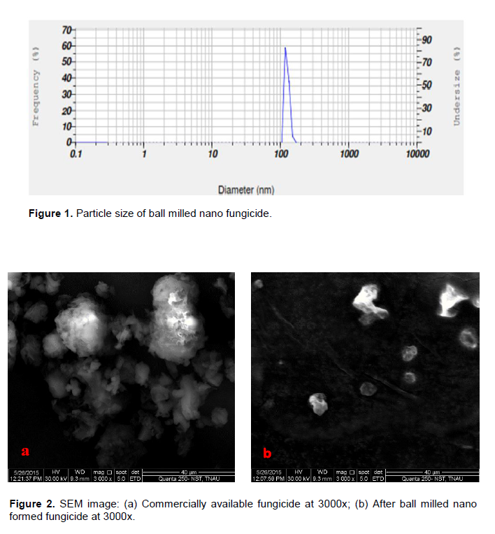
The surface morphology of the nanofungicides synthesized and examined under particle size analyzer (PSA) revealed that the particle size range from 108 to 130 nm diameter (Figure 1). But the size of traditionally available Trifloxystrobin 25% + Tebuconazole 50% (75 WG) was of 1.3 to 3.0 µm diameter.
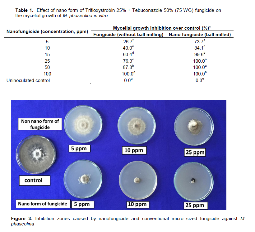
To confirm the results of PSA, the same nanoformulated Trifloxystrobin 25% + Tebuconazole 50% (75 WG) fungicide was characterized under SEM. Irregular shaped nanoparticles was observed and it ranged from 108 to 130 nm (Figure 2).
Antifungal activity of nanofungicide
The poisoned medium technique was employed to determine inhibitory efficacy of different concentrations of nanoform of Trifloxystrobin 25% + Tebuconazole 50% (75 WG) as compared to commercial fungicide against M. phaseolina (Table 1). Among the different concentrations, the maximum inhibition of the mycelial growth was obtained with 15 ppm concentration which accounted for 99.6% reduction of mycelial growth over control in M. phaseolina. This was followed by complete inhibition of mycelial growth at 25 ppm which accounted for 100% reduction of mycelial growth over control (Figure 3).
Exploitation of nanofungicides in future may pave the way for effective management of seed borne and soil borne pathogens like M. phaseolina; thereby may reduce the bulk pesticide use in the environment. Exploitation of nanoparticles as antifungal agents is relatively as being reported by recent workers (Kumar, 2011; Sridhar, 2012). Nanoparticles interactions with fungal pathogen are dependent on the size and shape of the nanoparticles (Pal et al., 2007). Metal nanoparticles are an obvious choice due to their effective antimicrobial effects (Duncan, 2011). The results of the present study clearly revealed that the maximum level of inhibition zone was observed with the increasing concentration of nanoparticles. Which was comparatively superior in its antifungal activity against non nanoform of Trifloxystrobin 25% + Tebuconazole 50% (75 WG).
Nanoparticles are highly antimicrobial and antioxidant to several species of bacteria, fungi and viruses. Antimicrobial property of nanoparticles may be due to penetration of the cell wall and modulation of the cellular level signaling by dephosphorylating putative key peptide substrates, which are critical for cell viability and cell division (Shrivastava et al., 2007). Nanoparticles are believed to inactivate microbial enzymes, facilitating production of reactive oxygen species that leads to microbial cell death (Allahverdiyev et al., 2011).
Morphological modification of mycelia
The mycelial fragment from nanoparticles treated and untreated plates were examined under SEM at various resolutions. Hyphal filament was smooth walled and equal in thickness throughout the length with blunt tips and found to bear sclerotial bodies (Figures 4a and 5a). However, in nanoparticles treated plates, hyphae were found broken and sclerotial formation either lacking or abnormal, if formed. In addition, the cell surface of hyphae was observed to be crinkled in M. phaseolina (Figures 4b and 5b).
The metallic nanoparticles are most promising as they have remarkable antibacterial properties due to their large surface area to volume ratio (Gong et al., 2007; Rai et al., 2009).These results lead us to consider that nanofungicide may be the most effective for controlling M. phaseolina. Size reductions involve the increase of contact surface, which is an important condition for the effects of nanofungicide. Another reason for considering nanofungicide as superior is its broad antimicrobial activity (Spacciapoli, 2001).
The present investigation is an offshoot of the main research on exploring the possibilities of nanofungicide for increasing the seed qualities especially in vegetables where the maintenance of seed viability is difficult. In the present work, it was demonstrated that, nanoformulation of Trifloxystrobin 25% + Tebuconazole 50% (75 WG) has significant fungicidal property against the fungi M.phaseolina.
Thus, it can be effectively explored against the soil borne pathogen M. phaseolina to protect various crop seeds, instead of using the commercially available synthetic fungicides, which shows higher toxicity to humans. Moreover, this report opens up for further research; field experiments are to be carried out in order to recommend the nanofungicide against the disease and the area of mode of action of nanocomposites against phytopathogenic fungi.
The authors have not declared any conflict of interests.
REFERENCES
Allahverdiyev AM, Emrah SA, Malahat B, Miriam R (2011). Antimicrobial effects of TiO2 and Ag2O nanoparticles against drug-resistant bacteria and leishmania parasites. Future Microbiol. 6:933-940.
Crossref |
|
|
|
Anandraj K (2013). Effect of nanoparticles for the maintenance of onion seed vigour and viability. M.Sc. Thesis, Tamil Nadu Agricultural University, Coimbatore. |
|
|
Diourte M, Starr JL, Jeger MJ, Stack JP, Rosenow DT (1995). Charcoal rot (Macrophomina phaseolina) resistance and the effects of stress on disease development in sorghum. Plant Pathol. 44:196-202.
Crossref |
|
|
Duncan TV (2011). Applications of nanotechnology in food packaging and food safety: barrier materials, antimicrobials and sensors. J. Colloid Interface Sci. 363:1-24.
Crossref |
|
|
|
Gogoi R, Dureja PS, Pradeep K (2009).Nanoformulations-A safer and effective option for agrochemicals. Indian Farming 59:7-12. |
|
|
Gogoi R, Singh PK, Kumar R, Nair KK, Alam I, Srivastava C, Yadav S, Gopal M, Choudhury SR, Goswami A (2013). Suitability of Nano-sulphur for Biorational Management of Powdery mildew of Okra (Abelmoschus esculentus Moench) caused by Erysiphe cichoracearum. J. Plant Pathol. Microbiol. 4:171
Crossref |
|
|
Gong P, Li H, He X, Wang K, Hu J, Zhang S, Yang X (2007). Preparation and antibacterial activity of Fe3O4@Ag nanoparticles. Nanotechnology 18:28.
Crossref |
|
|
|
Hall R (1991). Compendium of Bean Diseases. The American Phytopathological Society. St. Paul. Minnesota. USA. P 73. |
|
|
Javaid A, Saddique A (2011). Management of Macrophomina root rot of mungbean using dry leaves manure of Datura metel as soil amendment. Span. J. Agric. Res. 9:901905.
Crossref |
|
|
|
Kumar SS (2011).Customizing nanoparticles for the maintanence of seed vigour and viability in Blackgram (Vigna mungo) cv. VBN 4. M.Sc. Thesis, Tamil Nadu Agricultural University, Coimbatore. |
|
|
Mayek-Pérez N, Garcia-Espinosa R, LópezCasta-eda C, Acosta-Gallegos JA, Simpson J (2002). Water relations, histopathology, and growth of common bean (Phaseolus vulgaris L.) during pathogenesis of Macrophomina phaseolina under drought stress. Physiol. Mol. Plant Pathol. 60:185-195.
Crossref |
|
|
|
Packia Lekshmi NCJ, Benarcin Sumi S, Viveka S, Jeeva S, Raja Brindha J (2012). Antibacterial activity of nanoparticles from Allium sp. J. Microbiol. Biotechnol. Res. 2(1):115-119. |
|
|
Pal S, Tak YK, Song JM (2007). Does the antibacterial activity of silver nanoparticles depend on the shape of the nanoparticle? A study of the gram-negative bacterium Escherichia coli. Appl. Environ. Microbiol. 73:1712-1720.
Crossref |
|
|
Patolsky F, Zheng G, Lieber CM (2006). Nanowire sensors for medicine and life sciences. Nanomedicine 1:51-65.
Crossref |
|
|
Rai M, Yadav A, Gade A (2009). Silver nanoparticles as a new generation of antimicrobials. Biotechnol. Adv. 27(1):76-83.
Crossref |
|
|
Remesal E, Landa BB, Jimenez MD, Navas JA (2013). Sequence variation in two protein coding genes correlates with mycelial compatibility groupings in Sclerotium rolfsii. Pak. J. Phytopathol. 103:479-487.
Crossref |
|
|
|
Scrinis G, Lyons K (2007). The emerging nano-corporate paradigm: nanotechnology and the transformation of nature, food and agri-food systems.Int. J. Sociol. Food Agric.15:22-44. |
|
|
Shrivastava S, Bera T, Roy A, Gajendra S, Ramachandrarao P, Dash D (2007) Characterization of enhanced antibacterial effects of novel silver nanoparticles. Nanotechnology 18:9.
Crossref |
|
|
|
Shyla K, Natarajan N, Nakkeeran S. (2014). Antifungal Activity of Zinc Oxide, Silver and Titanium Dioxide Nanoparticles against Macrophomina phaseolina. J. Mycol. Plant Pathol. 44(3):269-274. |
|
|
Spacciapoli P, Buxton D, Rothstein D, Friden P (2001). Antimicrobial activity of silver nitrate against periodontal pathogens. J. Periodontal Res. 36:108-13.
Crossref |
|
|
|
Sridhar C (2012). Effect of nanoparticles for the maintenance of tomato seed vigour and viability. M.Sc. Thesis, Tamil Nadu Agricultural University, Coimbatore. |


