Full Length Research Paper

ABSTRACT
The interest of researchers in various areas has resulted in the investigation of different biofilm systems using a wide range of techniques. Biofilms are microbial communities consisting of mono or multi-species sessile cells, embedded in a matrix of extracellular polymers (exopolysaccharides-EPS) adhering to surfaces. In the food industry, the existence of biofilms is quite problematic, being responsible for the economic loss and contamination of food. Consequently, research involving the characterization of the ability of microbial biofilm formation is relevant for the subsequent studies using sanitizing and antibiotic agents for prevention or remediation of surfaces with already formed biofilms. This multidisciplinary study led to the description regarding the effect of antimicrobial solutions of essential oils of Syzygium aromaticum and Thymus vulgaris and their combination on biofilm formed by Staphylococcus aureus ATCC 25923 on AISI 304 stainless steel and polypropylene surfaces. All sanitizing solutions showed antibacterial potential, being effective in reducing bacterial biofilms on these surfaces. The solution containing the combination of essential oils was the most efficient by reducing 7.38 and 6.58 Log CFU.cm-2 of cells adhering on the surfaces of AISI 304 stainless steel and polypropylene, respectively, after 5 min of contact. Essential oils of S. aromaticum and T. vulgaris, alone or in combination, are new alternatives for disinfection of industrial stainless steel and polypropylene surfaces contaminated by S. aureus.
Key words: Antimicrobial effect, microorganism, Syzygium aromaticum, Thymus vulgaris.
INTRODUCTION
Microorganisms have been evolving for approximately 4 billion years, and up to 2 billion years ago they were the only life forms on Earth. They comprise a taxonomic definition that congregates varied groups of organisms of microscopic size that live in nature as isolated cells or cell aggregates (Manfio, 2003). Most bacteria, when in their natural habitat, live in communities of varying degrees of complexity, associated with a variety of biotic and, or abiotic surfaces, usually forming a biofilm.
Biofilm is defined as a community of sessile micro-organisms embedded in an extracellular polymeric matrix, that they produce themselves, characterized by cells irreversibly adhering to a surface or interface and which exhibits phenotypic alteration in relation to planktonic growth and gene transcription. A wide variety of micro-organisms are able to adhere and form biofilms on biotic and abiotic surfaces, presenting certain advantages as compared to planktonic cells (Garrett et al., 2008).
A common model for biofilm formation suggests that the process occurs in five stages. First, the reversible attack of planktonic bacterial cells occurs, as they approach the solid surface by fluid flow or motility, which have dominion over the repulsive forces between the cell and the surface. In the second stage, the transition from rever-sible to non-reversible attack occurs by the production of extracellular polymers by the bacterium itself, and, or specific adhesins localized on the pili or fimbriae that interact with the surface. The third stage consists in the initial development of the architecture of the biofilm. The fourth stage refers to the development of microcolonies within the mature biofilm; on the other hand, extracellular polymeric substances, that serve as an adhesive matrix and nutrients, may continue to be formed, as well as water channels and pores. In the final stage, there is a dis-persion of cells of the biofilm and the return of planktonic cells (Van Houdt and Michiels, 2005).
Despite these properties, the adhesion, activity and microbial growth is limited by chemical gradients resulting from the diffusion of nutrients and oxygen; characteristics of the microorganism, hydrophobicity, surface electric charge, flagellum, fimbriae and pili, the adherent material characteristics and the environment surrounding the micro-organism, such as pH, temperature, agitation time and a variety of other factors (Chen et al., 2005).
In the food industry, biofilm formation leads to serious health and economic losses due to food contamination and equipment damage. Biofilms can develop the most varied surfaces, and those most used in food production plants is the AISI 304 stainless steel (American Iron and Steel Institute) and polypropylene. The microorganisms present in the biofilm catalyze chemical and biological reactions causing corrosion of metal pipes and tanks, reducing heat transfer due to the thickness of the biofilm, among others (Shafahi and Vafai, 2009).
Among these microorganisms, Escherichia coli and Staphylococcus aureus are usually found in nature, their presence in food being the consequence of the often precarious sanitary conditions of food production, due to contaminated handling or surfaces. In the food industry, biofilm formation leads to serious hygiene problems and economic losses, mainly due to food contamination, spoi-lage and damage to equipment. Once established, biofilms act as points of constant contamination, releasing pathogenic and/or spoilage microorganisms (Boari et al., 2009).
Surveys have been documented involving food contact surfaces and various microorganisms, such as Listeria monocytogenes, Yersinia enterocolitica, Campylobacter jejuni, E. coli O157: H7, S. aureus and Pseudomonas aeruginosa, among others (Shi and Zhu, 2009).
In the quest for better understanding of the biofilm forma-tion process on different surfaces, the search for, and research on sanitizing agents and antimicrobial alternatives should be generated. In this context, it has been observed that the essential oils found in condiment plant extracts have antibacterial, antifungal and antioxidant properties, which has aroused the interest of food industries (Kalemba and Kunicka, 2003).
According to Sikkema et al. (1994), essential oils accu-mulate in the cytoplasmic membrane and cause damage such as loss of function of selective barrier. In recent years, several reports have been published on the composition and biological properties of essential oils of several condi-ment plants, among them Syzygium aromaticum, Thymus vulgaris, Cymbopogon citratus and Laurus nobilis (Fabio et al., 2007; Oliveira et al., 2010).
Some researches emphasize the existence of differences in the chemical composition among the extracted oils of different species or varieties. These variations tend to influence the antimicrobial activity of the oils and usually depend on factors such as genetically determined pro-perties, plant age, seasonal variation, water availability, environmental temperature at which the plant is found, nutrients available in the soil, altitude and UV radiation (Gobbo-Neto and Lopez, 2007).
The inhibitory effect of these oils on microorganisms is an alternative to reduce the use of chemical additives in food and for the formulation of new sanitizing agents. Various studies have shown that essential oils extracted from leaves and different parts of plant species have high antimicrobial activity (Gill and Holley, 2006).
Given the above, the objective of this study was to evaluate the action of sanitizing solutions formulated with essential oils of Clove (S. aromaticum) and Thyme (T. vulgaris) and their combinations, on bacterial biofilms formed by S. aureus on AISI 304 stainless steel and poly-propylene surface.
MATERIALS AND METHODS
Microbiological analyzes were performed in the Food Microbiology Laboratory, Food Science Department, Universidade Federal de Lavras (UFLA), MG.
Microorganism used, standardization, inoculum preparation and storage
The microorganism used in the development of this study was S. aureus ATCC 25923. The culture of S. aureus was maintained at -18°C in microcentrifuge tubes with freezing medium [glycerol (150 mL), peptone (5 g), yeast extract (3 g), NaCl (5 g), H2O (1.000 mL), pH 7.2 ± 7.4]. During the experiment, subcultures were made for the maintenance of cultures. Aliquots were transferred to the micro-centrifuge tubes containing tryptic soy broth (TSB) and incubated at 37°C/24 h. After culturing, 1 mL of the culture was dispensed into sterile microcentrifuge tubes and centrifuged at 6,000 xg for 8 min in a microcentrifuge. After removing the supernatant, the content was again coated with freezing medium and stored at -18°C.
For reactivation and use of the strain, 10 µL of the culture was inoculated in tubes containing 3 mL of TSB and incubated at 37°C/24 h. After incubation, 20 µL of the inoculum was removed and transferred to 200 mL of TSB. The number of cells per mL was quantified using standard curve and the growth monitored by spectrophotometry at 600 nm and then counting in TSA. The bacterial culture was standardized to a concentration of 108 CFU mL.
Experimental model of biofilm formation
Preparation and cleaning of coupons
The bacterial adhesion was conducted on AISI 304 stainless steel and polypropylene coupons with 1 mm thickness and dimensions of 10 x 20 mm.
The AISI 304 stainless steel coupons were cleaned individually with 100% acetone, submerged in detergent, rinsed with sterile distilled water, dried and cleaned with 70% ethanol (v/v). After cleaning, the coupons were again washed with sterile distilled water, dried for 2 h in oven at 60°C and autoclaved at 121°C/15 min (Rossoni and Gaylarde, 2000). As for the polypropylene coupons, they were initially immersed in a solution of commercial 0.3% peracetic acid for 30 min under stirring at 50 rpm at 50°C. They were then soaked in sterile distilled water at 80°C for 5 min and at room temperature for 1 min under agitation of 50 rpm. The coupons were dried at 40°C for 2 h and autoclaved for 15 min at 115°C (Oulahal et al., 2008).
Adhesion of bacterial cells to surfaces
In two Petri dishes (140 mm diameter) 45 AISI 304 stainless steel coupons and 80 mL TSB were added and inoculated with 108 CFU mL of culture. In two other Petri dishes, the same procedures were employed, with 45 polypropylene coupons, with the aim of promoting the formation of biofilms on these surfaces. The plates were incubated at 37°C with orbital agitation of 50 rpm. After 48 h of incubation, the coupons were collected, washed with peptone water (0.1% w/v) five times and immersed in TSB contained in sterile plates. This procedure was performed five times in order to complete the formation of the biofilm after 10 days of incubation (Joseph et al., 2001).
Enumeration of adhered cells
For enumerating the adherent cells, one AISI 304 stainless steel and one polypropylene coupon was removed from each Petri dish every two days of incubation. These were washed with peptone water (0.1% w/v) five times to remove planktonic cells and the sessile cells were collected using a standard sterile cotton swab. The swabs were transferred to tubes containing peptone water (0.1% w/v) then agitated in a vortex for 2 min. After this procedure, serial dilutions of the samples were carried out in which 0.1 mL aliquots were plated and the number of viable cells quantified in TSA (Triptic Soy Agar), using the surface seeding technique. The plates were incubated at 37°C/24 h, conducting a standard plate count at the end of this period, and results were expressed in CFU cm2 (Joseph et al., 2001).
Obtainment of essential oils
The essential oils of S. aromaticum and T. vulgaris were purchased through the Ferquima Ind. e Com. Ltda Company (Vargem Grande Paulista, São Paulo, Brazil), their physical and chemical parameters being described by the supplier, which produces and sells essential oils on an industrial scale.
Identification and quantification of chemical constituents
The essential oils chemical components were identified by gas chromatography coupled with mass spectrometry (GC–MS). A Shimadzu Gas Chromatograph (model GC 17A) equipped with a mass selective detector (model QP 5000) was operated under the following conditions: fused silica capillary column (30 m x 0.25 mm) coated with a DB-5 MS stationary phase; ion source temperature of 220°C; column temperature programmed at an initial temperature of 40°C, and increased by 3°C/min up to 240°C; helium carrier gas (1 ml/min); initial column pressure of 100.2 kPa; split ratio of 1:10 and volume injected of 1 μl (1% solution in dichloromethane). The following conditions were used for the mass spectrometer (MS): impact energy of 70 eV; decomposition velocity of 1000, decom-position interval of 0.50 and fragments of 45 Daltons and 450 Daltons decomposed. A mixture of linear hydrocarbons (C9H20; C10H22; C11H24;…C24H50; C25H52; C26H54) was injected under identical conditions. The mass spectra obtained were compared with those of the database (Wiley, 229), and the Kovat’s retention index (KI) calculated for each peak was compared with the values according to Adams (2007).
Determination of the minimum inhibitory concentration of the essential oils
The minimum inhibitory concentration (MIC) of essential oils was determined using the technique of disk diffusion in agar proposed by NCCLS (M7-A6) (2003) with modifications. The essential oils were diluted in Dimethyl Sulfoxide (DMSO) at different concen-trations (0.5, 1.5, 2.5, 5.0, 10.0, 15.0, 25.0, and 50.0%) and with DMSO control. The bacterial inoculum (in TSB) were added to vials containing TSA, the cell concentration was standardized to approxi-mately 108 CFU.mL–1, and the inocolum was poured directly into sterile Petri dishes (150 mm). After solidification, a volume of 5 μL of each essential oil was dispensed on filter paper discs 6 mm in diameter, which were placed on TSA inoculated. The plates were incubated in B.O.D. at 37°C for 24 h (Ogunwande et al., 2005). The diameters of the inhibition halos formed were measured using a caliper rule. The analyses were conducted in triplicate.
Preparation of sanitizing solutions
To perform the sessile cell sensitivity test, four sanitizing solutions were formulated containing saline (NaCl 0.85% w/v), ethanol (p.a.95% v/v) and essential oil as shown in Table 1.

All sanitizing solutions contained a total volume of 10 mL and the amount of essential oils used in the formulation of the sanitizing solutions was defined from the minimum inhibitory concentration test results previously performed by disk diffusion technique with modifications (Ogunwande et al., 2005).
Treatment of biofilms with sanitizing solutions containing essential oils at different contact times
On the tenth day of cultivation, polypropylene and steel coupons were taken from each Petri dish, rinsed in 0.1% peptone water (v/v) five times to eliminate non-adherent cells, and immersed in the above sanitizing solutions for 5 and 10 min at room temperature. After the sanitizing, the coupons were rubbed with standardized sterile swabs. The swabs were transferred to tubes containing 0.1% peptone water (v/v) and then agitated in a vortex for two minutes. After this procedure, serial dilution was conducted, 0.1 mL aliquots were plated and the number of viable cells determined in TSA medium, using the surface seeding technique. The plates were incubated at 37°C for approximately 24 h, conducting the standard plate count at the end of this period with, results expressed in CFU cm2 according to the method previously described by Joseph et al. (2001) with modifications.
Experimental design and statistical analysis
A completely randomized design (CRD) was used in a 2 x 5 factorial outline with 3 replicates, the surface factor having 2 levels: stainless steel and polypropylene, the time factor with 5 quantitative levels: 48, 96, 144, 192 and 240 h. The enumeration of adhered cells on the stainless steel and polypropylene coupons after treatment with these sanitizing solutions at different contact times, used the CRD in a factorial scheme (4 x 2 x 2) with 3 replicates with the factor agents at four qualitative levels: control, S. aromaticum, T. vulgaris and combination, the factor surfaces with two qualitative levels: stainless steel and polypropylene, and the time factor with 2 quantitative levels: 5 and 10 min. The statistical analyses were conducted utilizing the SISVAR program version 4.6 (Ferreira, 2008), R Development Core Team programs (R Development Core Team, 2004). For comparison of the averages, the Tukey test at the5% of probability level was used.
RESULTS AND DISCUSSION
Table 2 shows the counts of sessile cells that adhered to the surfaces of the AISI 304 stainless steel and poly-propylene coupons during the biofilm formation process.
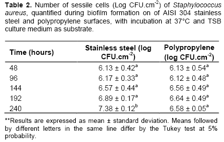
The adhesion of bacterial cells depends on factors such as physiology and cell morphology and physico-chemical properties of the contact surface. Gram nega-tive micro-organisms have greater ease of adhesion on surfaces as compared to Gram positive, as they have pili, flagella and fimbriae, as well as an outer membrane. Micro-organisms electrically charged with negative charges have more difficulty to link directly to surfaces. The participation of a conditioning film formed by various compounds and mole-cules from the aqueous phase, will be decisive (Boari et al., 2009; Van der Mei et al., 2003).
It can be observed that the microbial cells adhered similarly to both surfaces up to 192 h and differed signi-ficantly only at the end of the biofilm formation process, that is, at 240 h. The adhesion of bacterial cells depends on factors such as physiology and cell morphology and physicochemical properties of the contact surface.
The ability of S. aureus to adhere to solid surfaces pro-ducing compounds by multilayered cells embedded in a exopolysaccharide matrix is considered one of the relevant aspects of the epidemiology of this bacterium (Cucarella et al., 2001; Flach and Karnopp, 2005). This organization is extremely beneficial to all species of microorganisms, because it provides protection against adversity such as dehydration, colonization by bacteriophages and antimi-crobial resistance (Gilbert et al., 2003).
By the phenomenon known as passivation, chromium, due to its high affinity for oxygen, tends to combine with it, forming a thin layer of chromium oxide with an approxi-mate thickness of 40 Å. This passive layer is responsible for the corrosion resistance and the hydrophobicity of stainless steel. In this context, in the case of initial adhe-sion, the more hydrophobic the bacterial cell, the greater its ability to bind directly to this surface. Similar consi-derations were observed by Meylheuc et al. (2006) and Sheng et al. (2007). Thus, surfaces considered hydro-phobic, such as stainless steel, allow the adhesion to occur more easily than less hydrophobic or hydrophilic surfaces, which is evidenced by counts and electron micrographs, which show improved adhesion of the cells on the surface of stainless steel at the end of 240 h incubation as compared to the polypropylene surface.
In studies conducted by Boari et al. (2009), that consisted of evaluating S. aureus biofilm formation on stainless steel using milk as substrate and different growing conditions, biofilm formation by S. aureus was observed by scanning electron micrographs in all conditions tested, revealing the adhesion ability of this bacterium mainly to the stainless steel surface, which was also observed in electron micrographs of the present study. In a review by Chmielewski and Frank (2003), it is shown that a layer of organic matter on the surface can promote and facilitate bacterial adhesion. Moreover, these authors state that the time of contact between cells and surfaces also influence the bacterial adhesion. The irreversible cell adhesion to surfaces occurs between 20 min and a maximum of 4 h of contact. After this period, the removal of these cells requires the application of physi-cal force, chemicals or heat. In this present study, it is possible to observe that the bacterial cells have obtained adhesion to the stainless steel and polypropylene sur-faces from 48 h and increasing, to a small extent, up to 240 h.
The probability of cells remaining irreversibly attached
after sanitation procedures is high and corresponds to one of the main reasons for the formation of biofilms on surfaces that come into contact with food, becoming a constant source of contamination.
Table 3 presents the counts of sessile cells adhered to the surfaces of the AISI 304 stainless steel and poly-propylene coupons after treatment with the control sanitizing solution and the essential oil-based sanitizing solutions. Table 4 shows the reduction percentage of the number of sessile cells after treatment with the sanitizing solutions.
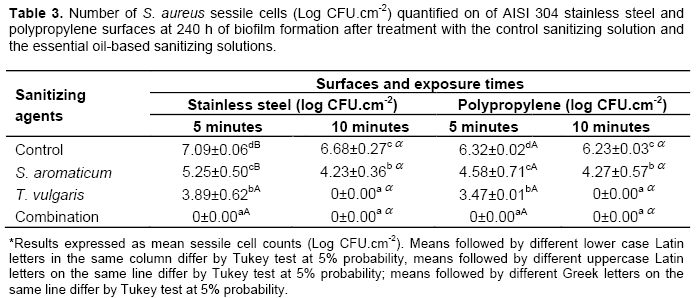
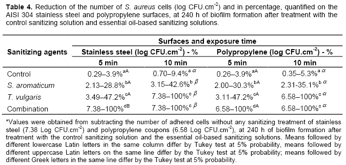
The effectiveness of the sanitizing solutions containing essential oils can be observed by the counts obtained after treatment of coupons on both surfaces and the reduc-tion percentage of these cells. A significant difference in the counts and reduction percentage of adhered sessile cells can be noted among the different treatments. All sanitizing solutions based on essential oils showed more superior antimicrobial activity than the control sanitizing solution.
The effectiveness of the sanitizing solutions based on S. aromaticum, T. vulgaris, and their combination differ significantly from each other, their combination being the most effective to reduce the number of sessile cells adhered to the surfaces. It can be observed that the five-minute exposure of the coupons containing the biofilm to the sanitizing solution based on the combination of oils was effective in promoting non-recovery of viable cells that adhered to both surfaces.
The sanitizing solution based on T. vulgaris was more effective as compared to S. aromaticum. This solution allowed the non-recovery of viable cells after exposure for 10 min to both surfaces. The sanitizing solution based on the essential oil of S. aromaticum was less effective, presenting a reduction in the number of sessile cells, but after 10 min of stainless steel and polypropylene coupon exposure to this solution, viable cells were still recovered.
As S. aureus is Gram positive, it is concluded that the cell wall does not serve as a barrier to the entrance of such antibacterial compounds through the cytoplasmic membrane. Since the cell wall of these bacteria is per-meable, usually it does not restrict the penetration of these sanitizing agents (Schaffer and Messner, 2005).
The difference between the performance of the sanitizing solutions within each phase of biofilm formation analyzed can be attributed to environmental and growth factors that are related to the concentration and nature of the chemical constituents, such as composition, func-tional groups and the structural configuration of the essential oil components (Chang et al., 2001).
The effect of the essential oil on the target micro-organism was considerably reduced when applied in the food model (as compared to in vitro studies). The appli-cation of essential oils for the control of pathogens and spoilage bacteria requires the evaluation of their effec-tiveness in food products or models that roughly simulate the composition of foods. Generally, the efficiency of some additives and natural antimicrobial agents can be reduced by certain components of foods. If higher concentrations of essential oils are generally required when added to food to maintain product safety, undesirable flavor and sensory changes may occur (Gutierrez et al., 2009). Researchers who have evaluated the effect of essential oil added to meat reported undesirable sensory changes caused by essential oil treatment in food samples (Govaris et al., 2010).
Brugnera (2011) evaluated the antibacterial effect of O. vulgare and S. officinalis against the growth and production of enterotoxin A by S. aureus inoculated in creamy ricotta, as well as the sonsorial acceptance of the ricottas with these spices added. As for the sensorial aspects, there was a higher preference for the ricottas with low spice concentrations.
The effects of colonization of surfaces where food is processed can result in various problems, because of an economic or public health nature. On the economic front spoilage bacteria can contaminate food by changing its characteristics and resulting in economic losses. The risk to public health is the most serious problem, because the biofilm can transport pathogenic microorganisms and be a source of chronic contamination (Ribeiro-Furtini, 2005).
This study led to the description of the sanitizing solutions essential oils of S. aromaticum and T. vulgaris and their combination on biofilm formed by S. aureus (ATCC 25923) on AISI 304 stainless steel and poly-propylene surfaces. All solutions showed potential antibacterial sanitizers, being effective in reducing bacterial biofilms on these surfaces. The solution containing the combination of essential oils was more efficient by reducing 7.38 and 6.58 Log CFU.cm-2 cells that adhered on the surfaces of AISI 304 stainless steel and polypropylene respectively, after 5 min of contact.
It was also observed that the total reduction in the number of surface-adhered cells presented by the disinfectant solution based on the combination of S. aromaticum and T. vulgaris essential oils at 240 h of biofilm formation (Table 4) emphasizes the synergistic action of the essential oils utilized. The term synergism can be defined as increase in the activity of compounds or factors when applied together, as compared to their individual activity (Ceylan and Fung, 2004). The study on synergism resulting from the combination of essential oils of different plant species was carried out in vitro, presenting promising results (Al-Bayati, 2008). However, no report has been found on the synergistic action of a combination of essential oils against surface-adhered bacteria.
S. aureus and L. monocytogenes are Gram positive bacteria, which can facilitate the action of the oils; in other words, there is high incorporation of the additive into the cell wall (Harpaz et al., 2003). In a study using the same test conducted in vitro, Dorman and Deans (2000) used essential oils of clove, oregano, geranium and pepper to evaluate their activity on 25 species of Gram positive and Gram negative bacteria. The authors observed that Gram positive bacteria were more susceptible to the essential oils studied than the Gram negative bacteria.
In conclusion, our findings suggested that S. aromaticum and T. vulgaris essential oils are new alternatives to sanitize industrial stainless steel surfaces contaminated by S. aureus. Their synergistic effect must not be ignored, as it can enhance the individual antibacterial activity of these compounds.
CONFLICT OF INTERESTS
The authors have not declared any conflict of interests.
ACKNOWLEDGMENTS
We acknowledge the financial support for this project by FAPEMIG, CNPq and CAPES.
REFERENCES
|
Adams RP (2007). Identification of essential oils components by gás chromatography/mass spectrometry. 4. ed Carol Stream: Allured Publishing Corporation, 804. |
|
|
Al-Bayati, FA (2008). Synergistic antibacterial activity between Thymus vulgaris and Pimpinella anisum essential oils and methanol extracts. J. Ethnopharmacol. 116(3):403-406. |
|
|
Boari CA, Alves MP, Tebaldi VMR, Savian, TV, Piccoli RH (2009). Biofilm formation by Aeromonas hydrophila and Staphylococcus aureus on stainless steel using milk and different conditions of cultivation. Ciênc. Tecnol. Aliment. 29:886-895. |
|
|
Brugnera DF (2011). Ricotta: microbiological quality and use of spices in the control of Staphylococcus aureus. 106 p. Dissertation (Master's in Food Science) – University of Lavras, Lavras, Brazil. |
|
|
Ceylan E, Fung DYC (2004). Antimicrobial activity of spices. J. Rapid Methods Autom. Microbiol. 12:1-55. |
|
|
Chang ST, Chen PF, Chang SC (2001). Antibacterial activity of leaf essential oils and their constituents from Cinnamomum osmophloeum. J. Ethnopharmacol. 77:123-127. |
|
|
Chen MJ, Zhang Z, Bott TR (2005). Effects of operating conditions on the adhesive strength of Pseudomonas fluorescens biofilms in tubes. Colloids Surf. B. 43:61-71. |
|
|
Chmielewski RAN, Frank JF (2003). Biofilm formation and control in food processing facilities. Comp. Rev. Food Sci. Food Saf. 2:22-32. |
|
|
Cucarella C, Solano C, Valle J, Amorena B, Lasa I, Penades JR (2001). Bap a Staphylococcus aureus surface involved in biofilm formation. J. Bacteriol. 183:2888-2896. |
|
|
Dorman HJ, Deans SG (2000). Antimicrobial agents from plants: antibacterial activity of plant volatile oils. J. Appl. Microbiol. 2(88):308-316. |
|
|
Fabio A, Cermelli C, Fabio G, Nicoletti P, Quaglio P (2007). Screening of the antibacterial effects of a variety of essential oils on micro-organisms responsible for respiratory infections. Phytother. Res. 21(4):374-377. |
|
|
Ferreira DF (2008). SISVAR - System analysis of variance for balanced data: Program analysis statistics and design of experiments. Version 4.6. Software. Lavras: DEX/UFLA. Rev. Symp. 6:36-41. |
|
|
Flach J, Karnopp C (2005). Biofilms formed on raw materials on contact with milk: factors virulence involved. Acta Sci. Vet. 33:291-296. |
|
|
Garrett TR, Bhakoo M, Zhang, Z (2008). Bacterial adhesion and biofilms on surfaces. Prog. Nat. Sci. 18:1049-1056. |
|
|
Gilbert P, Mcbain AJ, Rickard AH (2003). Formation of microbial biofilm in hygienic situations: a problem of control. Int. Biodeterior. Biodegrad. 51:245-248. |
|
|
Gill AO, Holley RA (2006). Disruption of Escherichia coli, Listeria monocytogenes and Lactobacillus sakei cellular membranes by plant oil aromatics. Int. J. Food Microbiol. 108:1-9. |
|
|
Gobbo-Neto L, Lopes NP (2007). Medicinal plants: factors.influence on the content of secondary metabolites. Quim. Nov. 30:374 381. |
|
|
Govaris A, Solomakos N, Pexara A, Chatzopoulou PS (2010). The antimicrobial effect of oregano essential oil, nisin and their combination against Salmonella enteritidis in minced sheep meat during refrigerated storage. Int. J. Food Microbiol. 137:175-180. |
|
|
Gutierrez J, Barry-Ryan C, Bourke P (2009). Antimicrobial activity of plant essential oils using food model media: efficacy, synergistic potential and interactions with food components. Food Microbiol. 26:142-150. |
|
|
Harpaz S, Glatman L, Drabkin V, Gelman A (2003). Effects of herbal essential oils used to extend the shelf life of freshwater-reared Asian sea bass fish (Lates calcarifer). J. Food Prot. 66(3):410-417. |
|
|
Joseph B, Ottas SK, Karunasagar I (2001). Biofilm formation by Salmonella spp. On food contact surfaces and their sensitivity to sanitizers. Int. J. Food Microbiol. 64:367-372. |
|
|
Kalemba D, Kunicka A (2003). Antibacterial and antifungal proprieties of essential oils. Curr. Med. Chem. 10:813-829. |
|
|
Manfio GP (2003). Review the state of knowledge of the biological diversity of Brazil. Microbiota. Multidisciplinary Center for Chemical, Biological and Agricultural/CPQBA. Division of Microbial Resources, UNICAMP, Brazil. |
|
|
Meylheuc T, Methivier C, Renault M, Herry JM, Pradier CM, Bellon-Fontaine MN (2006). Adsorption on stainless steel surfaces of biosurfactants produced by gram-negative and gram-positive bacteria: consequence on the bioadhesive behavior of Listeria monocytogenes. Colloids Surf. B. 52:128-137. |
|
|
NCCLS, National Committee for Clinical Laboratory Standards (2003). Methods for dilution antimicrobial susceptibility tests for bacteria that grow aerobically. Approved standard M7-A6, Wayne, Pa, USA. |
|
|
Ogunwande IA, Olawore NO, Ekundayo O, Walker TM, Schmidt JM, Setzer WN (2005). Studies on the essential oils composition, antibacterial and cytotoxicity of Eugenia uniflora. L. Int. J. Aromather. 15(3):147-152. |
|
|
Oliveira MMMD, Brugnera DF, Cardoso MDG, Alves E, Piccoli RH (2010). Disinfectant action of Cymbopogon sp. essential oils in different phases of biofilm formation by Listeria monocytogenes on stainless steel surface. Food Control 21:549‑553. |
|
|
Oulahal N, Brice W, Martial A, Degraeve P (2008). Quantitative analysis of survival of Spaphylococcus aureus or Listeria innocua on two types of surfaces: Polypropylene e strainless steel in contact with three different dairy products. Food Control 19:178-185. |
|
|
R Development Core Team (2004). R: a language and environment for statistical computing. Viena: R Foundation for Statistical Computing, 2004. http://www.R-project.org. Accessed 10 Jul 2013. |
|
|
Ribeiro-Furtini LL (2005). Characterization and isolation of adhering microorganisms in dairy tubing and its behavior toward detergency. Thesis (Ph.D. in Food Science) - University of Lavras, Lavras, Brazil. 80 p. |
|
|
Rossoni EMM, Gaylarde CC (2000). Comparison of sodium hypochlorite and peracet acid as sanitising agents for stainless steel food processing surfaces using epifluorescence microscopy. Int. J. Food Microbiol. 61:81-85. |
|
|
Schaffer C, Messner P (2005). The structure of secondary cell wall polymers: how gram-positive bacteria stick their cell walls together. Microbiology 151:643-651. |
|
|
Shafahi M, Vafai K (2009). Bioï¬lm affected characteristics of porous structures. Int. J. Heat Mass Transf. 52:574-581. |
|
|
Sheng X, Ting YP, Pehkonem SO (2007). Force measurements of bacterial adhesion on metals using a cell probe atomic force microscope. J. Colloid Interface Sci. 310:661-669. |
|
|
Shi X, Zhu X (2009). Biofilm formation and food safety in food industries. Trends Food Sci. Technol. 20:407-413. |
|
|
Van der Mei HC, Van de Belt-Gritter B, Pouwels PH, Martinez B, Busscher HJ (2003). Cell surface hidrophobicity is conveyed by S-layer proteins: a study in recombinant lactobacilli. Colloids Surf. B. 28(2):127-134. |
|
|
Van Houdt R, Michiels CW (2005). Role of bacterial cell surface structure in Escherichia coli biofilm formation. Res. Microbiol. 156:626-633. |
|
Copyright © 2024 Author(s) retain the copyright of this article.
This article is published under the terms of the Creative Commons Attribution License 4.0


