African Journal of
Microbiology Research

  • Abbreviation: Afr. J. Microbiol. Res.
  • Language: English
  • ISSN: 1996-0808
  • DOI: 10.5897/AJMR
  • Start Year: 2007
  • Published Articles: 5233

Full Length Research Paper

Antibiotics resistance patterns of Panton-Valentine leukocidin-positive methicillin-resistant staphylococci isolated from clinical samples in Abidjan, Côte d’Ivoire

Sylvie M. Kouamé-Sina
  • Sylvie M. Kouamé-Sina
  • Platform of Molecular Biology, Pasteur Institute, 01 BP 490 Abidjan 01, Côte d’Ivoire.
  • Google Scholar
N’Golo D. Coulibaly
  • N’Golo D. Coulibaly
  • Platform of Molecular Biology, Pasteur Institute, 01 BP 490 Abidjan 01, Côte d’Ivoire.
  • Google Scholar
Fernique Konan
  • Fernique Konan
  • National Reference Center for Antibiotics, Pasteur Institute, 01 BP 490 Abidjan 01, Côte d’Ivoire.
  • Google Scholar
Yakoura K. Ouattara
  • Yakoura K. Ouattara
  • Department of Food Science and Technology, Nangui Abrogoua University, Abidjan, Côte d’Ivoire.
  • Google Scholar
Kan S. Kouassi
  • Kan S. Kouassi
  • Platform of Molecular Biology, Pasteur Institute, 01 BP 490 Abidjan 01, Côte d’Ivoire.
  • Google Scholar
Solange Ngazoa-Kakou
  • Solange Ngazoa-Kakou
  • Platform of Molecular Biology, Pasteur Institute, 01 BP 490 Abidjan 01, Côte d’Ivoire.
  • Google Scholar
Nathalie K. Guessennd
  • Nathalie K. Guessennd
  • National Reference Center for Antibiotics, Pasteur Institute, 01 BP 490 Abidjan 01, Côte d’Ivoire.
  • Google Scholar
Adjéhi Dadié
  • Adjéhi Dadié
  • Department of Food Science and Technology, Nangui Abrogoua University, Abidjan, Côte d’Ivoire.
  • Google Scholar


  •  Received: 15 September 2018
  •  Accepted: 29 October 2018
  •  Published: 07 November 2018

 ABSTRACT

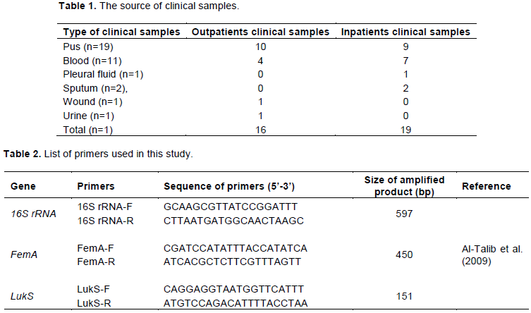
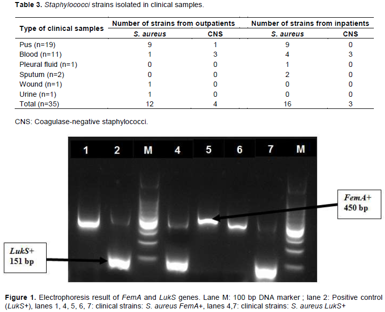
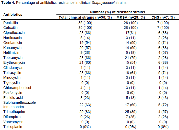
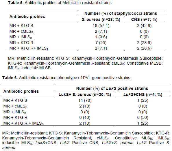
Methicillin-resistant staphylococci have emerged as significant pathogens which cause various infections and its multidrug resistance is a major concern. This study aimed to determine the prevalence of Panton-Valentine leukocidin (PVL) gene and antibiotic resistance patterns of staphylococci isolated from clinical infections in Abidjan. A total of 35 staphylococci strains was obtained from 35 clinical samples (pus, blood, pleural fluid, sputum, wound, and urine), then, characterized by polymerase chain reaction (PCR) to differentiate S. aureus from coagulase-negative staphylococci (CNS) and to detect the presence of PVL genes (LukS). The antimicrobial susceptibility was performed using disk diffusion method and the phenotype of resistance to macrolides-lincosamides-streptogramin B (MLSB) was detected. Out of 35 strains, 80% (28/35) were methicillin-resistant Staphylococcus aureus (MRSA) and 20% (7/35) were methicillin-resistant CNS (MR-CNS). S. aureus were isolated from 75% of outpatient samples and 84.2% of inpatient samples. However, CNS were isolated from 25% of outpatient samples and 15.8% of inpatient samples. LukS were detected in 68.6% of strains (20 MRSA and 4 MR-CNS) and both inpatients and outpatients. The highest resistance rates were observed for penicillin (100%), cefoxitin (100%), ciprofloxacin (66%), tobramycin (66%), tetracyclin (66%), sulphamethoxazole-trimethoprim (63%), erythromycin (60%), kanamycin (57%) and gentamicin (54%). In addition, S. aureus strains were subdivided into five antibiotics resistance phenotypes: 57.1% belonged to phenotype 1 (Methicillin-resistant and susceptible to Kanamycin-Tobramycin-Gentamicin) followed by 25% of phenotype 4 (Resistant to Methicillin-Kanamycin-Tobramycin-Gentamicin), 7.1% of phenotype 2 (MR with constitutive MLSB), 7.1% of phenotype 5 (MR and resistant to Kanamycin-Tobramycin-Gentamicin with inducible MLSB) and 3.6% of phenotype 3 (MR with inducible MLSB). CNS strains were grouped in three phenotypes (1, 4 and 5). 100% of LukS positive MRSA were multi-drug resistant, with 45% of strains resistant to 6 or more antibiotics. The high level of multi-drug resistance of clinical PVL positive staphylococci with inducible MLSB, suggest increasing the monitoring of these pathogens in Côte d’Ivoire.


Key words: Methicillin-resistant Staphylococcus aureus (MRSA), Panton-Valentine Leukocidin, inducible MLSB, multi-drug resistance.


 INTRODUCTION

Staphylococcus aureus and coagulase-negative staphylococci (CNS) are Gram-positive opportunistic pathogens that cause various diseases, ranging from localized mild infections to invasive life-threatening diseases (Pedroso et al., 2018; Tong et al., 2015). However, CNS have emerged as significant pathogens causing nosocomial infections (Lenart-Boron et al., 2016; Nanoukon et al., 2017). S. aureus, especially methicillin-resistant S. aureus (MRSA) strains increase the occurrence of serious infections (Skov et al., 2012) which are among the most frequent bacteria in healthcare-associated infections. Skin and soft tissue infections due to S. aureus are most common, whereas pneumonia, osteomyelitis, endocarditis, and sepsis, although less usual, account for greater morbidity and mortality (Tong et al., 2015; Dong et al., 2013). S. aureus possesses an arsenal of virulence factors that contribute to evasion of host defenses. These virulence factors, including toxins, exoenzymes, and adhesins, which are secreted or linked to cell membrane and fight against the action of antibiotics. Antibiotic resistance in both hospital-acquired methicillin resistant S. aureus and community-acquired MRSA strains has increased the difficulty to treat these infections (Messina et al., 2016; Shopsin et al., 2016). Most MRSA strains can also produce a leukotoxin as Panton-Valentine leukocidin (PVL), a bicomponent cytotoxin encoded by prophage that increases their virulence and can cause skin and soft tissue infection and necrotizing pneumonia (Shallcross et al., 2013; van der Meeren et al., 2014; Yanagihara et al., 2009). PVL is a member of pore forming toxins that targets host leukocytes. Two open reading frames are responsible for coding PVL, that is, LukS-PV and lukF-PV (Abdulgader et al., 2015). The MRSA strains are widely distributed around the world with quick evolution of antimicrobial resistance and it is a high priority according to WHO to found new antibiotics (Willyard, 2017). WHO global report on surveillance of antimicrobial resistance lacked information from the majority of the countries in sub-Saharan African countries or lacked data on priority pathogens such as MRSA (WHO, 2014). However, a recent retrospective review performed on wound infection between 2004 and 2016, had shown the prevalence of MRSA in Benin (34.6%), Congo (31.9%), Togo (14.3%) and Madagascar (14.5%) (Lai et al., 2018). S. aureus colonizes about one third of healthy humans and is most often found in the nose (Kaspar et al., 2016). A study in Burkina Faso, showed that the rate of S.aureus nasal carriage was 32.9% with 29% in healthy volunteers and 37% in hospital patients in  Bobo  Dioulasso.  In  addition, the percentage of MRSA strains isolated from hospital patients was very low (2.3%) with high prevalence of strains harboring PVL-encoding genes (45%) (Ouedraogo et al., 2016). Nosocomial infections are a major threat in most of the hospitals and as high as 19% in the developing countries, where number of direct contacts between the hands of Health Care Workers and the patient occurs, which mandates the strict adherence to infection control practices and standards (Maheshwari et al., 2014). In Ghana, Saba et al. (2017) isolated MRSA (17%) from handles and other points of contact in a public hospital environment. In the Burn Center of Korle Bu Teaching Hospital in Ghana, 50% of patients were infected with S. aureus including MRSA (Amissah et al., 2017). In Côte d’Ivoire, previous studies have reported the occurrence of S. aureus strains harboring PVL gene. During 2004 and 2005, two outbreaks caused by the same clone of  doxycycline resistant, PVL-positive methicillin-susceptible S. aureus (MSSA) were characterized in two French military companies (Lesens et al., 2007). In the Cocody University Teaching Hospital, Kacou et al. (2011) have reported high rate of PVL positive S. aureus strains (67.7%) from clinical samples in hospitalized patients. However, Zinzendorf et al. (2012) showed a high rate (77.4%) of clinical S. aureus strains from patients expressed PVL gene in Abidjan Military Hospital. The increasing prevalence of staphylococcal virulence factor, PVL, and MRSA strains isolated from patients is a growing problem. Constant surveillance and adequate infection control measures for S. aureus may reduce their roles in the incidence of nosocomial diseases and other infections. Update data on S. aureus resistances are particularly important to know the type of antibiotic resistance for establishing adequate therapeutic approaches. The objective of this study was to deternine the prevalence of PVL gene and antibiotic resistance patterns of Staphylococcus species isolated from patients in Abidjan, Côte d’Ivoire.


 MATERIALS AND METHODS

Sample collection
 
Pasteur Institute of Côte d’Ivoire is a laboratory where patients from different hospitals in Côte d’Ivoire are referred for medical tests. The clinical samples (n=35) were collected from 16 outpatients referred to Pasteur Institute and 19 inpatients of Cocody University Teaching Hospital., Samples of inpatients came from surgery (n=5), maternity/neonatal (n=2), neurology (n=1), obstetrics/gynecology (n= 2), pediatrics (n=3), pulmonology (n=2), emergency (n=3) and urology (n =1) services of Cocody University Teaching Hospital.
 
All samples were collected from Pasteur Institute between February and September, 2017 (Table 1).
 
Staphylococci isolation
 
Bacteria were isolated and identified in the Department of Bacteriology at Pasteur Institute of Côte d’Ivoire. The phenotypic identification of staphylococci species was carried out by standard microbiology methods including Gram staining, catalase activity, mannitol fermentation and the ability to coagulate rabbit plasma (Kateete et al., 2010; Amini et al., 2012). Molecular characterisation of strains was carried out in the Molecular Biology Laboratory at Pasteur Institute of Côte d’Ivoire.
 
Molecular characterization of strains
 
DNA extraction
 
Bacterial DNA was extracted by boiling method (Kacou et al., 2011; Oliveira et al., 2014). An overnight culture of staphylococci strains in Brain-Heart Infusion (1.5 mL) was centrifuged (12000 rpm, 5 min) and the supernatant was discarded. The pellet was dissolved in 400 µL deionized water and the suspension was heated at 100°C for 10 min and then immediatly frozen at -20°C for 10 min. Finally, after centrifugation for 10 min at 12000 rpm, the supernatant containing bacterial DNA was stored at -20°C for further analysis.
 
Detection of staphylococcal genes
 
All strains were screened for the presence of three Staphylococcus genes by modified method of PCR amplification previously described by Kacou et al. (2011). DNA extracts were used as template in the PCR. The multiplex PCR was performed  to  identify the Staphylococcus genus (16S rRNA gene), to differentiate S. aureus from CNS (FemA gene), and to detect PVL toxin (LukS) genes simultaneously. The PCR was performed in a final volume reaction of 25 μL containing 9.75 µL nuclease-free water (Ambion), 5 µL PCR buffer (5X), 1.5 µL magnesium chloride (MgCl2, 25 mM) (Promega Corporation, Madison, USA), 0.5 µL Deoxynucleotide Triphosphates (dNTPs; 10 mM), 0.5 μL of each primer (20 mM) (Table 2), 0.25 µL Go Tag®G2 Flexi DNA polymerase 5 U/µL (Promega Corporation, Madison, USA) and 5 μL of template DNA. The PCR method was performed according to the following program: initial denaturation (94°C, 5 min), 35 cycles each composed of initial denaturation (94°C, 30 s), primer annealing (60°C, 1 min) and extension (72°C, 1 min) and a final extension (72°C, 5 min). Primers used in this PCR were previously reported by Al-Talib et al. (2009) (Table 2). A previously known (Kacou et al., 2011) LukS gene positive S. aureus strain was used as a control strain. PCR amplification products were revealed on a gel Doc EZ® imager (Bio-Rad) after electrophoresis in 2% agarose gel containing Syber safe (Invitrogen).
 
 
Antimicrobial susceptibility
 
Susceptibilities were determined using the disk diffusion method in accordance with the performance standards for antimicrobial susceptibility testing, recommended by the Committee of Antibiogram of French Society of Microbiology (CA-SFM/EUCAST, 2016). The susceptibility testing was carried out by culturing strains on Mueller-Hinton agar (Bio-rad, Marne-la- coquette, France). Antibiotics used for susceptibility testing included penicillin 6 µg, cefoxitin 30 µg, ciprofloxacin 5 µg, norfloxacin 5 µg, gentamicin 10 µg, kanamycin 30 µg, tobramycin 10 µg, netilmicin 10 µg,

 erythromycin 15 µg, clindamycin 2 µg, tetracyclin 30 µg, minocyclin 30 µg, tigecyclin 15 µg, chloramphenicol 30 µg, fusidic acid 10 µg, sulphamethoxazole-trimethoprim 25 µg, rifampicin 30 µg, vancomycin 30 µg, and teicoplanin 30 μg. Susceptibility to methicillin was screened with the cefoxitin disk diffusion result. All antibiotics were obtained from Bio-rad. It should be indicated that the reference strain S. aureus ATCC 29213 were used as positive-control and provided by the National Reference Center for Antibiotics, Pasteur Institute of Côte d’Ivoire. The macrolide-lincosamide-streptogramin B (MLSB) group of antibiotics can be used to treat less severe skin and soft tissue infections (Liu et al., 2011). Strains resistant to both erythromycin and clindamycin in routine antibiotic susceptibility testing were considered constitutive MLSB phenotypes. Inducible MLSB phenotypes were identified using a double-disk diffusion test (D-test). Erythromycin 15 μg and clindamycin 2 μg disks were placed on a Mueller-Hinton agar plate containing a lawn culture of the test isolate at a distance of 15 mm edge to edge. After incubation at 35°C for 16 to 18 h, strains that showed no flattening of the inhibition zone around the clindamycin disk were reported as susceptible to clindamycin (negative D test); strains that showed flattening of the inhibition zone around the clindamycin disk adjacent to erythromycin disk (D zone) indicated inducible clindamycin resistance (positive D test). Strains with a D-shaped zone of inhibition were considered iMLSB Phenotype (resistant to erythromycin and susceptible to clindamycin) (Kumari et al., 2016; Pereira et al., 2016). Multidrug resistance (MDR) was defined as resistance to at least three distinct antimicrobial classes or being MRSA (Magiorakos et al., 2011).


 RESULTS

Clinical specimens and staphylococci strains
 
In this study, a total of 35 staphylococci were isolated from 35 patient samples. Patients were 7 days to 62 years old and 17 patients were female and 18 were male. S. aureus strains isolated from patients samples were identified by Gram-positive cocci, catalase positivity, mannitol fermentation, and coagulase production. However, strains that were Gram-positive cocci, catalase positive, and coagulase negative were considered as CNS. This phenotypic identification was confirmed by PCR results. Among 35 bacteria tested, 28 (80%) were S. aureus (16S rRNA positive and femA positive) and 7 (20%) were CNS (16S rRNA positive and femA negative).
 
S. aureus strains were recovered from 75% of outpatients samples (12/16) and 84.2% of inpatients samples (16/19). However, CNS were isolated from 25% of outpatients samples (4/16) and 15.8% of inpatients samples (3/19). S. aureus was reported in all type of clinical specimens but CNS were isolated only in blood (54.5%; 6/11) and pus (5.3%; 1/19) (Table 3). CNS strains were isolated from hospitalized patients provided from maternity/neonatal and pediatrics.
 
Detection of PVL gene
 
PVL-encoding gene (LukS) was detected in 24 (68.6%) of the 35 strains. The PVL gene was present in all type of clinical samples (blood, pus, pleural fluid, sputum, wound, and urine). Among all S. aureus strains (28), 71.4% carried the LukS gene (20/28) (Figure 1). In addition, the LukS gene was present in 57.1% (4/7) of CNS. LukS positive CNS (LukS+CNS) strains were only detected from blood samples of inpatients and outpatients. In contrast, LukS positive S. aureus strains were detected from blood (4/11), pus (12/19), pleural fluid (1/1), sputum (1/2), wound (1/1) and urine (1/1) samples.
 
Antibiotic susceptibility of bacteria isolated
 
Antibiotic susceptibility tests were performed on the 35 Staphylococcus spp. isolated from clinical samples. The highest resistance rates were observed for penicillin (100%) andcefoxitin (100%). Resistance rates of 50 70% were recorded for the antibiotics ciprofloxacin (66%), tobramycin (66%), tetracyclin (66%), sulphamethoxazole-trimethoprim (63%), erythromycin (60%), kanamycin (57%) and gentamicin (54%). The lower resistance rate <50% were observed for rifampicin (26%), netilmicin (26%), fusidic acid (23%), norfloxacin (14%), minocyclin (11%), clindamycin (11%) and chloramphenicol (11%). All the strains were susceptible to tigecyclin tgc (100%), vancomycin (100%) and teicoplanin (100%) (Table 4). The strains resistant to cefoxitin were classified  as  those strains methicillin-resistant staphylococci. In this study, 100% (35/35) of staphyloccocci strains were methicillin-resitant of which 80% were MRSA and 20% were methicillin-resistant CNS (Table 4).


Antibiotic phenotypes of methicillin-resistant strains
 
In this study, staphylococci strains were subdivided into five phenotypes  according  to  the  antibiotics  resistance
 
 pattern (Table 5): Phenotype 1 includes strains resistant to methicillin and susceptible to kanamycin, tobramycin, and gentamicin; Phenotype 2 includes strains resistant to methicillin with constitutive MLSB (cMLSB); Phenotype 3 includes strains resistant to methicillin with inducible MLSB (iMLSB); Phenotype 4 includes strains resistant to methicillin, kanamycin, tobramycin, gentamicin; Phenotype 5 includes strains resistant to methicillin, kanamycin, tobramycin, gentamicin with inducible MLSB (iMLSB).
 
The S. aureus strains present five antibiotics phenotypes: 57.1% of S. aureus belonged to phenotype 1 followed by phenotype 4 (25%), phenotype 2 (7.1%), phenotype 5 (7.1%) and phenotype 3 (3.6%). However, CNS strains were characterised by three phenotypes (1, 4 and 5).
 
Antibiotics resistance pattern of positive PVL strains
 
Among the 24 LukS positive strains, 20 were MRSA with four different phenotypes of antibiotic resistance. The majority of LukS positive strains (70%) were phenotype 1. Two (2/20) of LukS+S. aureus strains were phenotype 2, also two strains (2/20) were phenotype 4 and two (2/20) other were phenotype 5 (Table 6). Of the four LukS positive CNS (LukS+CNS), phenotype 4 was observed in two (50%) strains and phenotype 5 in one (25%) strain and also one other (25%) isolate was phenotype 1. The highest prevalence of resistance of LukS+MRSA strains was for penicillin (100%), cefoxitin (100%) followed by tobramycin (60%), ciprofloxacin (57%), erythromycin (47%), tetracyclin (43%), sulphamethoxazole-trimethoprim (40%), gentamicin (36%) and kanamycin (37%). Lower percentage of resistance was observed for rifampicin (21%), norfloxacin (15%), chloramphénicol (10%), fusidic acid (11%), minocyclin (7%) and netilmicin (6%). All the LukS+MRSA strains were susceptible to tigecyclin (100%), fosfomycin (100%), vancomycin (100%) and teicoplanin (100%).
 
In addition, multi-drug resistant strains were classified as those resistant to three or more antibiotics classes. All LukS+MRSA were multi-drug resistant (100%; 20/20), of which 9 (45%) strains were resistant to 6 or more antibiotics, 2 (10%) strains were resistant to 5 antibiotics, 7 (35%) were resistant to 4 antibiotics and 2 (10%) were resistant to 3 antibiotics.


 DISCUSSION

The present study reports the molecular characteristics and antibiotics resistance of staphycococci isolated from patients specimens in Abidjan, Côte d’Ivoire. A high prevalence of S. aureus was found in clinical samples (pus, blood, pleural fluid, sputum, wound, and urine) compared to the prevalence of CNS was lower. The strains of CNS was isolated in only 7 (20%) clinical samples. These prevalences were different than those reported in Nigeria where S. aureus and CNS were isolated at 56 and 43.9%, respectively in clinical infections (wounds, skin and soft tissue infections, osteomyelitis, burns, genitourinary tract infection, septicaemia, urinary tract infection, otitis media, and bronchitis) (Shittu et al., 2012). With regard to the virulence factors of these strains, PVL (a leukotoxin associated with human clinical diseases) encoding gene (LukS) was sought. This research showed that, the high rate of LukS gene prevalence in 68.6% of staphylococci strains was obtained from clinical infections of inpatients and outpatients. The LukS gene was detected at a high rate in S. aureus strains (71.4%) than in CNS strains (57%). The LukS positive CNS strains were detected only from blood. In the hospitalised patients, LukS+CNS strains were found in patients of neonatale and peditrics unit and maybe due to nosocomial infections. Indeed, CNS have emerged as one of the main microorganisms causing nosocomial infections and clinically (Nanoukon et al., 2017). The prevalence of PVL gene in S. aureus (71.4%) in the present study was high compared with the rate (67.8%) reported in a previous study conducted in Côte d’Ivoire in 2009 in clinical infections (Kacou et al., 2011). The prevalence of LukS carriage in S. aureus in clinical sample in Abidjan inceased from 67.8 to 71.4% between 2009 and 2017. The high frequency of PVL-positive S. aureus in patients  provide  important  insights to evaluate the risk of infection and dissemination of this bacteria in Abidjan, Côte d’Ivoire and in West Africa. In other sub-Saharan Africa countries, the frequency of PVL-positive S. aureus in clinical samples is high. In Ghana, PVL-encoding genes were detected in 75% (42/56) of S. aureus blood culture (Dekker et al., 2016) and in 27% (17/62) of carriage of PVL-positive S. aureus in burn patients (Amissah et al., 2017). This gene carriage rate was higher (90.7%; 68/75) in community-acquired-S. aureus isolated in Mozambique (van der Meeren et al., 2014), in the Democratic Republic of the Congo (49.1%) (Vandendriessche et al., 2017) and in Nigeria (33.3%; 17/51) in clinical infections (Shittu et al., 2012). The main findings of this study are a high prevalence of MRSA and PVL-positive gene strains. PVL is mostly associated with community-associated methicillin resistance in S. aureus infections. According the antibiotics resistance profile, 100% of staphylococci strains were methicillin-resistant, of which 80% were methicillin-resistant S. aureus (MRSA) and 20% were methicillin-resistant CNS. MRSA prevalence rate in the present study is very higher than MRSA prevalence rate (11.8%) obtained by Kacou et al. (2011) in 2009 in Abidjan. In addition, PVL gene (LukS) was detected only among methicillin-sensitive S. aureus (MSSA) strains. In contrast, in this present study, the PVL gene was commonly detected among MRSA as well as in methicillin-resistant CNS. The increase of prevalence of PVL-positive MRSA in Abidjan could present a significant challenge in disease management and infection control. The rate of PVL positive MRSA strains in Abidjan is higher than those reported from Ghana (63.8%) (Kpeli et al., 2016), Ouganda in pastoral communities (69.4%) (Asiimwe et al., 2017), Zambie (9.4%) (Samutela et al., 2017) and Nigeria (6.6%) (Shittu et al., 2012). However, the rate of PVL+MRSA was high than the rate (56.8%) reported by Bhatta et al. (2016) in MRSA isolated from clinical specimens in Nepal. The staphylococci isolated from patients in this study exhibit resistance to many antibiotics (Table 4). These results are in agreements with those reported by Chen et al. (2017) in China which the resistance rates of S. aureus to penicillin and cefoxitin were all 100%. However, all strains were susceptible (100%) to tigecyclin, vancomycin and teicoplanin. The MRSA strains collected from 13 different hospitals in Kuwait, were susceptible to vancomycin (100%), teicoplanin (100%) and linezolid (100%). Tigecyclin was not tested by the authors (Boswihi et al., 2018). The occurrence of MRSA is on the rise since the previous study (Kacou et al., 2011) in Abidjan. The threat posed by MRSA was vividly demonstrated in this study, as 100% of LukS+MRSA were multi-drug resistant and 45% of strains were resistant to 6 or more antibiotics tested. This might reflect the frequent and repeated administration of antibiotics, thus selecting for resistance and resulting in high frequencies of MDR. The MDR in staphylococci strains could lead to failure in treatment therapy, prolonged illnesses, increased expenses for health care, and in serious cases, risk of death (Tanwar et al., 2014). The prevalence of antimicrobial resistance in Abidjan clinical staphylococci strains was higher than those observed in other African countries. Indeed, the prevalence of antimicrobial resistance was below 5% for all antibiotics agents tested except for penicillin (97%), tetracycline (42%) and erythromycin (6%) in MRSA strains in Ghana (Egyir et al., 2014). Comparatively, the prevalence of MDR (9%) was lower than those reported in the present study. Bhatta et al. (2016) have reported overall 73% of MRSA from clinical specimens were multidrug reistant with 50% were PVL positive which indicates a lower prevalence than the present findings.
 
The high antimicrobial resistance in Abidjan is due to the overconsumption of antibiotics in the populations. Indeed, there is a huge prescription of antibiotics and because of poverty people turn to self-medication by sourcing from street drug sellers. It is important to note that there is a large informal market in Abidjan for medicines and antibiotics that are visible to everyone.
 
Indeed, the prevalence of antimicrobial resistance was below 5% for all antibiotics agents tested except for penicillin (97%), tetracycline (42%) and erythromycin (6%) in MRSA strains in Ghana (Egyir et al., 2014). Comparatively, the prevalence of MDR (9%) was lower than those reported in the present study. Bhatta  et  al.  (2016) have reported that overall 73% of MRSA from clinical specimens were multidrug reistant with 50% which were PVL positive indicating a lower prevalence than the present findings. Otherwise, according to resitance of antibiotics, clinical strains were grouped into 5 antibiotic phenotypes (Table 5). The frequency of cMLSB and iMLSB of the present  study was lower than those found in previous studies. Two other previous Brazil studies (Bottega et al., 2014; Pereira et al., 2016) have documented the rate of cMLSB and iMLSB in clinical MRSA strains ranging from 14.3 to 37.9% and 2.1 to 4.9%, respectively; while the KTG-R phenotype was not detected. In India, the MLSB phenotypes of MRSA in clinical samples were higher than those reported in this study. The iMLSB phenotype rate ranged from 19.1 to 35.2% and the cMLSB phenotype rate ranged from 11.4 to 31.9% in MRSA strains (Bhattacharya et al., 2015; Kumari et al., 2016). The antibiotic profile of CNS was not consitent with the results of Abdollahi et al. (2016) where 58.2% of 110 strains were methicillin resistant CNS to which 54.6% were cMLSB and 6.25% were iMLSB phenotype. It was important to know the type of MLSB resistance for establishing adequate therapy. The vancomycin is used for the treatment of serious infections and MLSB group are usually used to treat less severe skin and soft tissue infections (Liu et al., 2011). Reporting the susceptibility to clindamycin without verifying the presence of inducible resistance, may lead to inadequate therapy with this drug. In contrast, a negative result for inducible resistance to clindamycin, confirms the susceptibility of this antimicrobial, providing a very good therapeutic option (Pereira et al., 2016). Clindamycin is the preferred agent for the treatment of MRSA due to its excellent pharmacokinetic properties, such as optimal tissue penetration and accumulation in abscesses. However, the indiscriminate use of MLSB antibiotics has led to an increase in the number of Staphylococcus spp. strains that are resistant to these drugs (Bottega et al., 2014).


 CONCLUSION

The present analysis of S. aureus strains isolated from clinical specimens in Abidjan, showed a high level of multidrug resistance staphylococci strains both among inpatients than outpatients. These strains harboring a high rate of PVL gene with inducible resistance to MLSB antibiotics. Therefore, frequent monitoring of this pathogen, its antibiotic susceptibility and determining their virulence factors is of great importance in control and treatment of infections. This study provides an important data to increase the country-wide monitoring of methicillin resistant staphylococci.


 CONFLICT OF INTERESTS

The authors have not declared any conflict of interests.


 ACKNOWLEDGEMENTS

The authors are grateful to staff of Department of Bacteriology, Plateform of Molecular Biology and National Reference Center for Antibiotics of Pasteur Institute of Côte d’Ivoire for their cooperation while carrying out this work.


 REFERENCES

Abdollahi S, Ramazanzadeh R, Khiabani ZD, Kalantar E (2016). Epidemiological and Inducible Resistance in Coagulase Negative Staphylococci. Global Journal of Health Science 8(4):109.
Crossref


Abdulgader SM, Shittu AO, Nicol MP, Kaba M (2015). Molecular epidemiology of Methicillin-resistant Staphylococcus aureus in Africa: a systematic review. Frontiers in Microbiology 6:348.
Crossref


Al-Talib H, Yean CY, Al-Khateeb A, Hassan H, Singh K K, Al-Jashamy K, Ravichandran M (2009). A pentaplex PCR assay for the rapid detection of methicillin-resistant Staphylococcus aureus and Panton-Valentine Leucocidin. BMC Microbiology 9:113.
Crossref


Amini R, Abdulamir AS, Ling BP, Jahanshiri F, Hematian A, Zargar M, Sekawi Z, Jalilian FA (2012). Isolation and identification of methicillin-resistant Staphylococcus aureus from keys of college students using different detection methods. British Biotechnology Journal 2(1):13-25
Crossref


Amissah NA, van Dam L, Ablordey A, Ampomah O-W, Prah I, Tetteh CS, van der Werf TS, Friedrich AW, Rossen JW, van Dijl JM, Stienstra Y (2017). Epidemiology of Staphylococcus aureus in a burn unit of a tertiary care center in Ghana. PLoS ONE 12(7):e0181072.
Crossref


Asiimwe BB, Baldan R, Trovato A, Cirillo DM (2017). Molecular epidemiology of Panton-Valentine Leukocidin-positive community-acquired methicillin resistant Staphylococcus aureus isolates in pastoral communities of rural south western Uganda. BMC Infectious Diseases 17:24.
Crossref


Bhattacharya S, Bir R, Majumdar T (2015). Evaluation of Multidrug Resistant Staphylococcus aureus and their Association with Biofilm Production in a Tertiary Care Hospital, Tripura, Northeast India. Journal of Clinical and Diagnostic Research 9(9):DC01-DC04
Crossref


Bhatta DR, Cavaco LM, Nath G, Kumar K, Gaur A, Gokhale S, Bhatta DR (2016). Association of Panton Valentine Leukocidin (PVL) genes with methicillin resistant Staphylococcus aureus (MRSA) in Western Nepal: a matter of concern for community infections (a hospital based prospective study). BMC Infectious Diseases 16:199.
Crossref


Boswihi SS, Udo EE, Monecke S, Mathew B, Noronha B, Verghese T, Tappa SB (2018). Emerging variants of methicillin-resistant Staphylococcus aureus genotypes in Kuwait hospitals. PLoS One 13:e0195933.
Crossref


Bottega A, Rodrigues MA, Carvalho FA, Wagner TF, Leal IAS, dos Santos SO, Rampelotto RF, Hörner R (2014). Evaluation of constitutive and inducible resistance to clindamycin in clinical samples of Staphylococcus aureus from a tertiary hospital. Revista da Sociedade Brasileira de Medicina Tropical 47(5):589-592.
Crossref


Chen X, Wu Z, Zhou Y, Zhu J, Li K, Shao H, Wei L (2017). Molecular and virulence characteristics of methicillin-resistant Staphylococcus aureus in burn patients. Frontiers Laboratory Medecine 1:43-47.
Crossref


Committee of antibiogram of French Society of Microbiology (CA-SFM) /European Committee on Antimicrobial Susceptibility Testing (EUCAST). Recommandations.Version 1.0 February 2016.


Dekker D, Wolters M, Mertens E, Boahen K, Krumkamp R, Eibach D, Schwarz NG, Adu-Sarkodie Y, Rohde H, Christner M, Marks F, Sarpong N, May J (2016). Antibiotic resistance and clonal diversity of invasive Staphylococcus aureus in the rural Ashanti Region, Ghana. BMC Infectious Diseases 16:720-726.
Crossref


Dong J, Quiu J, Li H, Dai X, Zhang Y, Tan W, Niu X, Deng X, Zhao S (2013). Apigenin alleviates the symptoms of Staphylococcus aureus pneumonia by inhibiting the production of alpha-hemolysin. FEMS Microbiology Letters 338:124-131.
Crossref


Egyir B, Guardabassi L, Sørum M, Nielsen SS, Kolekang A, Frimpong E, Addo KK, Newman MJ, Larsen AR (2014). Molecular epidemiology and antimicrobial susceptibility of clinical Staphylococcus aureus from Healthcare Institutions in Ghana. PLoS ONE 9:e89716
Crossref


Kacou NA, Koffi KS, Ekaza E, Kouamé- Elogne C, Anne BJC, Dosso M (2011). Staphylococcus aureus infection and virulence genes in Abidjan (Côte d'Ivoire). European Journal of scientific Research 52:339-344.


Kaspar U, Kriegeskorte A, Schubert T, Peters G, Rudack C, Pieper DH, Wos-Oxley M, Becker K (2016). The culturome of the human nose habitats reveals individual bacterial Fingerprint patterns. Environmental Microbiology 18:2130-2142.
Crossref


Kateete DP, Kimani CN, Katabazi FA, Okeng A, Okee MS, Nanteza A, Joloba ML, Najjuka FC (2010). Identification of Staphylococcus aureus: Dnase and mannitol salt agar improve the efficiency of the tube coagulase test. Annals of Clinical Microbiology and Antimicrobials 9:23-29.
Crossref


Kpeli G, Otchere I D, Lamelas A, Buultjens A L, Bulach D, Baines SL, Seemann T, Giulieri S, Nakobu Z, Aboagye SY, Owusu-Mireku E, Pluschke G, Stinear TP, Yeboah-Manu D (2016). Possible healthcare-associated transmission as a cause of secondary infection and population structure of Staphylococcus aureus isolates from two wound treatment centres in Ghana. New Microbes and New Infections 13:92-101.
Crossref


Kumari J, Shenoy SM, Baliga S, Chakrapani M, Bhat GK (2016). Healthcare-Associated Methicillin-Resistant Staphylococcus aureus Clinical characteristics and antibiotic resistance profile with emphasis on macrolide-lincosamide-streptogramin B resistance. 2016. Sultan Qaboos University Medecine Journal 16:175-181.
Crossref


Lai PS, Bebell LM, Meney C, Valeri L, White MC (2018). Epidemiology of antibiotic-resistant wound infections from six countries in Africa. BMJ Global Health 2:e000475.
Crossref


Lenart-Boron A, Wolny-Koladka K, Stec J, Kasprowic A (2016). Phenotypic and molecular antibiotic resistance determination of airborne coagulase negative Staphylococcus spp. strains from healthcare facilities in Southern Poland. Microbial Drug Resistance 22:515-522.
Crossref


Lesens O, Haus-Cheymol R, Dubrous P, Verret C, Spiegel A, Bonnet R, Bes M, Laurichesse H, Beytout J, Etienne J, Migliani R, Koeck JL, the Working Group on Cutaneous Infections in the Army (2007). Methicillin susceptible, Doxycycline resistant Staphylococcus aureus, Côte d'Ivoire. Emerging Infectious Diseases 13:488-490.
Crossref


Liu C, Bayer A, Cosgrove SE, Daum RS, Fridkin SK, Gorwitz RJ, Kaplan SL, Karchmer AW, Levine DP, Murray BE, Rybak M J, Talan DA, Chambers HF (2011). Clinical practice guidelines by the Infectious Diseases Society of America for the treatment of methicillin-resistant Staphylococcus aureus infections in adults and children. Clinical Infectious Diseases 52:285-292.
Crossref


Magiorakos A, Srinivasan A, Carey RB, Carmeli Y, Falagas ME, Giske CG, Harbarth S, Hindler JF, Kahlmeter G, Olsson-Liljequist B, Paterson DL, Rice LB, Stelling J, Struelens MJ, Vatopoulos A, Weber JT, Monnet DL (2011). Multidrug resistant, extensively drug-resistant and pandrug-resistant bacteria: an international expert proposal for interim standard definitions for acquired resistance. Clinical Infectious Diseases 18:268-281.
Crossref


Maheshwari V, Kaore NCM, Ramani VK, Gupta SK, Borle A, Kaushal R (2014). A study to assess knowledge and attitude regarding hand hygiene amongst residents and nursing staff in a teritiary care setting of hospital city. Journal of Clinical and Diagnostic Research 8:DC04-07.


Messina JA, Thaden JT, Sharma-Kuinkel BK, Fowler Jr VG (2016). Impact of bacterial and human genetic variation on Staphylococcus aureus infections. PLoS Pathogens 12:e1005330.
Crossref


Nanoukon C, Argemi X, Sogbo F, Orekan J, Keller D, Affolabi D, Schramm F, Riegel P, Baba-Moussa L, Pre’vost G (2017). Pathogenic features of clinically significant coagulase-negative Staphylococci in hospital and community infections in Benin. International Journal of Medical Microbiology 307:75-82.
Crossref


Oliveira CF, Paim TGS, Reiter KC, Rieger A, D'azevedo PA (2014). Evaluation of four different DNA extraction methods in coagulase-negative staphylococci clinical isolates. Revista do Instituto de Medicina Tropical de São Paulo 56(1):29-33.
Crossref


Ouedraogo A-S, Dunyach-Remy C, Kissou A, Sanou S, Poda A, Kyelem CG, Solassol J, A-L Ba-uls, Van De Perre P, Ouédraogo R, Jean-Pierre H, Lavigne J-P, Godreuil S (2016). High Nasal Carriage Rate of Staphylococcus aureus Containing Panton-Valentine leukocidin- and EDIN-Encoding Genes in Community and Hospital Settings in Burkina Faso. Frontiers in Microbiology 7:1406.
Crossref


Pedroso S HSP, Sandes SHC, Filho RAT, Nunes A C, Serufo JC, Farias LM, Carvalho MAR, Bomfim MRQ, Santos SG (2018). Coagulase-Negative Staphylococci Isolated from Human Bloodstream Infections Showed Multidrug Resistance Profile. Microbial Drug Resistance 24:635-647.
Crossref


Pereira JN, Rabelo MA, Lima JL, Neto AM, Lopes AC, Maciel MA (2016). Phenotypic and molecular characterization of resistance to macrolides, lincosamides and type B streptogramin of clinical isolates of Staphylococcus spp. of a university hospital in Recife, Pernambuco, Brazil. The Brazilian Journal of Infectious Diseases 20:276-281.
Crossref


Saba CKS, Amenyona JK, Kpordze SW (2017). Prevalence and pattern of antibiotic resistance of Staphylococcus aureus isolated from door handles and other points of contact in public hospitals in Ghana. Antimicrobial Resistance and Infection Control 6:44.
Crossref


Samutela MT, Kalonda A, Mwansa J, Lukwesa-Musyani C, Mwaba J, Mumbula EM, Mwenya D, Simulundu E, Kwenda G (2017). Molecular characterisation of methicillin-resistant Staphylococcus aureus (MRSA) isolated at a large referral hospital in Zambia. PanAfrican Medical Journal 26:108.


Shallcross LJ, Fragasz E, Johnson AM, Hayward AC (2013). The role of the Panton-Valentine leucocidin toxin in staphylococcal disease: A systematic review and meta-analysis. Lancet Infectious Diseases 13:43-54.
Crossref


Shittu A, Oyedara O, Abegunrin F, Okon K, Raji A, Taiwo S, Ogunsola F, Onyedibe K, Elisha G (2012). Characterization of methicillin-susceptible and resistant Staphylococci in the clinical setting: a multicentre study in Nigeria. BMC Infectious Diseases 12:286.
Crossref


Shopsin B, Kaveri SV, Bayry J (2016). Tackling difficult Staphylococcus aureus infections: antibodies show the way. Cell Host Microbe 20: 555-557.
Crossref


Skov R, Christiansen K, Dancer SJ, Daum RS, Dryden M, Huang YC, Lowy FD (2012). Update on the prevention and control of community acquired meticillin-resistant Staphylococcus aureus (CA-MRSA). International Journal of Antimicrobial Agents 39:193-200.
Crossref


Tanwar J, Das S, Fatima Z, Hameed S (2014). Multidrug resistance: An emerging crisis. Interdisciplinary Perspectives on Infectious Diseases 2014:1-7.
Crossref


Tong SYC, Davis JS, Eichenberger E, Holland TL, Fowler Jr VG (2015). Staphylococcus aureus infections: epidemiology, pathophysiology, clinical manifestations, and management. Clinical Microbiology Reviews 28:603-661.
Crossref


van der Meeren BT, Millard PS, Scacchetti M, Hermans MH, Hilbink M, Concelho TB, Ferro JF, Wever PC (2014). Emergence of methicillin resistance and Panton-Valentine leukocidin positivity in hospital- and community-acquired Staphylococcus aureus infections in Beira, Mozambique. Tropical Medicine and International Health 19:169-176.
Crossref


Vandendriessche S, De Boeck H, Deplano A, Phoba M, Lunguya O, Falay D, Dauly N, Verhaegen J, Denis O, Jacobs J (2017). Characterisation of Staphylococcus aureus isolates from bloodstream infections, Democratic Republic of the Congo. European Journal of Clinical Microbiology and Infectious Diseases 36:1163-1171.
Crossref


Willyard C (2017). The drug-resistant bacteria that pose the greatest health threats. Nature 543:15.
Crossref


World Health Organization (WHO) (2014). Antimicrobial resistance: global report on surveillance. Geneva, Switzerland.


Yanagihara K, Kihara R, Araki N, Morinaga Y, Seki M, Izumikawa K, Kakeya H, Yamamoto Y, Yamada Y, Kohno S, Tsukamoto K, Kamihira S (2009). Efficacy of linezolid against Panton-Valentine leukocidin (PVL)-positive meticillin-resistant Staphylococcus aureus (MRSA) in a mouse model of haematogenous pulmonary infection. International Journal of Antimicrobial Agents 34:477-481.
Crossref


Zinzendorf NY, Krizo A, Baba-Moussa L, Edoh V, Loukou YG (2012). Molecular Characteristics of Staphylococcus aureus from Military Hospital in Abidjan, Côte d'Ivoire. Bulletin of Environment, Pharmacology and Life Sciences 1:54-58.


          */?>