It is estimated that about 80% of the world’s population use medicinal plants either in their crude unmodified form or partially in their modified semi-synthetic form for their medical care. The present study investigated the antibacterial activity of aqueous and methanol leaf extracts of Solanum incanum Linn. (Solanaceae) against multiple drug resistant (MDR) clinical isolates (Streptococcus pyogenes, Staphylococcus aureus, Klebsiella pneumoniae and Pseudomonas aeruginosa). The extraction was done by cold maceration. The antibacterial susceptibility of the bacteria was carried out using agar well diffusion method. The phytochemical screening revealed presence of cardiac glycosides, carbohydrates, reducing sugars and ketoses in both extracts. In addition, resin, flavonoid, terpenoids and steroids were found in the methanol extract while saponins and alkaloids were found in the aqueous extract. Evaluation of the antibacterial activities of S. incanum Linn. showed that the aqueous and methanol extracts have significant activities against S. pyogenes, S. aureus, K. pneumoniae and P. aeruginosa. Highest antibacterial activity was shown for both aqueous (MIC=2.62 mg/ml, MBC= 60 mg/ml) and methanol (MIC=7.50 mg/ml, MBC>80 mg/ml) extracts against P. aeruginosa, respectively. The least antibacterial activity was shown for both aqueous (MIC=0.05 mg/ml, MBC=20 mg/ml) and methanol (MIC=5.00 mg/ml, MBC=80 mg/ml) extracts against K. pneumonia. Thus, S. incanum Linn. (Solanaceae) can be said to have antibacterial activities against MDR bacterial isolates.
Plants have always been part of human cultures due to their usefulness in prevention and treatment of human and animal diseases (Anselem, 2004; Rios and Recio, 2005). According to experts, World Health Organization (WHO) estimated that 80% of the population of some Asian and African countries makes use of herbal medicine
especially in some aspect of primary health care (Augustine et al., 2017). The emergence and widespread of antibacterial resistant strain of bacteria has further compounded the global threat of infectious diseases. Medicinal plants have been reported as alternative treatment of disease in order to overcome the problem of antibacterial resistance by pathogenic micro-organisms (Emad, 2011; Ibrahim, 2014). The emergence and widespread of antibacterial resistant strain of bacteria has further compounded the global threat of infectious diseases. Alternative antimicrobial strategies are needed thus, this lead to re-evaluation of the therapeutic use of ancient remedies, such as plants (Mandal et al., 2010). The use of medicinal plants for medical treatment has become popular when people realized that the effective lifespan of antibiotics is limited and over prescription and misuse of traditional antibiotic are causing microbial resistance (Alam et al., 2009).
Solanum incanum L. (Solanceae) is a perenial wild shrub-like herb commonly called Sodom/bitter apple or bitter garden egg usually found in the middle East, East Asia and many regions in Africa (Nigeria inclusive). It is used traditionally in the treatment of sore throat, angina, stomach-ache, colic and headache (Kokwaro, 1993). The fruits of S. incanum has been reported to have marked antibacterial effect against several Gram positive and Gram negative bacteria such as Streptococcus pyogenes, Staphylococcus aureus, Clostridium perfringens, Bacillus anthracis, Brucella arbutus and Salmonella species (Alamri and Moustafa, 2012; Mwonjoria et al., 2014). Similarly, the leaf extracts showed antibacterial activity against Escherichia coli (Britto and Senthinkumar, 2001), S. pyogenes, S. aureus and P. aeuruginosa (Taye et al., 2011). The antifungal activities have also been demonstrated (Mwonjoria et al., 2014; Mbaya and Muhammed, 1976). However, the activities of this plant against multi drug resistance have not been adequately investigated; hence, the present study investigated the phytochemical constituents and antibacterial activities of leaf extracts of S. incanum L. against MDR bacterial isolates.
Plant collection and identification
Fresh leaves of S. incanum L. were collected from Biu Local Government Area, Borno State, Nigeria between January-March, 2017. Identification and authentication of the plant was done by plant taxonomist, Professor S. S. Sanusi of Department of Biological Science, Faculty of Sciences, University of Maiduguri and voucher specimen (Voucher No. 014) deposited in Pharmacology Laboratory, Department of Clinical Pharmacology and Therapeutics of the University.
Preparation of the leaf extracts
The leaves were washed in clean water, shade-dried at room temperature and pulverized using a blender. The powdered material was weighed and stored. Fifty (50) grams each of the powdered material was subjected to maceration using 500 ml of each of the solvents (99% methanol and water). The solutions were allowed to stand for 24 h with periodic shaking and then filtered. The filtrates were evaporated using a water bath at 56°C. The percent yields were determined using the formula below:
Percentage yield (%) = Final weight (g) / Initial weight (g) × 100
Phytochemical analysis of the leaf extracts
The extracts were subjected to phytochemical screening to determine the presence of the following constituents: alkaloids, carbohydrates, flavonoids, saponins, tanins, glycosides, (cardiac, steriodal), terpenes/terpenoids, fatty acids, resins using procedures described by Brian and Turner (1975), Vishnoi (1979), Markham (1982), Silva et al. (1998), Sofowora (2008), and Evans (2009) as described next.
Test for carbohydrates
General test (Molisch’s test): Few drops of Molisch’s reagent were added to the extract which was dissolved in distilled water. This was followed by the addition of 1 ml of concentrated tetraoxosulphate (IV) acid (H2SO4) by the side of the test tube, so that the acid formed a layer beneath the aqueous layer. The mixture was then allowed to stand for two minutes and then diluted with 5 ml distilled water. The formation of a dull violet colour at the interface of the layers showed a positive test (Evans, 2009).
Test for reducing sugar (Fehling’s test): Approximately 0.2 g of the extract was dissolved in distilled water and filtered. The filtrate was heated with 5 ml equal volumes of Fehling’s solutions A and B (which gives a deep blue coloration). Formation of a red precipitate of cuprous oxide (Cu2O) indicated the presence of reducing sugar (Evans, 2009).
Test for combined reducing sugars: Approximately 0.2 g of the extract was hydrolysed by boiling with 5 ml diluted hydrochloric acid and the resulting solution was neutralized with sodium hydroxide solution. Few drops of Fehling’s solution were added and then heated on a water bath for 2 min. Formation of cuprous oxide indicated the presence of combined reducing sugar (Evans, 2009).
Standard test for ketones (Salivanoff’s test): Few crystals of resorcinol and 2 ml of hydrochloric acid were added to a small quantity of the extract and the solution boiled for 5 min. A red colouration indicated the presence of ketoses (Vishnoi, 1979).
Test for monosaccharides (Barfoed’s test): Approximately 0.5 g of the extract was dissolved in water and filtered. One (1) ml of the filtrate was mixed with 1 ml of Barfoed’s reagent in a test tube. This was then heated on a water bath for 2 min. A red precipitate of cuprous oxide indicated the presence of monosaccharides (Brian and Turner, 1975).
Test for soluble starch: A small quantity of the extract was boiled with 1 ml of 5% potassium hydroxide (KOH), cooled and acidified with H2SO4. A yellow colouration showed the presence of soluble starch (Vishnoi, 1979).
Test for anthraquinones
Test for free anthraquinones (Bontrager’s test): The extract (0.5 g) was shaken with 10 ml of benzene and filtered. Then, 5 ml of 10% ammonia solution was added to the filtrate. The mixture was then shaken and appearance of a pink, red or violet colour in the ammonial (lower) phase indicated the presence of free anthraquinones (Evans, 2009).
Test for combined anthraquinones: The plant extract (0.5 g) was shaken with 10 ml of aqueous H2SO4 and then filtered while hot. The filtrate was the shaken with 5 ml of benzene, thereafter the benzene layer was separated and 10% ammonia solution on half of the benzene volume was added. The presence of pink, red or violet colouration in the ammonial (lower) phase indicated the presence of combined anthraquinones (Evans, 2009).
Test for cardiac glycosides
Test for steroidal nucleus (Salkowiski’s test): The extract (0.5 g) was dissolved in 2 ml of chloroform. Tetraoxosulphate (VI) acid was carefully added by the side of the test tube to form a lower layer. Appearance of a reddish brown coloration at the interphase indicated the presence of a steroidal ring (that is, aglycone portion of the cardiac glycoside structure) or methylated steroids (Silva et al., 1998).
Test for steroidal nucleus (Liebermann-Burchard’s test): Three millilitres (3 ml) of acetic anhydride was added to 0.5 g of the extract. After it dissolved, it was cooled in ice. Concentrated tetraoxosulphate (VI) acid was carefully added. Colour development from violet to blue or bluish-green indicated the presence of a steroidal ring (Silver et al., 1998).
Test for terpenoids: A small quantity of the extract was dissolved in ethanol. One millilitre (1 ml) of acetic anhydride was added, followed by the addition of concentrated tetraoxosulphate (VI) acid. The colour change from pink to violet indicated the presence of terpenoids (Silva et al., 1998).
Test for flavonoids
Ferric chloride test: The extract (small quantity) was boiled with distilled water and then filtered. To 2 ml of the filtrate, few drops of 10% ferric chloride were added. A green-blue or violet colouration indicated the presence of phenolic hydroxyl group (Evans, 2009).
Shinoda’s test: The extract (0.5 g) was dissolved in ethanol, then warmed and filtered. Few magnesium chips were added to the filtrate followed by few drops of concentrated hydrochloric acid. A pink coloration indicated the presence of flavonoids (Markham, 1982).
Lead ethanoate test: The extract (small quantity) was dissolved in water and then filtered. To 5 ml of the filtrate, 3 ml of lead ethanoate was added. The appearance of a buff-coloured precipitate indicated the presence of flavonoids (Brian and Turner, 1975).
Sodium hydroxide test: The extract (small quantity) was dissolved in water and filtered. To the filtrate, 2 ml of 10% aqueous sodium hydroxide was added to produce a yellow coloration. A change from yellow to colourless on addition of dilute hydrochloric acid indicated the presence of flavonoids (Evans, 2009).
Test for saponin glycoside (Frothing test)
The extract (1 g) was boiled with 5 ml of distilled water and filtered. The filtrate was divided into two portions. To the first portion, about3 ml of distilled water was added and shaken for about 5 min. Frothing which persisted on warming was an evidence for the presence of saponins (Sofowora, 2008). To the second portion, 2.5 ml of a mixture of equal volume of Fehling’s solution A and B was added. The appearance of brick-red precipitate was an indication for saponin glycosides (Vishnoi, 1979).
Test for phlobatannins
A small quantity of the extract was boiled with distilled water and then filtered. The filtrate was further boiled with 1% aqueous hydrochloric acid. The appearance of red precipitate indicated the presence of phlobatannins (Evans, 2009).
Test for tannins
To the extract (0.5 g), 10 ml of distilled water was added and stirred. The mixture was filtered and the filtrate was then used for the following test:
i) To 2 ml of the above filtrate, few drops of 1% ferric chloride solution was added. The occurrence of blue-black precipitate indicated the presence of tannins.
ii) A mixture of equal volume of 10% lead ethanoate was added to 2 ml of the filtrate. The formation of a white precipitate indicated the presence of tannins.
iii) The filtrate of the extract was boiled with 3 drops of 10% hydrochloric acid, and a drop of methanol. A red precipitate was an indication of the presence of tannins (Sofowora, 2008; Evans, 2009).
Test for alkaloids
The extract (0.5 g) was stirred with 5 ml of 1% aqueous hydrochloric acid on water bath and then filtered. Three millilitres (3 ml) of the filtrate was divided into two portions and used as follows:
i) To the first portion, few drops of Dragendoff’s reagent were added. The occurrence of orange red precipitate indicated the presence of alkaloid.
ii) To the second portion, 1 ml of Mayer’s reagent was added. The appearance of buff-coloured precipitate was an indication for the presence of alkaloids (Brian and Turner, 1975).
Test for resin
one (1) ml of the extract was dissolved in acetone and then 1 ml of distilled water is added. Turbidity indicates the presence of resin (Tripathi and Mishra, 2015).
Test for steroids
Approximately, 0.5 mls of each of the aqueous and methanol extracts of S. incanum L. was evaporated and dissolved in 2 ml chloroform, 2 ml of conc H2SO4 was carefully introduced by the side wall of the test-tube. Formation of red colour ring will confirm the presence of steroid (Pavitra et al., 2010).
Isolation of the multi-drug resistant bacterial isolates
A total of four isolates were used in this study: two Gram positive bacteria (S. aureus and S. pyogenes) and two Gram negative bacteria (K. pneumoniae and P. aeruginosa). The isolates were obtained from the samples analyzed in the Department of Microbiology, University of Maiduguri Teaching Hospital (UMTH). They were identified by morphological features on culture plates followed by biochemical analysis (Bonev et al., 2008). They were subjected to antibacterial sensitivity testing using Kirby-Bauer method (Bauer et al., 1966). Briefly, each isolate was suspended in peptone water to match McFarland turbidity standard. The Mueller-Hilton Agar plates were prepared by pouring 15 ml of molten media into sterile petri plates. The plates were allowed to solidify for 5 min and 0.1% inoculum suspension was swabbed uniformly using sterile L spreader on separate plate by lawn culture technique and the inoculum was allowed to dry for 5 min. Using a sterile forceps, antimicrobial discs were evenly distributed on the inoculated plate and lightly pressed. The plates were incubated aerobically at 37°C for 24 h. After incubation, the zone of inhibition was measured using a meter ruler and the results were interpreted as sensitive and resistance (Vineetha et al., 2015). The isolates resistant to at least three drugs of different classes were identified and used for subsequent analysis.
Determination of antibacterial activity of the leaf extracts
In the determination of antibacterial activity, the bacteria were preserved on Mueller-Hilton Agar plates at 37°C using Kirby-Bauer assay method (Bauer et al., 1966). Bacterial cultures were adjusted to 0.5 McFarland and incubated at 37°C for 24 h. Assessment of antibacterial activity of leaf extract of S. incanum L. of different concentrations (20, 40, 80 and 160 mg/ml) was based on measurement of the diameter of the inhibition zones around the discs after 24 h. Ciprofloxacin (10 μg) was used as standard antibiotic in this study. All tests were performed in triplicate and the mean values were determined.
Determination of minimum inhibitory concentration and minimum bactericidal concentration
The modified method used by Usman and Osuji (2007) was employed to determine minimum inhibitory concentration (MIC) and minimum bactericidal concentration (MBC). Briefly, 20, 40, 80 and 160 mg/ml of the extracts were prepared in microtitre plate in duplicates in sterile water. Selected plant extracts were subjected to serial dilution using sterile nutrient broth medium as a diluent. After 24 h of incubation at 37°C, the microtitre plate was observed for the presence of turbidity. The least concentration where no turbidity was observed was recorded as the MIC. For MBC, a portion of the liquid from the plates that exhibited no growth were inoculated and incubated at 37°C for 24 h. The lowest concentration that revealed no visible growth after sub-culture was taken as the MBC (Usman and Osuji, 2007).
Data analysis
The data were expressed as mean ± standard deviation and the analysis was done by One Way Analysis of Variance (ANOVA) using Statistical Package for Social Sciences version 21 (SPSS, 2006) followed by Student Newman-Keul post- hoc test. P<0.05 was considered significant.
Yield of the extracts
The yield of the aqueous and methanol leaf extracts of S. incanum L. appeared dark brown to greenish colour, pasty and sticky. The assessment of the percentage yield of the extracts indicated that aqueous extract yielded 34.51 g (69.0% w/w) while methanol extracts yielded 30.64 g (61.3% w/w) [p=0.999].
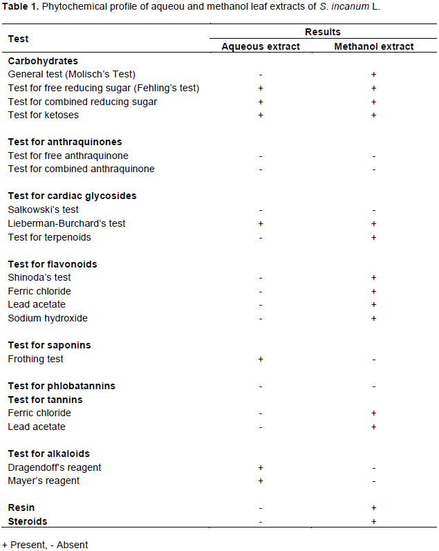
Phytochemical constituents of the extracts
The result of the phytochemical screening of the aqueous and methanol leaf extracts of S. incanum L. leaf extracts is summarized in Table 1. This study reveals the presence of phytochemicals considered as active medicinal chemical constituents. Important medicinal phytochemicals such as carbohydrate, reducing sugar, cardiac glycosides, alkaloids and saponin were found in aqueous leaf extract while carbohydrate, reducing sugar, cardiac glycosides, flavonoids, terpenoids, resin, steroid and tannins were found in methanol leaf extract.
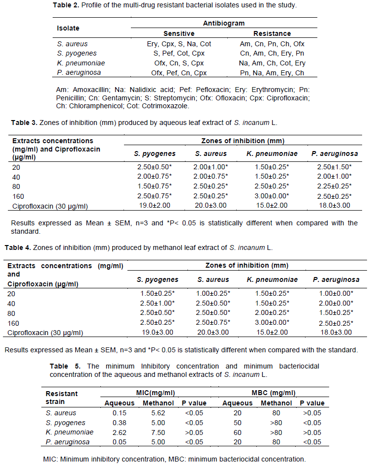
Profile of the multi-drug resistant bacterial isolates
The antibacterial susceptibility profile of the MDR bacterial isolates obtained from the samples of the patients seen at the Department of Microbiology, UMTH are shown in Table 2. The isolates were resistant to at least five (5) drugs. While all the isolates were resistant to amoxacillin, chloramphenicol and quinolones, they were all sensitive to aminoglycosides.
Antibacterial activity of S. incanum L. leaf extracts
The antibacterial activity of S. incanum L. was evaluated in vitro against MDR S. pyogenes, S. aureus, P. aeuruginosa and K. pneumoniae. In general, aqueous and methanol leaf extracts of S. incanum L. exhibited antibacterial activity (Tables 3 and 4).
The aqueous and methanol extracts demonstrated antibacterial activities against all the isolates; however, the activity was significantly lower than that of Ciprofloxacin even at highest concentration of 160 mg/ml (P<0.05).
Minimum inhibitory concentration and minimum bacteriocidal concentration
The MIC and MBC of the isolates were determined and presented in Table 5. The overall mean MIC for the aqueous and methanol leaf extracts were 0.8 mg/ml and 5.78 mg/ml while the overall mean MBC for the aqueous and methanol extracts was 37.5 and ≥320 mg/ml. Methanol extracts shows more activity against aqueous extracts.
Morbidity and mortality of bacterial infections are on the increase partly due to inadequacy and high cost of new generation antibacterials as well as widespread resistance to old generation antibacterials (Williams, 2000; Fair and Tor, 2014; Li and Webster, 2018). Therefore, there is need to look for new substances from other sources with proven antimicrobial activity.
Consequently, this has led to the search for effective antimicrobial agents of plant origin. The present study evaluated the antibacterial activities of aqueous and methanol leaf extracts of S. incanum L against MDR.
In this study, the antibiogram reflected all isolates sensitive to Ciprofloxacin but resistant to amoxacillin and chloramphenicol. Each of the isolate was resistant to at least five (5) drugs; therefore, the antibiogram of the bacterial isolates used in this study indicated that the isolates are multi-drug resistant. The resistance of strains of these bacterial (S. aureus, S. pyogenes, K. pneumoniae, P. aeruginosa) to numerous antibacterial drugs observed in this study is in accordance with previous studies that reported MDR among bacteria (Khan and Musharraf, 2004). Several factors may lead to increase in antimicrobial resistance which include; previous antibiotic used, inappropriate use of antimicrobial agents and inadequate adherence to infection control practice (Shu-Hui et al., 2011). Resistance of S. aureus, S. pyogenes and K. pneumoniae isolates to penicillins (amoxycillin) and chloramphenicol could be an indication of wide use of the drugs. Poor sensitivity to amoxycillin may be due to production of alpha-lactamase by resistant strains of isolates (Jhambh et al., 2012).
Secondary metabolites of plants have large economical value because of their involvement in production of colour or fragrance of flowers, taste and colour of food and resistance against diseases. The judicious use of different plant parts by folkloric medicine has played a key role in reducing human diseases (Mazid et al., 2012; Doss and Anand, 2012).
Flavonoids, resin, alkaloids, terpenoid, steroids and tannin have been reported to possess antimicrobial activities (Usman and Osuji, 2007; Doss and Anand, 2012; Sheel et al., 2014; Ghalem and Ali, 2017).
The results of this study suggested that the presence of potential pharmacologically active substances such as resin, flavonoids, alkaloids, steroids and saponins are responsible for antibacterial activities. The antibacterial activities of S. incanum L. showed that the aqueous and methanol leaves extract have activities against the bacterial tested (S. pyogenes, S. aureus, K. pneumoniae and P. aeruginosa) and this was in conformity with previous work done by Alamri and Moustafa (2012) in Saudi Arabia. Also, Owino et al. (2015) reported that the extract of S. incanum L. was found to be bacteriostatic and bacteriocidal against S. pyogenes, S. aureus, K. pneumoniae and P. aeruginosa.