ABSTRACT
Lichtheimia ramosa, a ubiquitous clinically important mould was isolated during a screen for thermotolerant fungi obtained from soil on a freshly burnt field in Ikorodu, Lagos State. In the laboratory, as expected it grew more luxuriantly on Potato Dextrose Agar than on size limiting Rose Bengal Agar. The isolate had mycelia with a white cottony appearance on both media. It was then identified based on morphological appearance, microscopy and by fungal Internal Transcribed Spacer ITS-5.8S rDNA sequencing. This might be the first report of molecular identification of L. ramosa isolate from soil in Lagos, as previously documented information could not be obtained.
Key words: Soil, thermotolerant, Lichtheimia ramosa, Internal Transcribed Spacer (ITS).
Zygomycetes of the order Mucorales are thermotolerant moulds that are ubiquitous in nature (Nagao et al., 2005). The genus Lichtheimia (syn. Mycocladus, Absidia) belongs to the Zygomycete class and includes saprotrophic microorganisms that can be isolated from decomposing soil and plant material (Alastruey-Izquierdo et al., 2010a). The awareness of the thermotolerant genus Lichtheimia increased markedly since its separation from the mesophilic genus Absidia (Hoffmann et al., 2007) and its taxonomic revision (Alastruey-Izquierdo et al., 2010b). Members of the genus Lichtheimia are considered to constitute thermotolerant fungi, because they grow at a wide range of temperatures, from 20 to 53°C, with optimum temperature for growth being 37°C, where it exhibits rapid growth (André et al., 2014).
L. ramosa is abundant in soil, decaying plant debris and foodstuff and is one of the causative agents of mucormycosis in humans (Barret et al., 1999). Mucormycosis is an opportunistic invasive infection caused by Lichtheimia, Mucor, and Rhizopus of the order Mucorales. Soil serves as a habitat and spore reservoir for Lichtheimia species. Several cases are known where traumatic injuries contaminated with soil resulted in Lichtheimia infections in immunocompetent patients (Belfiori et al., 2007, Corti et al., 2009, Blazquez et al., 2010). Mucormycosis (both rhino-orbital-cerebral and pulmonary) are acquired by the inhalation of their spores. Despite their low virulence, Lichtheimia species are currently regarded as emerging pathogens among Mucoralean fungi (Kutlu et al., 2014). There has been an increase in reports of L. ramosa infections among immunocompromised patients (Schwartze and Jacobsen, 2014).
L. ramosa is a microbe of clinical importance. Reported cases include L. ramosa isolated from a young patient’s infected wound after a road traffic accident (Neelaveni et al., 2017) and as a causal agent of primary cutaneous mucormycosis in a burn victim (Kaur et al., 2014). A fatal case of mucormycosis due to L ramosa affecting a 56-year-old male with diabetes mellitus and siderosis has also been reported (Mouronte-Roibás et al., 2016).
Genome sequence of L. ramosa and its close relative L. corymbifera have been published (Linde et al., 2014, Schwartze et al., 2014). Although Lichtheimia species tend to be morphologically and genetically distinct, they often share very similar antifungal drug susceptibilities.
The growth of the L. ramosa in media is rapid, with mycelia expanding to cover the entire plate in only a few (one to seven) days (Ziaee et al., 2016). Microscopically, L. ramosa is similar to L. corymbifera and differs in its ellipsoidal to cylindrical sporangiospores as compared to the subglobose to broadly ellipsoidal sporangiospores of L. corymbifera (Alastruey-Izquierdo et al., 2010a). In this study, a screen was undertaken to isolate and identify a thermotolerant fungus, L. ramosa from the environment by culture-based and molecular methods.
Sample collection
Soil samples were collected from a freshly burnt vegetation in Ikorodu Local Government, Lagos State with GPS coordinates 6°37'20.4"N 3°34'37.9"E. The farmland had been burnt following harvesting of farm produce. Samples were collected from the surface layer of the soil up to a depth of 7.5 to 10 cm.
Isolation of fungi
One gram of soil sample was placed in a test tube containing 9 ml of sterile distilled water and homogenized by shaking thoroughly. A ten-fold serial dilution scheme was made up to 10-5 dilutions. One milliliter aliquot of the homogenized solutions was taken from 10-3, 10-4 and 10-5 dilutions and plated on Potato Dextrose Agar supplemented with chloramphenicol and Rose Bengal chloramphenicol agar using the pour plate technique. Plating was done in triplicates for both Potato Dextrose Agar and Rose Bengal Agar. The plates were incubated at 30°C and the growth observed for 7 days.
Isolation of pure cultures and microscopy
Pure cultures of the white mycelial fungus were selected from the PDA plates and sub-cultured onto fresh PDA plates. The strain was stored on PDA slants at -20ºC for future use. For microscopic identification, a mycelial mat of the fungus was placed on a grease-free microscope glass slide and a drop of lactophenol cotton blue was added to the mycelia mat. A coverslip was placed on it with the aid of sterile forceps and the microscope slide was viewed under the microscope. Micrographs were thereafter obtained.
Molecular identification of the isolate
Genomic DNA was extracted from a five-day-old fungi culture grown on PDA using ZR Quick-DNA Fungal/Bacterial Miniprep™ extraction kit (Zymo Research, CA, USA) according to the manufacturer’s instructions. To check the DNA quality, it was run on a 1% ethidium bromide agarose gel (Figure 2A). Polymerase Chain Reaction (PCR) of the extracted genomic DNA was done in a GeneAmp PCR system 9700 PCR thermal cycler. ITS5F (GGAAGTAAAAGTCGTAACAAGG) and ITS4R (TCCTCCGCTTATTGATATG) primers were used for amplification. The PCR reaction mix (25 µl) contained 2.5 µl of 10x PCR buffer, 1 µl of 25 mM MgCl2, 1 µl each of forward and reverse primers, 1 µl of DMSO, 2 µl of 2.5 mM dNTPs, 0.1 µl of 5µg/ µl Taq DNA polymerase, 3 µl of 10ng/µl DNA and 13.4 µl Nuclease free water. The thermal cycler program used was as follows: Initial denaturation at 94ËšC for 5 min, followed by 36 cycles of denaturation at 94ËšC for 30 s, annealing at 54ËšC for 30 s, elongation at 72ËšC for 45 s, a ï¬nal elongation step at 72ËšC for 7 min and hold temperature at 10ËšC. The amplicons were electrophoresed on 1.5% agarose gels. The gel was prepared and run at 75 volts for 90 min and visualized with a UV Transilluminator (Figure 2B). The PCR product was Sanger sequenced using the BigDye 3.1 reaction protocol on 3130XL genetic analyzer (Applied Biosystems) at the Bioscience Center, IITA, Ibadan, Oyo. The sequences were checked for quality and assembled using BioEdit (version 7.2.5) Sequence Alignment Editor (Hall, 1999). The consensus sequence was compared to the GenBank nucleotide data library using the Basic Local Alignment Search Tool, BLAST software (Altschul et al., 1990)at the National Centre for Biotechnology Information (NCBI(http://www.ncbi.nlm.nih.gov). The sequences were submitted to the GenBank database and an accession number was assigned to the isolate.
Isolation of thermotolerant fungus from the soil sample
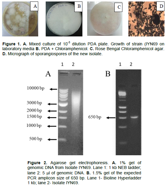
After 7 days of incubation, mixed culture PDA plates of few fungi were obtained (Figure 1A). From the 10-3 dilution plate, a dominant whitish fungus that appeared on all the other serial dilution plates was selected and sub-cultured. This selected strain (IYN69) had a more luxuriant growth on PDA plates than on Rose Bengal agar plate when grown in pure culture (Figures 1B and C). The strains grew at room temperature up to 37°C. The micrograph showed sporangiospores ellipsoidal to cylindrical in shape (Figure 1D).
Molecular identification of Isolate IYN69
In order to confirm the identity of the isolate IYN69 by molecular techniques, DNA product was visualized on 1% agarose gel stained with ethidium bromide (Figure 2A) and then the PCR product amplified by the pair of ITS4-R and ITS5-F primers were confirmed on a 1.5% agarose gel (Figure 2B).
The sequencing results from ITS5-ITS4 amplification of the ITS regions of isolate IYN69 was edited using BioEdit (version 7.2.5) Sequence Alignment Editor (Hall, 1999) and compared with the GenBank database using NCBI Basic Local Alignment Search Tool (BLASTn). The mould was identified as Lichtheimia ramosa with 100% identity to Lichtheimia ramosa isolate PUR 3 internal transcribed spacer 1 on the NCBI platform.
Nucleotide sequence
The partial sequence of the internal transcribed spacer (ITS) region of this isolate has been submitted to GenBank and can be found under accession number MT373684.
L. ramosa was the dominant fungus among others from a screen of thermotolerant fungi from recently burnt vegetation. Its isolation under such conditions is an indication that it can withstand high temperatures. Its thermotolerance has been linked to its virulence (Schwartze et al., 2012). Morphological and microscopic characteristics and phylogenetic identification were sufficient to confirm that the isolate was L. ramosa strain. Its features are similar to L. ramosa strain H71D and mycelia were identical to the type species of L. ramosa described by Hoffmann (2010) and Alastruey-Izquierdo et al. (2010a).
It is likely that this report might be the first documenting soil isolation of L. ramosa from Lagos and its molecular identification as no existing records were found. The BLAST hits showed many L. ramosa species that had significant sequence alignments with the query sequence. Based on the available molecular data, L. ramose isolate IYN69 most closely matched another isolate PUR 3 (GenBank Accession Number: MF033505) associated with a study on soil fungi and L. ramosa clone 7 (Alastruey-Izquierdo et al., 2010a). L. ramosa isolate IYN69 ITS 1, partial sequence; 5.8S ribosomal RNA gene, complete sequence; and ITS 2, partial sequence can be found in GenBank under accession number MT373684.
Incidences of L. ramosa infection in patients have been linked to road traffic accidents (RTAs) suggesting contamination by this soil fungus (Neelaveni et al., 2017; Bibashi et al., 2012). This is of great significance because RTAs are a major public health concern and third leading cause of death in Nigeria (Dixie, 1999; Onyemachi and Ofoma, 2016). The next steps would be to do whole genome sequencing and compare its genome with the existing reference genome (Linde et al., 2014) especially regions responsible for sensitivity to antifungals. This strain would be tested for its sensitivity to existing antifungal drugs (Amphotericin B, posaconazole, itraconazole) that are known to be effective against Lichtheimia species. Although, there are not many cases of L. ramosa infections worldwide (Mouronte-Roibás et al., 2016); for an emerging pathogen, the information from such studies would help to put in place good treatment options should burn, accident victims or diabetics get infected with this strain from this locality. Since L. ramosa is also known for the production of enzymes like xylanase (Alvarez-Zuniga et al., 2017), and mannase (Xie et al., 2019), an assay for these and other thermophilic enzymes with biotechnological applications will be conducted on this new isolate.
The authors have not declared any conflict of interests.
The authors would like to express their appreciation to Ayodeji A. Odunsi and Gabriel B. Adeloye for their assistance and Amarachi Anyanwu for providing the micrograph.
REFERENCES
|
Alastruey-Izquierdo A, Hoffmann K, de Hoog GS, Rodriguez-Tudela JL, Voigt K, Bibashi E, Walther G (2010a). Species recognition and clinical relevance of the zygomycetous genus Lichtheimia (syn. Absidia Pro Parte, Mycocladus). Journal of Clinical Microbiology 48:2154-2170.
Crossref
|
|
|
|
Alastruey-Izquierdo A, Cuesta I, Walther G, CuencaEstrella M, Rodr'ıguez-Tudela JL (2010b). Antifungal susceptibility profile of human-pathogenic species of Lichtheimia. Antimicrobial Agents and Chemotherapy 54(7):3058-3060.
Crossref
|
|
|
|
|
Alvarez-Zuniga MT, Santiago-Hernandez A, Rodriguez-Mendoza J, Campos, J E, Pavon-Orozco P. Trejo-Estrada S, Hidalgo-Lara ME (2017). Taxonomic identification of the thermotolerant and fast-growing fungus Lichtheimia ramosa H71D and biochemical characterization of the thermophilic xylanase LrXynA. AMB Express 7:194.
|
|
|
|
|
André AL, Hoffmann K, Lima D X, de Oliveira RJ, Vieira HE, Malosso E, Maia LC, da Silva GA (2014). A new species of Lichtheimia (Mucoromycotina, Mucorales) isolated from Brazilian soil. Mycological Progress 13:343-352.
Crossref
|
|
|
|
|
Altschul SF, Gish W, Miller W, Myers EW, Lipman DJ (1990). Basic Local Alignment Search Tool. Journal of Molecular Biology 215:403-410.
Crossref
|
|
|
|
|
Barret JP, Ramzy PI, Heggers JP, Villareal C, Herndon DN, Desai MH (1999). Topical nystatin powder in severe burns: a new treatment for angioinvasive fungal infections refractory to other topical and systemic agents. Burns 25:505-508.
Crossref
|
|
|
|
|
Belfiori R, Terenzi A, Marchesini L, Repetto A (2007). Absidia corymbifera in an immune-competent accident victim with multiple abdominal injuries: Case report. BMC Infectious Diseases 7:46-50.
Crossref
|
|
|
|
|
Bibashi E, de Hoog GS, Pavlidis TE, Symeonidis N, Sakantamis A, Walther G (2012). Wound infection caused by Lichtheimia ramosa due to a car accident. Medical Mycology Case Reports 2:7-10.
Crossref
|
|
|
|
|
Blazquez D, Ruiz-Contreras J, Ferna'ndez-Cooke E, Gonza'lez-Granado I, Delgado, MD, Menendez MT, Rodriguez-Gil Y, Ballen A, Del Palacio A (2010). Lichtheimia corymbifera subcutaneous infection successfully treated with amphotericin B, early debridement, and vacuum-assisted closure. Journal of Pediatric Surgery 45:13-15.
Crossref
|
|
|
|
|
Corti G, Mondanelli N, Losco M, Bartolini L, Fontanelli A, Paradisi F (2009). Posttraumatic infection of the lower limb caused by rare Enterobacteriaceae and Mucorales in a young healthy male. International Journal of Infectious Diseases 13:57-60.
Crossref
|
|
|
|
|
Dixie RA (1999). 'Fatalism', accident causation and prevention: issues for health promotion from an exploratory study in a Yoruba town, Nigeria. Health Education Research: Theory and Practice 14(2):197-208.
Crossref
|
|
|
|
|
Hall TA (1999). BioEdit: A user-friendly biological sequence alignment editor and analysis program for Windows 95/98/NT. Nucleic Acids Symposium Series 41:95-98.
|
|
|
|
|
Hoffmann K, Discher S, Voigt K (2007). Revision of the genus Absidia (Mucorales, Zygomycetes) based on physiological, phylogenetic, and morphological characters; Thermotolerant Absidia spp. form a coherent group, Mycocladiaceae fam. nov. Mycological Research 111:1169-1183.
Crossref
|
|
|
|
|
Hoffmann K (2010). Identification of the genus Absidia (Mucorales, Zygomycetes): a comprehensive taxonomic revision. In: Gherbawy Y, Voigt K (eds) Molecular identification of fungi. Springer, Berlin, pp. 439-460.
Crossref
|
|
|
|
|
Kaur R, Bala K, Ahuja RB, Srivastav P, Bansal U (2014). Primary cutaneous mucormycosis in a patient with burn wounds due to Lichtheimia ramosa. Mycopathologia 178:291-295.
Crossref
|
|
|
|
|
Kutlu M, Ergin C, Hilmioglu-Polat S, Gumral R, Necan C, Kocyigit A, Sayn-Kutlu S (2014). Pulmonary mucormycosis due to Lichtheimia ramosa in a patient with HIV infection. Mycopathologia 178(1-2):111-115.
Crossref
|
|
|
|
|
Linde J, Schwartze V, Binder U, Lass-Flörl C, Voigt K, Horn F (2014). De novo whole-genome sequence and genome annotation of Lichtheimia ramosa. Genome Announcement 5:e00888-14.
Crossref
|
|
|
|
|
Mouronte-Roibás C, Leiro-Fernandez, V, Botana-Rial M, Ramos-Hernandez C, Lago-Preciado G, Fiano-Valverde C, Fernandez-Villa A (2016). Lichtheimia ramosa: A fatal case of mucormycosis. Canadian Respiratory Journal 2:1-4.
Crossref
|
|
|
|
|
Nagao K, Ota T, Tanikawa A, Takae Y, Mori T, Udagawa S, Nishikawa T (2005). Genetic identification and detection of human pathogenic Rhizopus species, a major mucormycosis agent, by multiplex PCR based on internal transcribed spacer region of rRNA gene. Journal of Dermatological Science 39(1):23-31.
Crossref
|
|
|
|
|
Neelaveni V, Tupaki-Sreepurna A, Thanneru V, Jyoti-Kindo A (2017). Lichtheimia ramosa isolated from a young patient from an infected wound after a road traffic accident. Journal of The Academy of Clinical Microbiologists 19(1):59.
|
|
|
|
|
Onyemachi NOC, Ofoma UR (2016). The Public Health Threat of Road Traffic Accidents in Nigeria: A call to Action. Annals of Medical and Health Science Research 6(4):199-204.
Crossref
|
|
|
|
|
Schwartze VU, Hoffmann K, Nyilasi I, Papp T, Vágvölgyi C, de Hoog S, Voigt K, Jacobsen ID (2012). Lichtheimia species exhibit differences in virulence potential. PLoS One 7:40908.
Crossref
|
|
|
|
|
Schwartze VU, Jacobsen ID (2014). Mucormycoses caused by Lichtheimia species. Mycoses 57:73-78.
Crossref
|
|
|
|
|
Schwartze VU, Winter S, Shelest E, Marcet-Houben M, Horn F, Wehner S, Linde J, Valiante V, Sammeth M, Riege K, Nowrousian M, Kaerger K, Jacobsen ID, Marz M, Brakhage AA, Gabaldón T, Böcker S, Voigt K (2014). Gene expansion shapes genome architecture in the human pathogen Lichtheimia corymbifera: An evolutionary genomics analysis in the ancient terrestrial Mucorales (Mucoromycotina). PLoS Genetics 10:e1004496.
Crossref
|
|
|
|
|
Xie J, Pan L, He Z, Liu W, Zheng D, Zhang Z, Wang B (2019). A novel thermophilic β-mannanase with broad-range pH stability from Lichtheimia ramosa and its synergistic effect with α-galactosidase on hydrolyzing palm kernel meal. Process Biochemistry 88:51-59.
Crossref
|
|
|
|
|
Ziaee A, Zia M, Bayat M, Hashemi J (2016). Molecular identification of Mucor and Lichtheimia species in pure cultures of zygomycetes. Jundishapur Journal of Microbiology 9:1-8.
Crossref
|
|