African Journal of
Microbiology Research

  • Abbreviation: Afr. J. Microbiol. Res.
  • Language: English
  • ISSN: 1996-0808
  • DOI: 10.5897/AJMR
  • Start Year: 2007
  • Published Articles: 5233

Full Length Research Paper

The bacterial diversity in infected tissue pus of an East Asian finless porpoise

Caiwen Wu
  • Caiwen Wu
  • Jiangsu Key Laboratory for Biodiversity and Biotechnology, College of Life Science, Nanjing Normal University, Wenyuan Road 1, Qixia District, Nanjing 210023, China.
  • Google Scholar
Zhe Hong
  • Zhe Hong
  • College of Life Science, Shenyang Normal University, Huanghebei Street 253, Shenyang, 110034, China.
  • Google Scholar
Aamir Ibrahim
  • Aamir Ibrahim
  • Jiangsu Key Laboratory for Biodiversity and Biotechnology, College of Life Science, Nanjing Normal University, Wenyuan Road 1, Qixia District, Nanjing 210023, China.
  • Google Scholar
Di Zhang
  • Di Zhang
  • Jiangsu Key Laboratory for Biodiversity and Biotechnology, College of Life Science, Nanjing Normal University, Wenyuan Road 1, Qixia District, Nanjing 210023, China.
  • Google Scholar
Lin Wang
  • Lin Wang
  • Jiangsu Key Laboratory for Biodiversity and Biotechnology, College of Life Science, Nanjing Normal University, Wenyuan Road 1, Qixia District, Nanjing 210023, China.
  • Google Scholar
Ting Li
  • Ting Li
  • Jiangsu Key Laboratory for Biodiversity and Biotechnology, College of Life Science, Nanjing Normal University, Wenyuan Road 1, Qixia District, Nanjing 210023, China.
  • Google Scholar
Bingyao Chen
  • Bingyao Chen
  • Jiangsu Key Laboratory for Biodiversity and Biotechnology, College of Life Science, Nanjing Normal University, Wenyuan Road 1, Qixia District, Nanjing 210023, China.
  • Google Scholar


  •  Received: 22 September 2019
  •  Accepted: 30 October 2019
  •  Published: 30 November 2019

 ABSTRACT

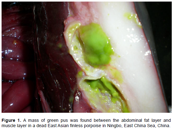
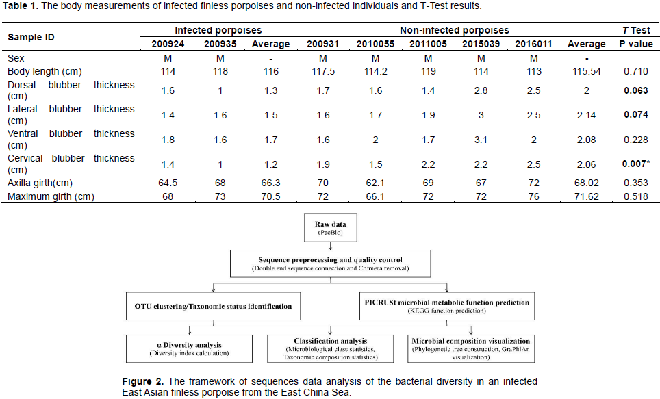
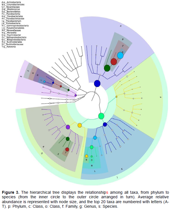
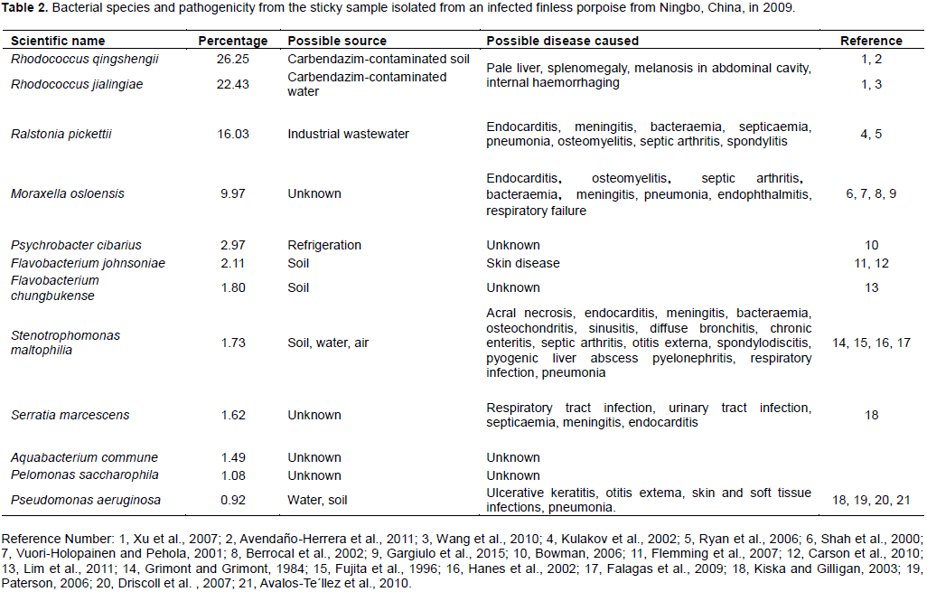
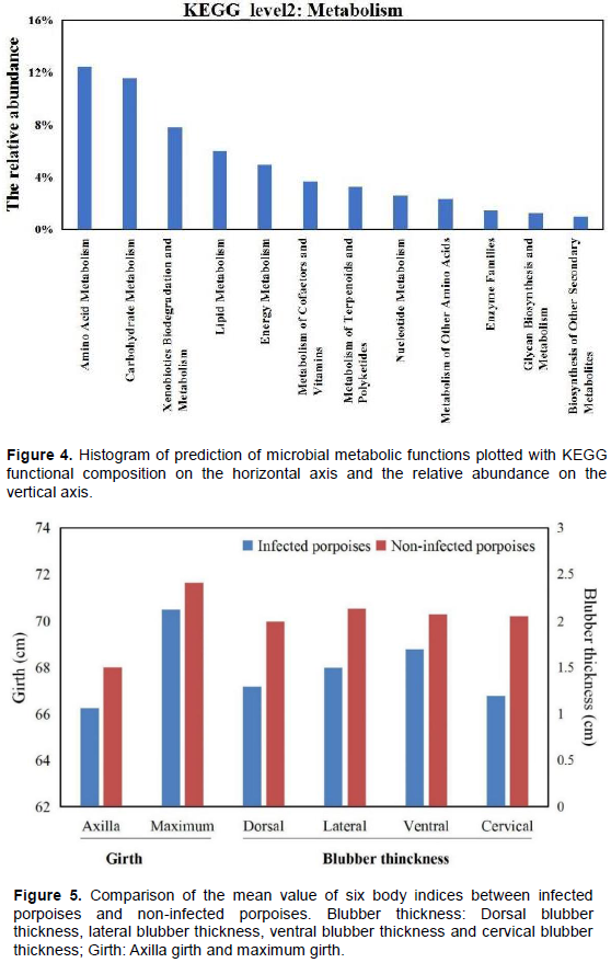
Two East Asian finless porpoises (Neophocaena asiaeorientalis sunameri) in Ningbo, East China Sea, China, were observed to be bacterially infected between the fat layer and muscle layer. The microbial communities in pus samples were identified by metagenome sequencing 16S in the PacBio platform and explored the relationship between emaciation and bacterial infection. The present paper was the first report on bacterial diversity in infected tissue pus of finless porpoise. In total, 101 bacterial species were identified, and the top nine species were Rhodococcus qingshengii (26.25%), Rhodococcus jialingiae (22.43%), Ralstonia pickettii (16.03%), Moraxella osloensis (9.97%), Psychrobacter cibarius (2.97%), Flavobacterium johnsoniae (2.11%), Flavobacterium chungbukense (1.80%), Stenotrophomonas maltophilia (1.73%) and Serratia marcescens (1.62%). These main bacteria could cause various diseases or metabolic disorder, such as endocarditis, meningitis, bacteraemia, septicaemia, and so on; they also involve microbiome metabolism of amino acids (12.47%), carbohydrates (11.58%), xenobiotic biodegradation (7.81%), lipids (6.01%) and energy (4.98%). Regardless of the type of disease metabolic disorder will firstly cause body weight loss. In conclusion, these bacteria could cause diseases or metabolic disorders, resulting in emaciation of East Asian finless porpoises. Admittedly, pathogenic assay is furthermore needed to determine the mechanisms regarding the pathological phenomena.


Key words: Bacterial diversity, East Asian finless porpoises, Metagenome sequencing 16S.


 INTRODUCTION

Infectious diseases caused by bacteria were thought to be the primary cause of mortalities in aquatic mammals (Dunn et al., 2001). These bacteria contributed to different degrees of pathogenicity. For example, a Helicobacter sp. isolated from stomachs may play a role in the etiopathogenesis of  gastritis  and  gastric ulcers  in dolphins (Harper et al., 2000). Pathogenic vibrios have been found, which might lead to, secondary infections in debilitated (e.g., stranded) marine mammals was a distinct possibility (Buck and Stephen, 2010). Brucella sp. infections and associated lesions were described in a harbour porpoise (Phocoena  phocoena) (Jauniaux et al.,
 
2010). 20 bacterial pathogens were identified in captive and free-ranging killer whales or sympatric odontocetes species, and they were considered to affect fecundity or reproductive success (Gaydos et al., 2004).
 
Studies on infectious disease in marine mammals have seldom been conducted in China. Although nearly 45 marine mammal species harbor in Chinese waters (Zhou, 2004), the specimens of most animals were difficult to obtain except for finless porpoise. The East Asian finless porpoise (Neophocaena sunameri) was one of three finless porpoises species (Zhou et al., 2018), and most common marine mammal was found stranded or by caught. 170 porpoises were accidentally caught in Bohai and the Northern Yellow Sea from 2004 to 2008 (Liu, 2009). At present, bacterial infections in wild finless porpoises has rarely been reported due to scarce infected specimens and limitation of technology.
 
To identify these bacteria/microorganism species, 16S ribosomal RNA (rRNA) gene sequencing was commonly used as these genes comprise of highly conserved domains, interspersed with variable regions (You, 2005; Pootakham et al., 2017). To begin with, 16S rRNA gene profiling was performed using clone-based Sanger sequencing, which provides accurate, full-length or almost full-length sequences. Due to the high cost and low-output nature of the approach, the number of 16S rRNA sequencing used in Sanger-based bacterial profiling studies were often lower than 200 sequences per sample, which was insufficient to cover the complete diversity of the bacterial community (Sharp et al., 2012). Currently, various next generation sequencing platforms such as Roche 454 (Bayer et al., 2013), Illumina (Schmidt et al., 2013) and Ion Torrent PGM (Salipante et al., 2014) have gradually replaced the use of the Sanger sequencing method. Of these new sequencing platforms, the Pacific Biosciences (PacBio) single-molecule, real-time (SMRT) sequencing technology was recommended to be capable of analyzing the bacterial profiles based on the full length 16S rRNA gene (Amir et al., 2013; Pootakham et al., 2017). This technology favours metagenomic analysis and affords the researcher a relatively unbiased view of the microbial communities at different taxonomic levels of a sample (Charuvaka and Rangwala, 2011).
 
In Ningbo, East China Sea, China, two East Asian finless porpoises were observed to be bacterial infections between the fat and the muscle layers. The present paper focused on the bacterial diversity in the tissue pus sample, and the relationship between bacterial composition and infections symptoms. The recommended SMRT sequencing of the full-length 16S rRNA gene were applied.


 MATERIALS AND METHODS

Sample collection
 
Within February and May 2009, 12 dead East Asian finless porpoises  from   fisheries  by  catch  accidentally,  in  Ningbo,  East China Sea were collected. The collection and research on specimens were authorized by Ningbo Ocean and Fisheries Bureau. Then pathological anatomy was conducted later on. During the necropsy, the green pus was found between the abdominal fat and muscle layers of two dead finless porpoises (Figure 1). Except of green pus, the fat and muscle tissues were normal, no lesions were found. The pus samples of one finless porpoise were collected in a sterile valve bags and stored at -20°C until its DNA was extracted.
 
Both infected porpoises were male with a body length of 114 and 118 cm. To explore the differences in nutritional status between infected porpoises and non-infected porpoises, six morphological characteristics, that is, dorsal blubber thickness, lateral blubber thickness, ventral blubber thickness, cervical blubber thickness, axilla girth and maximum girth were chosen. Meanwhile, to minimize the influence of aging and gender, data on five dead male finless porpoises with similar body lengths (113 to 119 cm) were collected in the same period, used for comparison (Table 1).
 
16S rRNA gene amplification and sequencing
 
Genomic DNA was extracted using the MoBio PowerSoil® DNA Isolation Kit (MoBio Laboratories Inc., Carlsbad, CA, USA) according to the manufacturer’s instructions. The DNA quality was assessed on 0.8% agarose gel to ensure no degradation or rRNA contamination. The DNA samples were subsequently quantified using a spectrophotometer (Eppendorf, model RS-232C, Hamburg, Germany).
 
The diversity of bacterial communities was analyzed using single molecule real-time PacBio sequencing technology (Pacific Biosciences, Menlo Park, CA, USA). The full-length 16S ribosomal RNA gene was amplified from genomic DNA using the bacterial-specific primer. The primer sequences were 5’-AGAGTTTGATCMTGGCTCAG-3’ and 5’- ACCTTGTTACGACTT-3’.
 
To obtain barcoded 16S rRNA amplicons, the amplifications in two steps was performed. A first round of PCR was performed in a 25 μL PCR solution containing 5 μL NEB Q5 High Fidelity DNA polymerase (New England Biolabs, Ipswich, MA), 5 μL GC Enhancer (New England Biolabs, Ipswich, MA), 2 μL DNTP mixture (2.5 mmol/L-1), 1 μL DNA template (20 ng/μL-1), 1 μL of each primer (10 μmol/L-1), and 10 μL sterilized distilled water. The thermal cycling conditions were as follows: 98°C for 2 min; 25 cycles of 98°C for 30 s; 50°C for 30 s; and a final extension time of 90 s at 72°C. The primer of the second round of PCR contained a set of 16-nucleotide barcodes for the barcoded SMRT sequencing of the full-length 16S rRNA gene. The primary PCR products were diluted to 2 ng/μL-1, and the diluted products were used as templates for the secondary amplification. A second round of PCR was performed in a 25 μL PCR solution containing 5 μL NEB Q5 High Fidelity DNA polymerase (New England Biolabs, Ipswich, MA), 5 μL GC Enhancer (New England Biolabs, Ipswich, MA), 2 μL DNTP mixture (2.5 mmol/L-1), 1 μL DNA template (2 ng/μL-1), 1 μL of each primer (10μmol/L-1), and 10 μL sterilized distilled water. The conditions used for amplification in the thermocycler were as follows: 98°C for 2 min; 15 cycles of 98°C for 30 s; 62°C for 30 s; and 72°C for 90 s; and a final extension time of 5 min at 72°C.
 
A PacBio library was constructed using the Template Prep Kit 1.0 (Pacific Biosciences, Menlo Park, CA, USA) containing a pool of barcoded amplicons from the bacterial sample. The full-length 16S rRNA gene was sequenced using P6-C4 chemistry on a PacBio RS II instrument (Pacific Biosciences, Menlo Park, CA, USA). 
 
 
Sequence data analysis and microbial population identification
 
Raw data were processed by the SMRT Portal, Version 2.7 (PacBio) to  obtain  effective  sequences  with  a  minimum  of  3 full  passes and a minimum predicted accuracy of 90 (Hou et al., 2015). The sequencing data were processed using the software package QIIME version 1.8.0 (Caporaso et al., 2010). Sequences shorter than 500 bp were removed prior to downstream analyses. Chimeric sequences were detected and removed using USEARCH version 5.2.236 (Edgar, 2010). The remaining sequences were clustered into OTUs (Blaxter et al., 2005) based on an “open-reference” OTU-selecting method at 97% identity using UCLUST (Edgar, 2010) (Figure 2). Taxonomy was assigned to the representative sequence of each OTU using the Greengenes database version 13.8 (Desantis et al., 2006), Ribosomal Database Project (RDP) database version 11.1 (Cole et al., 2009) and Silva database version 115 (Quast et al., 2013). The OTUs occurring only once or twice were discarded. After the sequencing data were rarefied, the following alpha diversity measures were calculated: Number of OTUs, Chao1 estimate of species richness (Chao, 1984), ACE estimate of species richness (Dawid, 1993), Shannon diversity (Shannon, 1948), and Simpson diversity (Simpson, 1949).
 
GraPhlAn is a new tool for compact and publication-quality representation of circular taxonomic and phylogenetic trees with potentially rich sets of associated metadata (Asnicar et al., 2015), and it was used to quickly detect the dominant microbe groups from the complex community data in this study. Then, KEGG PATHWAY Database (http://www.genome.jp/kegg/pathway.html) in PICRUSt (Phylogenetic Investigation of Communities by Reconstruction of Unobserved States) (Langille et al., 2013) was applied to predict the functional composition of the metagenome of the microbial community from its 16S profile. 


 RESULTS

Bacterial diversity
 
Following    the    removal    of   chimeras    and    circular consensus sequencing reads shorter than 500 bp, a total of 30768 reads of processed full-length 16S rRNA sequences were obtained for their samples. In total, 101 bacterial species were identified, and they belonged to six phyla: Actinobacteria (50.62%), Proteobacteria (42.24%), Bacteroidetes (5.06%), Firmicutes (1.82%), Cyanobacteria (0.22%) and Acidobacteria (0.05%). Four alpha diversity indices were calculated; the Chao1 estimator (307) was similar to the ACE estimator (307) while the Shannon diversity index and Simpson index were 4.2 and 0.86, respectively. Various colours on the GraPhlAn circular taxonomic and phylogenetic trees were used to distinguish among different taxa and their abundance were reflected through the node size (Figure 3).
 
Among the bacterial species, Rhodococcus qingshengii (26.25%), Rhodococcus jialingiae (22.43%), Ralstonia pickettii (16.03%) and Moraxella osloensis (9.97%) were the most prevalent (Table 2). Of these bacteria, two bacteria, Stenotrophomonas maltophilia and Pseudomonas aeruginosa, can produce green pigmentations consistent with the green pus in the two dead finless porpoises.
 
Prediction of microbial metabolic function
 
The KEGG analysis indicated that 12 metabolic functions were associated with the microbial samples isolated from the infected finless porpoise. The major functions were amino acid metabolism (12.47%), carbohydrate metabolism (11.58%),   xenobiotics   biodegradation   and metabolism (7.81%), lipid metabolism (6.01%) and energy metabolism (4.98%) (Figure 4).
 
Nutritional status comparison
 
The mean value of the six  bodies  indices  (dorsal blubber thickness, lateral blubber thickness, ventral blubber thickness, cervical blubber thickness, axilla girth and maximum girth) of the infected porpoises were less than in the non-infected porpoises (Figure 5). One sample t-test was performed using IBM SPSS Statistics version 21 (SPSS, Inc., Chicago,  Ill., USA), and it showed significantly different cervical blubber thickness (p=0.007) and nearly significant lateral blubber thickness (p=0.074) and dorsal blubber thickness (p=0.063) (Table 1).
 
This indicated that the bacteria-infected individuals were more emaciated than the non-infected individuals.


 DISCUSSION

In this paper, single molecule real-time (SMRT) sequencing, developed by Pacific BioSciences (PacBio) were used to overcome the limitations of first- and second-generation sequencing (Rhoads and Au, 2015), which were hardly enough to capture the complete diversity of the bacteria (Pootakham et al., 2017). Therefore, this paper provided a more precise and reliable sequences than first- and second-generation technology.
 
The infected porpoises generally showed clear notable marasmus unlike the non-infected porpoises. This indicated that the porpoises might have been suffering from infections, which resulted in consumption of fats and body marasmus. Although these assumed diseases have not been determined, the analysis of the pathogenicity of the bacteria provides us with some insights.
 
A total of 101 bacteria were identified, among them, observations of R. qingshengii (26.25%) and Rhodococcus jialingiae (22.43%)  have been  reported  in the sludge of a carbendazim wastewater treatment facility and carbendazim-contaminated soil (Wang et al., 2010). Ralstonia pickettii has been identified in biofilm formation in ultrapure water in industrial systems (Kulakov et al., 2002). The pathogenicity of these species has been reported in some aquatic species, e.g., genus Rhodococcus and Flavobacterium johnsoniae for freshwater and marine fish (Olsen et al., 2006; Flemming et al., 2007) and a  West  African  dwarf crocodile that died from acute Stenotrophomonas maltophilia septicaemia (Harris and Rogers, 2001). The finless porpoise as a top predator in marine environments very likely to contacted these bacteria along the marine food cycle.
 
Meanwhile, Moraxella osloensis (9.97%), Flavobacterium johnsoniae (2.11%), Stenotrophomonas maltophilia (1.73%), Serratia marcescens (1.62%) and Pseudomonas aeruginosa (0.92%) were conditioned  pathogens. Most of them are widely distributed in nature and exist in animals as normal flora. When this symbiotic relationship turns into parasitism caused by bacterial translocation, as in dysbacteriosis or host-lowered immunity, these normal floras might damage their hosts (Sun et al., 2011).
 
Additionally, species of the genus Psychrobacter have been isolated from various low-temperature habitats or sources, including sea ice, fish, chilled meat  and  blood products, krill stomach, Antarctic ornithogenic soil and contaminants on lab media (Bowman, 2006), and Psychrobacter cibarius (2.97%) has never been detected as an  animal  pathogen.  It  can be presumed that the possible origin in the sample was refrigeration in the refrigerator.
 
Microorganisms   and    their   hosts   share   the  same environment, and the activities of the microbiomes and their metabolic products influence a variety of aspects in metazoans (Lee and Hase, 2014). This was supported by the KEGG analysis in this study, that is, the microbial communities in finless porpoise involved in major metabolism functions which were the most fundamental requirements for survival, and metabolic disorders may lead to various health problems.
In fact, the main bacterial species have been proven to be related to various pathogenicities, skin diseases, respiratory diseases, diverse inflammations, hematologic diseases, and visceral lesions. For example, Flavobacterium johnsoniae and Pseudomonas aeruginosa were known as opportunistic skin pathogens (Driscoll et al., 2007; Carson et al., 2010), and they cause soft tissues infections in fish species (Carson et al., 2010). Moraxella osloensis was a pathogen that causes respiratory failure (Gargiulo et al., 2015). Stenotrophomonas maltophilia could cause endocarditis, meningitis, osteochondritis, sinusitis, diffuse bronchitis, chronic enteritis, septic arthritis, otitis externa, spondylodiscitis, pyogenic liver abscess pyelonephritis (Falagas et al., 2009) and pneumonia (Hanes et al., 2002). Pseudomonas aeruginosa has been reported to cause chronic bronchopneumonia in bottlenose dolphins (Avalos-Te´llez et al., 2010). Ralstonia pickettii has been associated with unusual clinical situations such as bacteraemia and septicaemia (Ryan et al., 2006). Infection from Rhodococcus qingshengii could cause severe visceral lesions, e.g., inducing the presence of the pseudo-membranes on the spleen, liver and heart. Histopathology revealed perivascular leucocytic infiltration, marked granulomas and increased fibroblastic reticular cells in spleen (Avendaño-Herrera et al., 2011).
 
Based on the analysis above, it was speculated that bacterial infections cause some diseases and metabolic disorders, which might impact the health of finless porpoises to some extent. Regardless of the type of diseases and metabolic disorders would first cause of body weight loss. Further study on pathogenicity experiment was needed in the future.


 CONFLICT OF INTERESTS

The authors have not declared any conflict of interests.


 ACKNOWLEDGEMENTS

The present study was financially supported by the Priority Academic Program Development of Jiangsu Higher Education Institutions (PAPD).


 REFERENCES

Amir A, Zeisel A, Zuk O, Elgart M, Stern S, Shamir O, Turnbaugh PJ, Soen Y, Shental N (2013). High-resolution microbial community reconstruction by integrating short reads from multiple 16S rRNA regions. Nucleic Acids Research 41(22):e205.
Crossref


Asnicar F, Weingart G, Tickle TL, Huttenhower C, Segata N (2015). Compact graphical representation of phylogenetic data and metadata with GraPhlAn. Peer Journal 3:e1029.
Crossref


Avalos-Te'llez R, Sua'rez-Gu¨emes F, Carrillo-Casas EM, Herna'ndez-Castro R (2010). Bacteria and yeast normal microbiota from respiratory tract and genital area of bottlenose dolphins (Tursiops truncatus). Current Research, Technology and Education Topics in Applied Microbiology and Microbial Biotechnology 2:666-673.


Avendaño-Herrera R, Balboa S, Doce A, Ilardi P, Lovera P, Toranzo AE, Romalde JL (2011). Pseudo-membranes on internal organs associated with Rhodococcus qingshengii infection in Atlantic salmon (Salmo salar). Veterinary Microbiology 147:200-204.
Crossref


Bayer T, Neave MJ, Alsheikh-Hussain A, Aranda M, Yum LK, Mincer T, Hughen K, Apprill A, Voolstra CR (2013). The microbiome of the Red Sea coral Stylophora pistillata is dominated by tissue-associated Endozoicomonas bacteria. Applied and Environmental Microbiology 80:4759-4762.
Crossref


Blaxter M, Mann J, Chapman T, Thomas F, Whitton C, Floyd R, Abebe E (2005). Defining operational taxonomic units using DNA barcode data. Philosophical Transactions of the Royal Society 360:1935-1943.
Crossref


Bowman JP (2006). The genus Psychrobacter. In: Dworkin M, Falkow S, Rosenberg E, Schleifer KH, Stackenbrandt E, eds. The Prokaryotes. New York: Springer pp. 920-930.
Crossref


Buck JD, Stephen S (2010). The occurrence of potentially pathogenic vibrios in marine mammals. Marine Mammal Science 2:319-324.
Crossref


Caporaso JG, Kuczynski J, Stombaugh J, Bittinger K, Bushman FD, Costello EK, Fierer N, Peña AG, Goodrich JK, Gordon JI, Huttley GA, Kelley ST, Knights D, Koenig JE, Ley RE, Lozupone CA, McDonald D, Muegge BD, Pirrung M, Reeder J, Sevinsky JR, Turnbaugh PJ, Walters WA, Widmann J, Yatsunenko T, Zaneveld J, Knight R (2010). Qiime allows analysis of high-throughput community sequencing data. Nature Methods 7:335-336.
Crossref


Carson J, Schmidtke LM, Munday BL (2010). Cytophaga johnsonae: a putative skin pathogen of juvenile farmed barramundi, Lates calcarifer Bloch. Journal of Fish Diseases 16:209-218.
Crossref


Chao A (1984). Nonparametric Estimation of the Number of Classes in a Population. Scandinavian Journal of Statistics 11:265-270.


Charuvaka A, Rangwala H (2011). Evaluation of short read metagenomic assembly. BMC Genomics 12:S8.
Crossref


Cole JR, Wang Q, Cardenas E, Fish J, Chai B, Farris RJ, Kulam-Syed-Mohideen AS, McGarrell DM, Marsh T, Garrity GM, Tiedje JM (2009). The Ribosomal Database Project: improved alignments and new tools for rRNA analysis. Nucleic Acids Research 37:D141-D145.
Crossref


Dawid AP (1993). Stopping rules and estimation for recapture debugging with unequal failure rates. Biometrika 80:193-201.
Crossref


Desantis TZ, Hugenholtz P, Larsen N, Rojas M, Brodie EL, Keller K, Huber T, Dalevi D, Hu P, Andersen GL (2006). Greengenes, a chimera-checked 16S rRNA gene database and workbench compatible with ARB. Applied and Environmental Microbiology 72:5069-5072.
Crossref


Driscoll JA, Brody SL, Kollef MH (2007). The epidemiology, pathogenesis and treatment of Pseudomonas aeruginosa infections. Drugs 67:351-368.
Crossref


Dunn JL, Buck JD, Robeck TR (2001). Bacterial Diseases of Cetaceans and Pinnipeds. CRC Handbook in Marine Mammal Medicine, Second edition pp. 309-335.
Crossref


Edgar RC (2010). Search and clustering orders of magnitude faster than BLAST. Bioinformatics 26:2460-2461.
Crossref


Falagas ME, Kastoris AC, Vouloumanou EK, Dimopoulos G (2009). Community-acquired Stenotrophomonas maltophilia infections: a systematic review. European Journal of Clinical Microbiology and Infectious Diseases 28:719-730.
Crossref


Flemming L, Rawlings D, Chenia H (2007). Phenotypic and molecular characterisation of fish-borne Flavobacterium johnsoniae-like isolates from aquaculture systems in South African. Research in Microbiology 158:18-30.
Crossref


Fujita J, Yamadori I, Xu G, Hojo S, Negayama K, Miyawaki H, Yamaji Y, Takahara J (1996). Clinical features of Stenotrophomonas maltophilia pneumonia in immunocompromised patients. Respiratory Medicine 90:35.
Crossref


Gargiulo C, Pham VH, Hai NT, Nguyen KCD, Davey KF, Abe K (2015). A case report of an elderly patient with respiratory failure caused by Moraxella osloensis infection. Anaesthesia 1(1):17-21.


Gaydos JK, Iii KCB, Osborne RW, Dierauf L (2004). Evaluating potential infectious disease threats for southern resident killer whales, Orcinus orca: a model for endangered species. Biological Conservation 117:253-262.
Crossref


Grimont PAD, Grimont F (1984). Family I. Enterobacteriaceae. Genus VIII. Sermtia Bizio 1823. In: Krieg NR, Holt JG, eds. Bergey's Manual of systematic bacteriology, Baltimore: Williams and Wilkins 1:477-484.


Hanes SD, Demirkan K, Tolley E, Boucher BA, Croce MA, Wood GC, Fabian TC (2002). Risk factors for late-onset nosocomial pneumonia caused by Stenotrophomonas maltophilia in critically ill trauma patients. Clinical Infectious Diseases 35:228-235.
Crossref


Harper CMG, Dangler CA, Xu S, Feng Y, Shen Z, Sheppard B, Stamper A, Dewhirst FE, Paster BJ, Fox JG (2000). Isolation and characterization of a helicobacter sp. from the gastric mucosa of dolphins, Lagenorhynchus acutus and Delphinus delphis. Applied and Environmental Microbiology 66:4751-4757.
Crossref


Harris NB, Rogers DG (2001). Septicemia associated with Stenotrophomonas maltophilia in a West African dwarf crocodile (Osteolaemus tetraspis subsp. tetraspis). Journal of Veterinary Diagnostic Investigation 13:255-258.
Crossref


Hou Q, Xu H, Zheng Y, Xi X, Kwok LY, Sun Z, Zhang H, Zhang W (2015). Evaluation of bacterial contamination in raw milk, ultra-high temperature milk and infant formula using single molecule, real-time sequencing technology. Journal of Dairy Science 98:8464-8472.
Crossref


Jauniaux TP, Brenez C, Fretin D, Godfroid J, Haelters J, Jacques T, Kerckhof F, Mast J, Sarlet M, Coignoul FL (2010). Brucella ceti infection in harbor porpoise (Phocoena phocoena). Emerging Infectious Diseases 16:1966-1968.
Crossref


Kiska DL, Gilligan PH (2003). Pseudomonas. In: Murray PR, Baron EJ, Jorgensen JH, Pfaller MA, Yolken RH, eds. Manual of clinical microbiology. 8th ed. Vol. 1. Washington, DC: American Society for Microbiology Press pp. 719-728.


Kulakov LA, Mcalister MB, Ogden KL, Larkin MJ, O'Hanlon JF (2002). Analysis of bacteria contaminating ultrapure water in industrial systems. Applied and Environmental Microbiology 68:1548-1555.
Crossref


Langille MGI, Zaneveld J, Caporaso JG, McDonald D, Knights D, Reyes JA, Clemente JC, Burkepile DE, Thurber RLV, Knight R, Beiko RG, Huttenhower C (2013). Predictive functional profiling of microbial communities using 16S rRNA marker gene sequences. Nature Biotechnology 31(9):814-821.
Crossref


Lee WJ, Hase K (2014). Gut microbiota-generated metabolites in animal health and disease. Nature Chemical Biology 10:416-424.
Crossref


Lim CS, Oh YS, Lee JK, Park AR, Yoo JS, Rhee SK, Roh DH (2011). Flavobacterium chungbukense sp. nov., isolated from soil. International Journal of Systematic and Evolutionary Microbiology 61:2734-2739.
Crossref


Liu Y (2009). The analyses of genetic diversity MHC-DQB exon 2 of finless porpoises in Bohai and north Yellow Sea. Shandong University Master Dissertation pp. 1-57.


Olsen AB, Birkbeck TH, Nilsen HK, MacPherson HL, Wangel C, Myklebust C, Laidler LA, Aarflot L, Thoen E, Nygård S, Thayumanavan T, Colquhoun DJ (2006). Vaccine-associated systemic Rhodococcus erythropolis infection in farmed atlantic salmon Salmo salar. Diseases of Aquatic Organisms 72:9-17.
Crossref


Paterson DL (2006). The epidemiological profile of infections with multidrug-resistant Pseudomonas aeruginosa and Acinetobacter species. Clinical Infectious Diseases 43:43-48.
Crossref


Pootakham W, Mhuantong W, Yoocha T, Putchim L, Sonthirod C, Naktang C, Thongtham N, Tangphatsornruang S (2017). High resolution profiling of coral-associated bacterial communities using full-length 16S rRNA sequence data from PacBio SMRT sequencing system. Scientific Reports 7:2774.
Crossref


Quast C, Pruesse E, Yilmaz P, Gerken J, Schweer T, Yarza P, Peplies J, Glöckner FO (2013). The SILVA ribosomal RNA gene database project: improved data processing and web-based tools. Nucleic Acids Research 41(Database issue), pp. 590-596.
Crossref


Rhoads A, Au KF (2015). PacBio Sequencing and its applications. Genomics Proteomics Bioinformatics 13:278-289.
Crossref


Ryan MP, Pembroke JT, Adley CC (2006). Ralstonia pickettii: a persistent gram-negative nosocomial infectious organism. Journal of Hospital Infection 62:278-284.
Crossref


Salipante SJ, Kawashima T, Rosenthal C, Hoogestraat DR, Cummings LA, Sengupta DJ, Harkins TT, Cookson BT, Hoffman NG (2014). Erratum to Performance comparison of Illumina and ion torrent next-generation sequencing platforms for 16S rRNA-based bacterial community profiling. Applied and Environmental Microbiology 80:7583-7591.
Crossref


Schmidt PA, Bálint M, Greshake B, Bandow C, Römbke J, Schmitt I (2013). Illumina metabarcoding of a soil fungal community. Soil Biology and Biochemistry 65:128-132.
Crossref


Shah SS, Ruth A, Coffin SE (2000). Infection due to Moraxella osloensis: case report and review of the literature. Clinical Infectious Diseases 30:179-181.
Crossref


Shannon CE (1948). A Mathematical Theory of Communication. The Bell System Technical Journal 27:379-423.
Crossref


Sharp KH, Distel D, Paul VJ (2012). Diversity and dynamics of bacterial communities in early life stages of the Caribbean coral Porites astreoides. The ISME Journal 6:790-801.
Crossref


Simpson EH (1949). Measurement of Diversity. Nature 163:688.
Crossref


Sun JX, Wang Y, Liu D, Wang LX (2011). An ambiguous concept: clarification on conditioned pathogen. Microbiology China 38:1584-1586.


Vuori-Holopainen E, Pehola H (2001). Reappraisal of lung tap: Review of an old method for better etiologic diagnosis of childhood pneumonia. Clinical Infectious Diseases 32(5):715-726.
Crossref


Wang Z, Xu J, Li Y, Wang K, Wang Y, Hong Q, Li WJ, Li SP (2010). Rhodococcus jialingiae sp. nov., an actinobacterium isolated from sludge of a carbendazim wastewater treatment facility. International Journal of Systematic and Evolutionary Microbiology 60:378-381.
Crossref


Xu JL, He J, Wang ZC, Wang K, Li WJ, Tang SK, Li SP (2007). Rhodococcus qingshengii sp. nov., a carbendazim-degrading bacterium. International Journal of Systematic and Evolutionary Microbiology 57:2754-2757.
Crossref


You SJ (2005). The microorganism community in an aerobic denitrification membrane bioreactor by 16S rDNA based molecular biotechnology. Environmental Technology 26:767-772.
Crossref


Zhou K (2004). Fauna Sinica, Mammlia. Vol. 9: Cetacea, Carnivora: Phocoidea, Sirenia.Beijing: Science Press.


Zhou X, Guang X, Sun D, Xu S, Li M, Seim I, Jie W, Yang L, Zhu Q, and Xu J. Gao Q, Kaya A, Dou Q, Chen B, Ren W, Li S, Zhou K, Gladyshev VN, Nielsen R, Fang X, Yang G (2018). Population genomics of finless porpoises reveal an incipient cetacean species adapted to freshwater. Nature Communications 9:1276.
Crossref


          */?>