Full Length Research Paper

ABSTRACT
The effects of sub-chronic administration of Pterocarpus santalinoides methanol leaf extract (PSME) on haematology, serum biochemistry and histology of albino rats were evaluated. Twenty female albino rats, randomly assigned into four groups (A-D) of five rats each, were used for the study. Groups A-C were treated orally with 500, 250 and 50 mg/kg PSME, respectively, while Group D was given distilled water as placebo at the dose of 10 ml/kg (untreated control). Treatment was done daily for 3 months after which blood samples were collected for haematology and serum biochemistry. After blood sample collection, the rats were weighed and humanely euthanized. The liver, kidneys, heart and spleen were eviscerated and weighed, and relative organ weights calculated. Thin slices of the liver and kidneys were processed for histopathology. Results showed no significant (p > 0.05) differences between the groups in all the haematological parameters assayed. The PSME (50 mg/kg) led to significantly (p < 0.05) higher serum albumin level, and at 250 and 50 mg/kg, it led to significantly (p < 0.05) lower serum creatinine level and mild random vacoulation of the hepatocytes. The PSME at all doses used in the study led to significantly (p < 0.05) higher body weights and significantly (p < 0.05) lower relative spleen and heart weights. It also caused moderate hyperaemia of the renal cortex at 500 mg/kg. It was concluded that oral sub-chronic administration of graded doses of methanol leaf extract of P. santalinoides to albino rats is non-toxic.
Key words: Pterocarpus santalinoides, methanol extract, sub-chronic toxicity, haematology, serum biochemistry, histopathology.
INTRODUCTION
Medicinal plants are widely used for the prevention and treatment of various diseases and ailments worldwide, and more especially in Africa and other developing countries, where a reasonable percentage of the population rely on indigenous plants for the treatment of various ailments (Acary and Anshu, 2008; Obidah et al., 2009). The rich floral biodiversity of Africa has provided herbal health practitioners with an impressive pool of ‘natural pharmacy’ from which plants are selected as remedies or as ingredients in the preparation of herbal medicines for the treatment, management and/or control of an array of disorders (Salawu et al., 2008). These herbs are generally accessible, affordable and acceptable to the consumers (Sofowora, 1985). However, intake of herbal medicines or drugs could cause alteration of the normal haematological and blood biochemical parameters of the body (Ayman, 2013). Blood acts as a pathological reflector of the status of exposed animals to toxicants and other conditions (Aderemi, 2004; Afolabi et al., 2010; Olafedehan et al., 2010).
Blood constituents change in relation to health conditions. These changes are of value in assessing the response of animals and humans to various physiological and pathological challenges and situations (Sancho et al., 2000). These alterations may also serve as indicators of adverse effects of these substances to the body system, thus, haematology and serum biochemistry parameters, and histopathology can be used as indicators of toxicity (Iwu, 1993; Ojiako and Nwanjo, 2006). The evaluation of toxic potentials of medicinal plants is thus crucial when considering such plants for public use.
Haematological, biochemical and histomorphological changes are often used to evaluate the health status of the body and to determine the impact of stresses due to nutritional and/or pathological factors (Amaechi, 2009). In practice, evaluation of toxicity typically includes the assessment of acute, sub-chronic, chronic, carcinogenic and teratogenic effects (Subramanion et al., 2011; Asante-Duah, 2002). The problem with claims about some medicinal plants has been that of insufficient scientific data on the efficacy and safety of the plants to back up the claims (Briejer et al., 2001). Therefore, there is need to validate/establish scientific bases for the safe ethno-medical uses of medicinal plants and to determine their toxicological profiles.
Pterocarpus santalinoides DC (Figure 1) is a plant indigenous in tropical Western and Southern Africa, and South America. It belongs in the Family Papilionaceae (Fabaceae) (Keay, 1989; Ogan, 2004; Adetunji, 2007). It is an evergreen small tree up to 15 metres tall (Keay, 1989). Pterocarpus santalinoides is commonly known in English language as “red sandal wood” (Adetunji, 2007; Anowi et al., 2012), and as nturukpa in Igbo language of Eastern Nigeria (Anowi et al., 2012). In Eastern Nigeria, the tender leaves are used as vegetable in soups, while the leaf extracts are used ethno-medically in the treatment of various ailments which include diarrhoea and vomiting, dysentery, elephantiasis, malaria, cold, cough, heart and liver diseases (Adesina, 1982; Okwu and Ekeke, 2003). Phytochemical analysis of the leaf extract of P. santalinoides DC showed the presence of flavonoids, tannins, terpenes, alkaloids, glycosides, saponins, carbohydrates, phenol and sterols, in varying concentrations (Anowi et al., 2012; Eze et al., 2012; Odeh and Tor-Anyim, 2014; Ihedioha et al., 2017; 2018; 2019; Enemali et al., 2019). The traditional use of the leaves of P. santalinoides both as a vegetable and medicament in humans, and as fodder or medicine for livestock has not been reported to be associated with any form of toxicity (Poppenga, 2007; Tiwari and Sinha, 2010; Anowi et al., 2012). Previous experimental studies have shown that methanol leaf extract of P. santalinoides is acutely non-toxic, has lipid and glucose lowering properties, and also possesses hepatoprotective and antioxidant activities (Anowi et al., 2012; Offor et al., 2015; Ihedioha et al., 2017; 2018; 2019; Obi et al., 2019). In view of the numerous traditional uses of P. santalinoides leaves in the treatment and management of various ailments (Adesina, 1982; Okwu and Ekeke, 2003) and the results of these previous studies (Anowi et al., 2012; Offor et al., 2015; Ihedioha et al., 2017; 2018; 2019; Obi et al., 2019), the purpose of this present study was to evaluate the effects of sub-chronic administration of graded doses of methanol leaf extract of P. santalinoides DC on haematology, serum biochemistry and histopathology of albino rats (Rattus norvegicus).

MATERIALS AND METHODS
Assay kits, chemicals and reagents
The serum biochemistry assay kits for the evaluation of the serum enzyme activity concentrations of alkaline phosphatase (ALP), aspartate aminotransferase (AST), alanine aminotransferase (ALT), serum levels of total cholesterol, total proteins and albumins, and kidney function test kits for serum creatinine determination were sourced from Quimica Clinica Applicada (QCA), Spain. The test kit for the evaluation of serum total bilirubin was sourced from Randox Laboratories Ltd, County Antrim, United Kingdom, while that for serum urea evaluation was procured from DIALAB, Neudorf, Austria. The glucometer and blood glucose test strips used for the evaluation of fasting blood glucose levels were sourced from Roche Diagnostics GmbH, Mannheim, Germany. Sodium thiopentone was obtained from Chandra Bhagat Pharma Pvt., Ltd., Mumbai, India. All other routine chemicals and reagents used for the study were of analytical grade.
Collection and identification of plant material and preparation of plant extract
Fresh leaves of P. santalinoides DC were collected from Diodu Nru in Nsukka Local Government Area of Enugu State, Nigeria, in January 2018. The plant was identified by Mr. A.O. Ozioko, a plant taxonomist at the Department of Plant Science and Biotechnology, University of Nigeria, Nsukka. A voucher specimen (UNH/2018 No. 2) was deposited at the University of Nigeria herbarium. The leaves were spread under shade to dry, and then ground into powder. Five hundred grams (500 g) of the powdered leaves were extracted with 80% methanol using the cold maceration extraction technique, with intermittent shaking at 2-h interval for 48 h. The extract obtained was filtered with Whatman size 1 filter paper, and the filtrate was evaporated to dryness in a rotary evaporator (Buchi, Switzerland). The dried extract was stored in a refrigerator at 4°C until time of use, and was referred to as P. santalinoides methanol extract (PSME).
Experimental animals
Twenty female albino rats (R. norvegicus) of twelve weeks of age, and body weight between 220 - 240 g, were used for the study. They were sourced from the Laboratory Animal Unit of the Department of Veterinary Physiology and Pharmacology, University of Nigeria, Nsukka. The albino rats were housed in stainless steel cages in a fly proof animal house at room temperature between 24 – 28°C, and allowed 3 weeks to acclimatize before the commencement of the study. They were fed commercial rat pellets (Grand Cereals Nig. Ltd, Jos, Nigeria), which comprised 2600 Kcal/kg metabolizable energy, 8% fat, 13% crude protein, 0.35% phosphorus, 15% crude fiber and 0.9% calcium. Clean drinking water was made available to the rats ad libitum. The albino rats were humanely handled and well cared for all through the study period. Guidelines for the use of animals for laboratory experiments were strictly adhered to (Zimmermann, 1983; Ward and Elsea, 1997). The protocol for the laboratory animal experiment was approved by the Faculty of Veterinary Medicine Institutional Animal Care and Use Committee, University of Nigeria, Nsukka, (Approval No: FVM-UNN-IACUC/2018/0814).
Effects of PSME on haematology, serum biochemistry and histology of albino rats
The twenty female albino rats used for the study were randomly assigned into four (4) groups (A – D) of 5 rats each. Groups A – C were treated daily with 500, 250, and 50 mg/kg PSME, respectively, while Group D was given distilled water as placebo at the dose of 10 ml/kg as untreated control. Treatment was done orally for 3 months. Blood samples were collected at the end of the study for haematology and serum biochemistry. Blood sample collection was by the orbital technique (Bolliger and Everds, 2010).
Blood sample for haematology was collected and dispensed into sample bottles containing ethylene diamine tetra-acetic acid (EDTA) to prevent clotting. Packed cell volume (PCV) was determined by the microhaematocrit method (Thrall and Weiser, 2002). Red blood cell (RBC) count was determined by haemocytometer method (Thrall and Weiser, 2002). Total white blood cell (WBC) count was also determined by the haemocytometer method (Schalm et al., 1975; Thrall and Weiser, 2002). Differential leukocyte counts were done on air-dried blood smears made on glass slides and stained with Leishman stain; the cells were enumerated using the longitudinal counting method (Schalm et al., 1975; Thrall and Weiser, 2002). The haemoglobin concentration of the blood samples was determined by the cyanomethaemoglobin method (Higgins et al., 2008a).
Blood for serum biochemistry was collected into clean test tubes and allowed to stand at room temperature for 30 minutes to clot. It was then centrifuged at 3000 rpm for 10 min, after which the serum was aspirated with a syringe into clean labeled test tubes, and used immediately for the following tests based on the specified standard procedures. Serum alanine aminotransferase (ALT) and aspartate aminotransferase (AST) activities were determined by the Reitman-Frankel method (Reitman and Frankel, 1957; Colville, 2002), while the serum alkaline phosphatase (ALP) activity was determined by the phenolphthalein monophosphate method (Klein et al., 1960; Babson et al., 1966; Colville, 2002).
Serum total cholesterol was determined by the enzymatic colorimetric method (Rifai et al., 2008), while serum total bilirubin was determined by the Jendrassik-Grof method (Higgins et al., 2008b). Serum total protein was determined by the direct Biuret method, serum albumin was determined by the bromocresol green method and the serum globulin was calculated as the difference between the serum total protein and serum albumin (Johnson, 2008). The serum urea was determined by the modified Berthelot-Searcy method for the in-vitro determination of urea in serum (Fawcett and Scott, 1960; Searcy et al., 1967; Lamb and Price, 2008), while the serum creatinine level was determined by the modified Jaffe method (Blass et al., 1974; Lamb and Price, 2008). Fasting blood glucose levels were determined on whole blood, using Accu-Chek Active® glucometer and test strips (Roche Diagnostics, Mannheim, Germany), which was based on the glucose oxidase method (Sacks, 2008).
After blood sample collection, the rats were weighed and humanely sacrificed by intra-peritoneal injection of 250 mg/kg sodium thiopentone (AVMA, 2013). The liver, kidneys, heart and spleen samples were eviscerated and weighed, and relative organ weights were calculated. Thin slices of the liver and kidneys of rats from Groups A, B, C and D were fixed in 10% buffered formal saline and processed for histopathology (Winsor, 1994; Nowacek, 2010). The slides were examined under a light microscope. Photomicrographs were captured using a Moticam Images Plus 2.0 digital camera (Motic China Group Ltd.) attached to the microscope.
Data analysis
Data obtained from the study were analyzed using a one way analysis of variance (ANOVA). Variant means were separated post-hoc using the least significant difference (LSD) method. Significance was accepted at p < 0.05. Summary of the results were presented as tables of means with standard error and bar charts with standard error bars.
RESULTS
Results showed no significant (p > 0.05) differences between the groups in all the haematological parameters assayed (Table 1). The serum total protein levels of rats in Group C (50 mg/kg PSME) was significantly (p < 0.05) higher than that of rats in Group A (500 mg/kg PSME), while the serum albumin levels of rats in Group C was significantly (p < 0.05) higher than that of rats in Group B (250 mg/kg PSME) and Group D (untreated control) (Table 2). The serum creatinine levels of rats in Groups B and C were significantly (p < 0.05) lower than that of rats in Group D (Table 2). There were however no significant (p > 0.05) variations amongst the groups in their serum ALT, AST and ALP activities, and serum levels of globulins, bilirubins, cholesterol and urea, and blood glucose (Table 2).


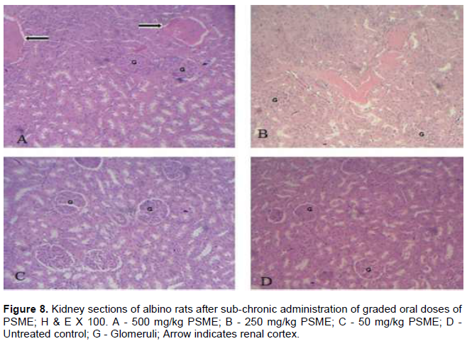
The mean body weight of Groups B and C were significantly (p < 0.05) higher than that of Group D (Figure 2). No outward signs of abnormalities were recorded for each of the rat groups all through the study, and no mortality was recorded in any of the groups all through the study. There were no significant (p > 0.05) differences in relative liver and kidney weights amongst the groups (Figures 3 and 4). However, the relative spleen and heart weights of rats that received PSME at all the doses used in the study [500, 250 and 50 mg/kg (Groups B and C)] respectively, were significantly (p < 0.05) lower than those of rats in the untreated control (Group D) (Figures 5 and 6). Liver section from rats in Group A showed normal architecture of the liver with normal portal vein (PV), hepatic artery (HA) and bile duct (BD). Group B and Group C showed mild random vacoulation of the hepatocytes around the portal vein (PV), while Group D (untreated control) showed normal liver section, showing chords of hepatocytes around the central vein (CV) (Figure 7). Kidney section from Group A rats showed normal glomeruli and renal tubules with moderate hyperaemia of the renal cortex (arrow). Group B rats showed normal glomeruli and renal tubules with hyperaemic area at the center, while Group C and Group D rats showed normal architecture of the kidneys as indicated in normal glomeruli and renal tubules (Figure 8).


DISCUSSION
The results of haematology showed no variations amongst the rat groups in all the haematological parameters assayed. This implies that PSME at the doses used in the study had no significant effect on the hematopoietic system of the rats. The haematopoietic system is very sensitive to toxic compounds and serves as an important index of the physiological and pathological status in both animals and humans (Adeneye et al., 2006; Kulkarni and Veeranjaneyulu, 2012). This finding of no significant effect on haematology is in agreement with reports by Salawu et al. (2008) who also recorded no significant effect of ethanol stem bark extract of Pterocarpus erinaceus (a related plant of the same genus) on all the haematological parameters studied. However, Offor and Ogbugo (2015) reported increases in the levels of haemoglobin and PCV in albino rats treated with ethanol leaf extract of P. santalinoides for two weeks. This elevation in the levels of haemoglobin and PCV after 2 weeks of administration, reported by Offor and Ogbugo (2015) was attributed to an immediate response to PSME administration because elevations in some haematological parameters often occur naturally in response to some medications (Kulkarni and Veeranjaneyulu, 2012).
The higher serum total protein level recorded in Group C (50 mg/kg PSME), and the higher serum albumin level recorded at the doses of 50 mg/kg and 500 mg/kg PSME implied that PSME enhanced total protein and albumin synthesis. This result implied that treatment with PSME especially at 50 mg/kg enhanced hepatic synthetic ability. It is thought that the earlier reported hepatoprotective activity of P. santalinoides methanol leaf extract (Ihedioha et al., 2017, 2019) may partly be due to this enhanced hepatic synthetic ability, as most liver toxicities and disorders are usually associated with poor hepatic synthetic ability (Thapa and Walia, 2007).
The significantly lower serum creatinine levels in the treated groups suggests that treatment with PSME as used in this study enhanced renal clearance of creatinine and thus kidney function. Creatinine, a product of muscle metabolism (Thrall et al., 2012), is freely filtered through the glomeruli, and is neither reabsorbed nor secreted by the renal tubules. It’s concentrations in serum is an index of renal excretory function and is used to diagnose impairment of renal function. Elevation of serum levels of creatinine and urea is known as azotemia and indicates renal failure (Uchino et al., 2012).
The significantly higher body weight recorded in all the PSME-treated groups may be attributed to improved feed conversion efficiency and possibly the presence of some micronutrients, vitamins and phytochemicals in the extract which may have acted as growth promoters. The fact that no sign of morbidity or mortality was recorded in all the treated groups all through the experimental period indicates that PSME was well tolerated by the rats. The findings in this study of no significant variation in relative liver weight between all the PSME-treated groups and the
untreated control suggests that the extract did not cause any adverse effect on the liver, which was also corroborated by it having no significant effect on the serum enzyme markers of hepatocellular (ALT and AST) and hepatobilliary function (ALP). This suggests that sub-chronic administration of PSME is not hepatotoxic. Ihedioha et al. (2017, 2019) had earlier reported that methanol leaf extract of P. santalinoides is hepatoprotective. Also the finding that the relative kidney weight showed no variation between all the PSME-treated groups and the untreated control indicates that sub-chronic administration of PSME as used in the study did not cause any adverse effect on the kidneys. This was corroborated by the significantly lower serum creatinine levels in the treated groups, and the no effect it
had on the serum urea level in albino rats treated with the extract at all the doses used in the study. The relatively lower spleen and heart weights of all the PSME-treated groups is a possible indication that prolonged administration of PSME may cause a decrease in the relative organ percentage of the spleen and heart, though this is not thought to possibly be an adverse effect.
The mild random vacoulation of hepatocytes observed in the PSME-treated groups at the doses of 250 mg/kg (Group B) and 50 mg/kg (Group C) is not thought to be pathologically significant, as it did not concur with the activities and levels of biochemical markers of liver function (ALT, AST and ALP), serum total cholesterol and bilirubin, which were not significantly altered at these doses. This result implies that the mild vacoulation was not significant as to cause a negative impact on the hepatocellular integrity and functional capacity of the liver. The mild to moderate hyperaemia of the glomeruli and renal tubules seen in rats given 500 mg/kg PSME (Group A) and 250 mg/kg PSME (Group B) respectively, however, did not result to higher relative kidney weight and higher serum creatinine and serum urea levels; rather these values were lowered. This suggests that long term (sub-chronic) therapy with PSME at high doses did not cause any significant damage to the kidneys, but may have acted as a nephroprotective agent.
In summary, sub-chronic oral administration of P. santalinoides methanol leaf extract to albino rats at the doses of 500, 250 and 50 mg/kg led to no significant effects on the haematopoietic system and the hepatocellular integrity of the albino rats, it enhanced serum total protein and albumin synthesis, and also enhanced kidney function of the albino rats. The general results of this present study concurs with earlier reports of acute toxicity studies on leaf extracts of P. santalinoides (Anowi et al., 2012; Ihedioha et al., 2017, 2018, 2019; Obi et al., 2019) which showed that leaf extracts of P. santalinoides were acutely safe up to the dose of 5000 mg/kg.
CONCLUSION
Based on the results of this study, it was concluded that sub-chronic oral administration of graded doses of methanol leaf extract of P. santalinoides DC to albino rats is nontoxic and may be safely used for the treatment and management of diseases for which it is effective.
CONFLICT OF INTERESTS
The authors have not declared any conflict of interests.
ACKNOWLEDGMENTS
The authors appreciate the support of the Tertiary Education Trust Fund Institution Based Research (Grant Number: TETFUND/DESS/UNI/NSUKKA/2017/RP/Vol.1/11), and the Biomedical Research Support Unit of the Foundation for Education and Research on Health (FERH), Nsukka, Nigeria.
REFERENCES
|
Acary E, Anshu PS (2008). Herbal preparations. Traditional Medicine Preparations 61:142-170. |
|
|
Adeneye AA, Ajagbonna OP, Adeleke TI, Bello SO (2006). Preliminary toxicity and phytochemical studies of the stem bark aqueous extract of Musanga cecropioides in rats. Journal of Ethnopharmacology 105:374-379. |
|
|
Aderemi FA (2004). Effects of replacement of wheat bran with cassava root unsupplemented with enzyme on the haematology and serum biochemistry of pullet chicks. Tropical Journal of Animal Science 7:147-153. |
|
|
Adesina SK (1982). Studies on the Nigerian herbal medicinal plants. International Journal of Crude Drug Research 20(2):93-100. |
|
|
Adetunji JA (2007). Reviewing Pterocarpus species and their distribution. African Journal of Traditional, Complementary and Alternative Medicine 4(2):23-36. |
|
|
Afolabi KD, Akinsoyinu AO, Olajide R, Akinleye SB (2010). Haematological parameters of the Nigerian local grower chickens fed varying dietary levels of palm kernel cake. Proceedings of the 35th Annual Conference of Nigerian Society for Animal Production, pp. 247. |
|
|
Amaechi NC (2009). Nutritive and Anti-Nutritive Evaluation of wonderful Kola Seeds. Pakistan Journal of Nutrition 8(8):1120-1122. |
|
|
Anowi CF, Okonkwo C, Agbata CA, Ezeokafor E (2012). Preliminary phytochemical screening, evaluation of acute toxicity and antipyretic activity of methanolic extract of Pterocarpus santalinoides (Fabaceae). International Journal of Pharmaceutical and Phytopharmacological Research 1(6):343-346. |
|
|
Asante-Duah K (2002). Public health risk assessment for human exposure to chemicals (illustrated). Kluwer Academic Publishers, Dordrecht, The Netherlands, pp. 234-258. |
|
|
AVMA (2013). AVMA guidelines for the euthanasia of animals: 2013 edition. American Veterinary Medical Association (AVMA), Illinois, USA, pp. 48-50. |
|
|
Ayman MM (2013). Hematological alterations in diabetic rats-role of adipocytokines and effects of citrus flavonoids. Experimental and Clinical Sciences Journal 12:647-657. |
|
|
Babson AL, Greeley SJ, Coleman CM, Philips GE (1966). Phenolphthalein monophosphate as a substrate for serum alkaline phosphatase. Clinical Chemistry 12(18):482-490. |
|
|
Blass KG, Thiebert RJ, Lam LK (1974). A study of the mechanism of the Jaffe reaction. Journal of Clinical Chemistry and Clinical Biochemistry 12:336-343. |
|
|
Bolliger AP, Everds NE (2010). Hematology of laboratory rodents: mouse (Mus musculus) and rat (Rattus norvegicus). In: Weiss DJ and Wardrop KJ (eds), Schalm's Veterinary Hematology, 6th edn. Wiley-Blackwell, Iowa pp. 852-862. |
|
|
Briejer MR, Bosmans JP, Van Dade P, Jurzaak M, Heylen L, Leysen JE, Prins NH, Schuurkes JA (2001). The in vitro pharmacological profile of procalopride, a novel enterokinetic compound. European Journal of Pharmacology 423(1):71-83. |
|
|
Colville J (2002). Blood chemistry. In: Hendrix CM (ed), Laboratory procedures for Veterinary Technicians, 4th edn. Mosby, St Louis pp. 75-103. |
|
|
Enemali MO, Udedi SC, Ubaoji KI, Haruna GS, Augustine E (2019). Modulatory effect of leaf extracts of Pterocarpus santalinoides Hook F. (red sandal wood) on acetamoniphen-induced hepatotoxicity in albino rats. FUW Trends in Science and Technology 4(2):366-371. |
|
|
Eze SO, Cornelius C, Okereke HC (2012). Phytochemical and antimicrobial analyses of the stem bark of Pterocarpus santalinoides (Nturu ukpa). Asian Journal of Natural and Applied Sciences 1(3):26-32. |
|
|
Fawcett JK, Scott JE (1960). A rapid and precise method for the determination of urea. Journal of Clinical Pathology 13:156-159. |
|
|
Higgins T, Beutler E., Doumas BT (2008b). Analytical methodology - Serum bilirubin. In: Burtis CA, Ashwood ER, Bruns DE (eds), Tietz Fundamentals of Clinical Chemistry, 6th edn. Saunders Elsevier, Missouri, pp. 524-525. |
|
|
Higgins T, Beutler E, Doumas BT (2008a). Measurement of haemoglobin in blood. In: Burtis CA, Ashwood ER, Bruns DE (eds), Tietz Fundamentals of Clinical Chemistry, 6th edn. Sanders Elsevier, Missouri, pp. 524-525. |
|
|
Ihedioha TE, Onwuegbuka LU, Ihedioha JI (2017). Hepatoprotective effects of methanol leaf extract of Pterocarpus santalinoides DC on acetaminophen-induced liver damage in albino rats (Rattus norvegicus). Nigerian Journal of Animal Production 44(5):34-39. |
|
|
Ihedioha TE, Asuzu IU, Anaga AO, Ihedioha JI (2019). Hepatoprotective and antioxidant activities of Pterocarpus santalinoides methanol leaf extract. African Journal of Pharmacy and Pharmacology 13(18):359-373. |
|
|
Ihedioha TE, Okechukwu VN, Ihedioha JI (2018). Effects of aqueous leaf infusion of Pterocarpus santalinoides DC on serum lipid profile of guinea pigs (Carvia porcellus). Journal of Complementary Medicine Research 7(2):154-160. |
|
|
Iwu MM (1993). Handbook of African medicinal plants, 1st edn. Chemical Rubber Company Press Incorporation, Boca Raton, U.S.A. pp. 133-135. |
|
|
Johnson AM (2008). Amino acids and proteins. In: Burtis CA, Ashwood ER, Bruns DE (eds), Tietz Fundamentals of Clinical Chemistry, 6th edn. Saunders Elsevier, Missouri pp. 206-316. |
|
|
Keay RWJ (1989). Trees of Nigeria. A revised version of Nigeria trees (1960 - 1964). Keay RWJ, Onochie CFA, Stanfied DP (eds). Claredon Press, Oxford, United Kingdom, p. 476. |
|
|
Klein B, Read PA, Babson AL (1960). Rapid method for the quantitative determination of serum alkaline phosphatase. Clinical Chemistry 6:269-275. |
|
|
Kulkarni YA, Veeranjaneyulu A (2012). Toxicological evaluation of the methanol extract of Gmelina arborea Roxb. bark in mice and rats. Toxicology International 19:2. |
|
|
Lamb EJ, Price CP (2008). Creatinine, urea and uric acid. In: Burtis CA, Ashwood E R, Bruns DE (eds), Tietz Fundamentals of Clinical Chemistry, 6th edn. Saunders Elsevier, Missouri, pp. 363-372. |
|
|
Nowacek JM (2010). Fixation and tissue processing. In: Kumar GL and Kierman JA (eds), Pathology Education Guide: Special Stains and H & E, 2nd edn. Dako, California, North America pp. 141-152. |
|
|
Obi CF, Nzeakor TA, Okpala MI, Ezeh IO, Nwobi LG, Omeje MO, Ezeokonkwo RC (2019). Evaluation of anti-trypanosomal activity of Pterocarpus santalinoides L'Herit ex DC hydroethanol leaf extract in rats experimentally infected with Trypanosoma brucei. Journal of Ethnopharmacology, 243, 112085. |
|
|
Obidah N, Sa'ad UA, Wurochekke AU (2009). Toxic effects of aqueous stem bark extract of Cassia sieberiana on some biochemical parameters in rats. African Journal of Biochemistry Research 3(5):229-231. |
|
|
Odeh IC, Tor-Anyim A (2014). Phytochemical and antimicrobial evaluation of leaf-extracts of Pterocarpus santalinoides. European Journal of Medicinal Plants 4(1):105-110. |
|
|
Offor CE, Ogbugo SO (2015). Effects of ethanol leaf extract of Pterocarpus santalinoides on haematological parameters in rats. Academic Journal of Oral and Dental Medicine 2(2):19-22. |
|
|
Offor CE, Nwankwegu NJ, Ugwu-Okechukwu PC, Aja PM (2015). The effects of ethanol leaf extract of Pterocarpus santalinoides on liver enzymes of albino rats. American-Eurasian Journal of Agriculture and Environmental Sciences 15(5):920-922. |
|
|
Ogan MT (2004). Trees of Nigeria. Journal of Complementary and integrative medicine 123(1):125-129. |
|
|
Ojiako OA, Nwanjo HV (2006). Is Vernonia amygdalina hepatotoxic or hepatoprotective? Response from biochemical and toxicity studies in rats. Journal of African Biotechnology 5:1648-1659. |
|
|
Okwu DE, Ekeke T (2003). Phytochemical screening and medicinal composition of chewing sticks in South Eastern Nigeria. Global Journal of Pure and Applied sciences 9 (1):235-238. |
|
|
Olafedehan CO, Obun AM, Yusuf MK, Adewumi OO, Oladefedehan AO, Awofolaji AO, Adeniji AA (2010). Effects of residual cyanide in processed cassava peal meals on haematological and biochemical indices of growing rabbits. Proceedings of 35 Annual Conference of Nigerian Society for Animal Production, pp. 212-218. |
|
|
Poppenga RH (2007). Herbal medicine: potential for intoxication and interactions with conventional drugs. In: Wynn S, Fougere B (eds.) Veterinary Herbal Medicine. Mosby Inc., Missouri USA pp. 183-207. |
|
|
Reitman S, Frankel S (1957). A colorimetric method for determination of serum glutamic oxaloacetic and glutamic pyruvic transaminases. American Journal of Clinical Pathology 28:56-62. |
|
|
Rifai N, Warnick GR, Remaley AT (2008). Analysis of lipids, lipoproteins and apolipoproteins. In: Burtis CA, Ashwood ER, Bruns DE (eds), Tietz Fundamentals of Clinical Chemistry, 6th edn. Saunders Elsevier, Missouri, pp. 422-427. |
|
|
Sacks DB (2008). Measurement of glucose in body fluids. In: Burtis CA, Ashwood ER, Bruns DE (eds.), Tietz Fundamentals of Clinical Chemistry, 6th edn. Saunders Elsevier, Missouri, pp. 389-393. |
|
|
Salawu OA, Aliyu M, Tijani AY (2008). Haematological studies on the ethanolic stem bark extract of Pterocarpus erinaceus poir (fabaceae). African Journal of Biotechnology 7(9):1212-1215. |
|
|
Sancho E, Cerón JJ, Ferrando MD (2000). Cholinesterase activity and haematological parameters as biomarkers of sub-lethal molinate exposure in Anguilla anguilla. Ecotoxicology Environmental Safe 46:81-86. |
|
|
Schalm OW, Jain NC, Carrol EJ (1975). Veterinary haematology, 3rd edn. Lea and Febieyer, Philadelphia pp. 55 - 77. |
|
|
Searcy RL, Reardon JE, Foreman JA (1967). A new photometric method for serum urea nitrogen determination. American Journal of Medical Technology 33:15-20. |
|
|
Sofowora EA (1985). Medicinal plants and traditional remedies in Africa. University of Ife Press, Nigeria. |
|
|
Subramanion LJ, Zuraini YC, Yee LL, Lachimanan YL, Sreenivasan S (2011). Acute oral toxicity of methanolic seed extract of Cassia fistula in mice. Molecules 16. |
|
|
Thapa BR, Walia A (2007). Liver function tests and their interpretation. Indian Journal of Pediatrics 74:663-671. |
|
|
Thrall MA, Weiser MG (2002). Hematology. In: Hendrix CM (ed), Laboratory Procedures for Veterinary Technicians, 4th edn. Mosby, Missouri pp. 29-74. |
|
|
Thrall MA, Weiser MG, Allison R, Campbell TW (2012). Veterinary Hematology and Clinical Chemistry, 2nd edn. John Wiley and Sons, Wiley-Blackwell, USA pp. 762. |
|
|
Tiwari RH, Sinha M (2010). Veterinary Toxicology. Oxford Book Company, Jaipur, India, pp. 133-174. |
|
|
Uchino S, Bellomo R, Goldsmith D (2012). The meaning of the blood urea nitrogen/creatinine ratio in acute kidney injury. Clinical Kidney Journal 5(2):187-191. |
|
|
Ward JW, Elsea JR (1997). Animal care and use in drug fate and metabolism. Methods and techniques Vol.1, Edward RG, Jean LH (eds), Marcel Dekker, New York pp. 372-390. |
|
|
Winsor L (1994). Tissue processing. In: Woods A and Ellis R (eds), Laboratory histopathology. Churchill Livingstone, New York pp. 4.2-1-4.2-39. |
|
|
Zimmermann M (1983). Ethical guidelines for investigations of experimental pain in conscious animals. Pain 16(2):109-110. |
|
Copyright © 2024 Author(s) retain the copyright of this article.
This article is published under the terms of the Creative Commons Attribution License 4.0


