African Journal of
Pharmacy and Pharmacology

  • Abbreviation: Afr. J. Pharm. Pharmacol.
  • Language: English
  • ISSN: 1996-0816
  • DOI: 10.5897/AJPP
  • Start Year: 2007
  • Published Articles: 2283

Full Length Research Paper

Phenytoin: Is it genotoxic in isolated cultured human lymphocytes without metabolic activation by S9?

Mahmoud A. Naga
  • Mahmoud A. Naga
  • Department of Clinical Pharmacology, Mansoura University, Mansoura, Egypt,
  • Google Scholar
Ahmad Shata
  • Ahmad Shata
  • Department of Clinical Pharmacology, Mansoura University, Mansoura, Egypt,
  • Google Scholar
Hany Ahmed El kattawy
  • Hany Ahmed El kattawy
  • Department of Medical Physiology, Zagazig University, Zagazig, Egypt.
  • Google Scholar


  •  Received: 22 July 2016
  •  Accepted: 28 September 2016
  •  Published: 08 November 2016

 ABSTRACT

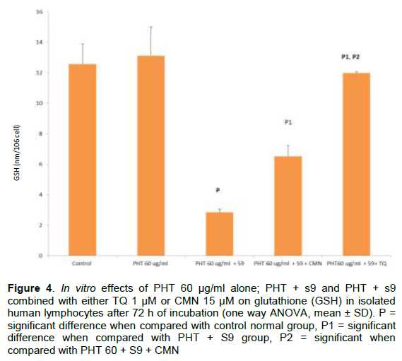
There are many conflicting reports around the phenytoin (PHT)-induced genotoxic effect especially in the in-vitro studies. PHT was claimed to cause genotoxic effect by the oxidative stress of its metabolic intermediates. However, by reviewing the distribution and activity of the enzymes responsible for PHT metabolism, we found that PHT is rarely metabolized by human lymphocytes. So that, we will use isolated cultured human lymphocytes to determine which is genotoxic, PHT itself or its metabolites? PHT 60 μg/ml were added to lymphocytes before and after metabolic activation by S9. Also, this study will investigate the possible antioxidant genoprotective effects of Thymoquinone (TQ) 1 μM and Curcumin (CMN) 15 μM on the chromosomal injury induced by PHT or its metabolites. After the end of culture period, the effects of PHT on the lymphocytes were investigated by measuring levels of chromosomal aberrations (CAs); mitotic index (MI); reduced glutathione (GSH); malondialdehyde (MDA); and 8-hydroxydeoxyguanosine (8-OH-dG). Only PHT after metabolic activation caused oxidative genotoxic effects which were significantly ameliorated by TQ more than CMN. Hence, the present study is the first to record that PHT without metabolic activation in isolated human lymphocytes from non epileptic donors cause dose dependant direct toxic effect rather than genotoxic effect.

Key words: Phenytoin, thymoquinone, curcumin, genotoxic


 INTRODUCTION

Genotoxins are compounds causing chemical or physical alterations in DNA structure leading to inaccurate replication of that region of the genome (Bajpayee et al., 2005). Approximately 30% of all marketed drugs, exhibit genotoxic effect when tested by the standard genetic toxicology tests (Snyder, 2009).
 
Phenytoin (PHT), the well known antiepileptic drug has been suspected for teratogenic and mutagenic effects during pregnancy (Kaul et al., 2001). However, there are many conflicting reports observed around its genotoxic effect especially in the in-vitro studies (NTP, 1993; IARC, 1996; Snyder and Greenb, 2001; Snyder, 2009). PHT was
 
claimed to cause genotoxic effect by the oxidative stress of its metabolic intermediates. However, by reviewing the distribution and activity of the enzymes responsible for PHT metabolism, we found that PHT is rarely metabolized by human lymphocytes (Basta-Kaim et al., 2008).
 
For testing indirect chemical mutagens, human lymphocyte was exposed directly to an Ames-type microsomal (S9) activation system (Sbrana et al., 1984).
 
Many naturally occurring compounds have been reported to have anti-mutagenic activities (El Hamss et al., 2003). Curcumin (CMN), the active constituent in the rhizomes of Curcuma longa, is a nutriceutical compound with antioxidant (Iqbal et al., 2009) and antimutagenic effects (Corona-Rivera et al., 2007). In addition, Thymoquinone (TQ), is another known antioxidant (Badary et al., 2003) have been reported to exert antimutagenic activity in few studies (Badary et al., 2007; Abou Gabal et al., 2007). However, neither TQ nor CMN were tried yet to protect against PHT-induced genotoxicity in isolated human lymphocytes.
 
The aim of the present study is to use isolated cultured human lymphocytes to determine which genotoxic, PHT itself is or its metabolites after metabolic activation by S9? Also, this study will investigate the possible antioxidant genoprotective effects of Thymoquinone and Curcumin on the chromosomal injury induced by either PHT or its metabolites.


 MATERIALS AND METHODS

Human blood samples
 
10 ml fresh venous blood samples were taken from 30 adult donors after consent. All donors were of either sexes between the ages of 20-45 years, apparently healthy, non-smoking, non-alcoholic and they did not take any medications recently. The donors were obtained from the blood-banking Center of Mansoura University Hospital, Mansoura Faculty of Medicine, Egypt. All blood samples were taken on heparin to prevent clotting.
 
Chemicals
 
All chemicals and reagents used in this study were of the highest analytical grade from Sigma-Aldrich (St. Louis, MO, USA). Phenytoin was purchased as sodium salt ≥ 99%, 25 g soluble in water.
 
Isolation and culture of human lymphocytes
 
Lymphocytes were isolated from whole blood samples and cultured as described by Durante et al. (1998) with minor modification. All blood samples were collected in an isolation tube for blood cells. The sample was centrifuged at 1600 g (2900 rpm) for 20 min, and the layer of mononuclear cells and platelets was collected by a pipette and transferred to 10 ml centrifuge tube. PRMI 1640 medium was added up to 10 ml. and the sample was centrifuged at 390 g (1500 rpm) for 10 min. After the removal of the supernatant, the cell pellet was re-suspended in 10 ml RPMI 1640 medium at a density of 1.0 X 10⁶ cells/ml. Isolated lymphocytes from each blood sample were cultured in  10 ml  RPMI  1640  culture  medium  for  a total period of 72 h at 37°C in the dark in a 5% CO2 humidified atmosphere (Watson, 1992).
 
Plan of the study and grouping of isolated lymphocytes
 
Isolated lymphocytes from each blood samples were divided randomly into 5 groups, each of 6 samples: the 1st group was none treated; the 2nd treated with PHT 60 ug/ml (Ponnala et al., 2009); the 3rd treated with PHT 60 ug/ml plus S9 (Sbrana et al., 1984, Ponnala et al., 2009); the 4th treated with PHT 60 ug/ml (Ponnala et al., 2009) + S9 + TQ (Khader et al., 2009); the 5th treated with PHT 60 ug/ml + S9 + CMN (Siddique et al, 2010). The potential genotoxic drugs were added twice, at 24 and 48 h from the start of culture period and after stimulation of mitotic division with phytohaemagglutinin that was added at the start of culture period to induce mitosis within 24 h according to standard protocol of Poddar et al. (2004). The genoprotective drugs were added as a prophylactic therapy 2 h prior to addition of the genotoxic drug.
 
In a pilot study we tried to increase PHT dose above 60 ug/ml (90 & 120), but these doses were very toxic and fatal to isolated lymphocytes as indicated by MI, so that we did not include these fatal doses in this study.
 
Evaluation of the drug effects
 
To investigate the chromosomal effect induced by PHT before and after metabolic activation cells were harvested at the end of the culture period (72 h) for screening CAs following the standard protocol (Carrano and Natarajan 1988) in order to avoid heterogenecity of cycle stage of the treated cells and to score only the first division mitotic cells. Colcemid 0.1 ml was added to stop mitosis and prevent spindle formation and was left 1.5 h. The isolated lymphocytes after recovery from the incubator were investigated for chromosomal aberrations (CAs), mitotic index (MI), 8-hydroxy-2'-deoxyguanosine (8-OH-dG), reduced glutathione (GSH) and malondialdehyde (MDA).
 
Assay of chromosomal aberrations (Karyotype) in isolated lymphocytes using Gimsa stain
 
It was done according to the protocol of Poddar et al. (2004). Cells were stained using 10% Giemsa for 12 minutes immersed in distilled water for washing and air-dried. Analysis of cytogenetic data was performed using light microscopy. Slides were scored blind and individual aberrations were recorded. Fifty metaphases were examined for each sample in the different groups (300 metaphase for each group), searching for any chromosomal anomalies either structurally or numerically.
 
Determination of mitotic index (MI) as a measure of cytotoxicity
 
The mitotic index (MI) was used as indicators of adequate cell proliferation. Its inhibition could be considered as cellular death, or delay in the cell proliferation kinetics (EroÄŸlu, 2011). The mitotic index evaluates the cytotoxicity of chemical agents (Calderón-Ezquerro et al., 2007). MI, is easily assessed when CAs are performed. The number of lymphocytes in metaphase was counted in 2000 lymphocytes per sample to determine the mitotic index (Kannan et al., 2006).
 
Measurement of intracellular reduced glutathione (GSH)
 
Intracellular    reduced    GSH   in   the   isolated   lymphocytes  was extracted according to the method described by Anderson, (1985), and then reduced GSH was measured according to the method described by Beulter et al. (1963) employing colorimetric method using spectrophotometer determination method (JENWAY 6405, spectrophotometer).
 
Measurement of malondialdehyde level (MDA)
 
Lipid peroxidation products (MDA) were released from isolated lymphocytes by sonication according to the method described by Stacey and Klaassen (1981). Then MDA was measured by thiobarbituric acid (TBA) test according to the method described by Draper and Hadley (1990), employing colorimetric method using spectrophotometer determination method (JENWAY 6405, spectrophotometer).
 
Measurement of 8-hydroxy-2-deoxy Guanosine (8-OH-dG)
 
The 8-OH-dG was assayed using Cayman 8-hydroxy-2-deoxy Guanosine enzyme-linked immunosorbent assay (elisa = EIA) Kit (Cayman Chemical’s ACE™, USA). Cayman’s 8-OH-dG EIA is a competitive assay that can be used for the quantification of 8-OH-dG in urine, cell culture, plasma, and other sample matrices. This assay is based on the competition between 8-OH-dG and 8-OH-dG-acetylcholinesterase (AChE) conjugate (8-OH-dG Tracer) for a limited amount of 8-OH-dG Monoclonal Antibody (Pradelles et al., 1985; Maclouf et al., 1987).
 
Statistical analysis of the data
 
All statistical calculations of the data were performed with SPSSÒ
version 21. Multiple comparisons of the data for each biochemical parameter were performed using one-way analysis of variance (ANOVA) followed by post-Hoc test for comparing the different groups with each other. P-value of ≤ 0.05 was considered significant.


 RESULTS

The assessment of the types of chromosomal aberrations (CAs) in the isolated cultured human lymphocytes in all groups of this study caused only structural CAs and no numerical CAs were found.
 
In vitro effects of Phenytoin (PHT) 60 μg/ml: There were insignificant changes in the CAs, MI, MDA, GSH, or 8-OH-dG levels when compared to the non-treated normal group (Figures 1, 2, 3, 4, 5).
 
In vitro effects of Phenytoin (PHT) 60 μg/ml plus S9: They caused significant increase in the structural CAs when compared to control normal group (Figure 2). They caused also cytotoxicity and decrease in lymphocyte proliferation  indicated  by  significant  decrease  in the MI when compared to control normal group (Figure 1). In addition, there was significant increase in the MDA level, 8-OH-dG level and significant decrease in the GSH level when compared to control group (Figures 1, 2, 3, 4, 5).


The effects of combined use of PHT +S9 with Thymoquinone (TQ) or Curcumin (CMN): The combined use of PHT
+S9 with TQ 1 μM, or CMN 15 μM caused significant decrease in the structural CAs when compared to PHT +S9 treated group (Figure 2). It caused also improvement in lymphocyte proliferation indicated by significant increase in the MI when compared to PHT + S9 group (Figure 1). In addition, there was significant decrease in the MDA and 8-OH-dG levels together with significant increase in the GSH level when compared to PHT + S9 group alone (Figures 3, 4, 5). However, the protective effects of PHT + S9 with TQ treated group on CAs, MI, MDA, GSH, and 8-OH-dG were more significant than the protective effects of PHT + S9 with CMN (Figures 1, 2, 3, 4, 5). This means that PHT + S9 -induced toxic changes were significantly ameliorated by TQ more than CMN.


 DISCUSSION

In vitro effects of phenytoin (PHT) and PHT + S9 on the isolated cultured human lymphocytes
 
To the best of our knowledge, this study  was  the  first  to record that PHT is only genotoxic in isolated human lymphocytes after its metabolic activation by S9, and without this activation genotoxicity doesn't occur. This result was collaborated by other studies that reported PHT genotoxic effect after metabolic activation in the presence of an exogenous metabolic activation system (S9) in bacteria (Sezzano et al., 1982) and Chinese hamster ovary cells (Galloway et al., 1987). In addition this result was consistent with some other studies that hold PHT without metabolic activation is not genotoxic. Witczak et al. (2008) assessed the potential genotoxic effect of PHT therapy in pregnancy on DNA of umbilical cord blood lymphocytes using Micronucleus (MN) assay. They did not show any significant differences between the MN rates of PHT-treated patients and controls, indicating a lack of genotoxicity of the PHT. In addition, Schaumann et al. (1990), tested the potential genotoxic effect of PHT using sister chromatid exchange (SCE) assay in isolated cultured lymphocytes from adult epileptic patients treated with PHT. He did not show any significant differences between the SCE rates of PHT-treated patients and controls, indicating a lack of mutagenicity of the PHT.  Also, the negative tests for PHT genotoxicity were observed in germ cells of male Drosophila melunogaster (Woodruff et al., 1985), many strains of Salmonella typhimurium (Leonard et al., 1984), and cultured Chinese hamster ovary cells (Kindig et al., 1992). Large body of evidence supports this notation.
 
But in contrary to our study, there were other studies that    reported     PHT   genotoxicity    without   metabolic activation in Chinese hamster ovary cell (Winn et al., 2003),  rodents  (Kim  et   al.,  1997),  and  some  isolated human cells (Ponnala et al., 2009; Al-Jassabi and Azirun, 2010). This  debate  around  the  ability of  PHT to induce  genotoxic effects was not observed only by our group, but was also observed in many other reports (NTP, 1993; IARC, 1996; Snyder and Greenb, 2001; Snyder, 2009).
 
This debate may be solved if we can understand the mechanism of PHT induced-genotoxic effects. Most of the genotoxic effects of PHT were due to PHT metabolic intermediates not the PHT itself, mainly the para hydroxyphenyl phenyl hydantion (p-HPPH) metabolite (Kaul et al., 2001). These metabolic intermediates induce production of ROS leading to exhaustion of the cellular antioxidant systems (Jacobsen et al., 2008) with subsequent oxidation of DNA, proteins, and lipids (Zegura et al., 2004). This toxic effect will lead to oxidative DNA base modification with DNA strand breaks, lipid peroxidation and decreased GSH-mediated cytoprotection (Al-Jassabi and Azirun, 2010). Our study supports this explanation because when we used PHT without metabolic activation by S9, its genotoxic effect was insignificant. However, when S9 was added to PHT, its genotoxic effect was significant indicating that PHT genotoxic effect could be achieved only after metabolic activation to reactive metabolic intermediates. This conclusion may be true if we were able to prove that the isolated lymphocytes do not contain any of the enzymes responsible for PHT metabolism.
 
There  are   three   main   metabolic   pathways  for  the conversion of PHT to reactive metabolic intermediates. The first include the bioactivation of about 80% of PHT to para hydroxyphenyl phenyl hydantion (p-HPPH) (Soga et al., 2004), a process catalyzed mainly by the CYP2C9 and to much lesser by CYP2C18 and CYP2C19 (Al-Jassabi and Azirun, 2010). The second pathway includes the hydroperoxidase component of Prostaglandin endoperoxide synthetase pathway (Parman et al., 1998). Last pathway included the bioactivation of PHT to reactive free radical intermediates through the myeloperoxidase enzyme commonly present in leukocytes (Mays et al., 1995). However, by reviewing the distribution and activity of the enzymes responsible for PHT metabolism, we found that the CYP450s present in lymphocytes are mainly CYP1A1, CYP1B1, CYP 2E1 and CYP3A4 (Anzenbacher and Anzenbacherová, 2001) while those included in PHT metabolism are CYP2C9, CYP2C18 and CYP2C19 (Al-Jassabi and Azirun, 2010). The prostaglandin endoperoxide synthetase was found in relatively high concentrations in lymphocytes (Dailey and Imming, 1999), but this enzyme can not metabolize PHT without metabolic activation through addition of high amounts of arachidonic acid (Kubow and Wells, 1989). The last enzyme myeloperoxidase was found to be distributed unequally between leucocytes where it is excess in  neutrophils  and  very little in lymphocytes (Tay et al., 1998). These facts about the metabolism of PHT in isolated human lymphocytes support the results of our study and previous studies with similar results and give an alert for contrary studies to reconsider their results specially when used PHT in isolated non liver cells without metabolic activation.
 
Poojan et al. (2015) hold that curcumin combined with selenite in utero may prevent the disruption of Skin Stem Cell through different mechanisms including de novo GSH biosynthesis. In the same direction, Sankar et al. (2014) support protective effects of curcumin on genotoxic effects exerted by Arsenic in bone marrow cells through attenuation of its drawbacks on chromosomal aberrations, micronuclei formation and DNA damage. Tawfik et al. (2013) studied protective effects of curcumin on irradiated mice and found that it had significant radio-protective and radio-recovery activities.
 
Malhotra et al. (2012) reported that combined treatment with curcumin and resveratrol stimulate apoptosis and hence, they modulates mitotic injury in benzo(a)pyrene -treated mice. Curcumin normalize mRNA expression levels and ameliorate progression of diabetic nephropathy (Ibrahim et al., 2016). Nicotinamide phosphoribosyl transferase and sirtuin proteins play crucial roles in threshold of cell death modulation and curcumin can increase their levels, so it can be potentially used to reduce chemotherapy-induced nephrotoxicity (Ugur et al., 2015).
 
Badary et al. (2007) studied daily intake of TQ to rats after and before benzo(a)pyrene exposure, that significantly reduced the frequencies of CAs and damaged cells. Aboul-Ela (2002) also found that TQ can protect against chromosomal aberrations in mouse cells infected with schistosomiasis. El-Sheikh et al. (2016) found that TQ can reverse intestinal microscopic changes induced by methotrexate and improve oxidative/ nitrosative stress, inflammatory and apoptotic markers in intestine. Gökce et al. (2016) stated that TQ improved decreased levels of oxidative products Like MDA and proinflammatory cytokines, and reduce motor neuron apoptosis. Hepatic level of MDA in mice exposed to Aflatoxin B1, was reduced by TQ pre-treatment (Daba et al., 1998).
 
In conclusion, TQ more than CMN can significantly ameliorate oxidative genotoxic effects exerted by PHT. PHT without metabolic activation in isolated human lymphocytes from non-epileptic donors cause dose dependant direct toxic effect rather than genotoxic effect.


 CONFLICT OF INTERESTS

The authors have not declared any conflict of interests.


 REFERENCES

Abou Gabal AA, Essawy AE, Abdel-Moneim AM, Hame SS, Elzergy AA (2007). The protective effect of black seed (Nigella sativa) against carbon tetrachloride-induced chromosomal aberrations and ultrastructural changes of bone marrow cells. Arab J. Biotechnol. 10(2):275-288.


Aboul-Ela EI (2002). Cytogenetic studies on Nigella sativa seeds extract and thymoquinone on mouse cells infected with schistosomiasis using karyotyping. Mutat. Res. 516(1-2):11-7.
Crossref


Al-Jassabi S, Azirun MS (2010). Phenytoin-Induced Hepatic 8-Hydroxy-deoxyguanosine in DNA of Balb/C Mice and Its Reduction by Curcumin. Mohd . Am-Eurasian J. Toxicol. Sci. 2(3):129-133.


Anderson ME (1985). Tissue glutathione. In: Greenwald, R., A., ed. Handbook of methods for oxygen radical research. Boca Raton: CRC Press. pp. 317-323.


Anzenbacher P, Anzenbacherová E (2001). Cytochromes P450 and metabolism of xenobiotics. Cell Mol. Life Sci. 58(5-6):737-47.
Crossref


Badary OA, Abd-Ellah MF, El-Mahdy MA, Salama SA, Hamada FM (2007). Anticlastogenic activity of thymoquinone against benzo(a)pyrene in mice. Food Chem. Toxicol. 45(1):88-92.
Crossref


Badary OA, Taha RA, Gamal el-Din AM, Abdel-Wahab MH (2003). Thymoquinone Is a Potent Superoxide Anion Scavenger. Drug Chem. Toxicol. 26:87-98.
Crossref


Bajpayee M, Pandey AK, Parmar D, Dhawan A (2005). Current Status of Short-Term Tests for Evaluation of Genotoxicity, Mutagenicity, and Carcinogenicity of Environmental Chemicals and NCEs. Toxicol. Mech. Methods 15(3):155-80.
Crossref


Basta-Kaim A, Budziszewska B, Lasoń W (2008). Effects of antiepileptic drugs on immune system. Przegl Lek. 65(11):799-802.


Beulter E, Duron O, Kelly BM (1963). Improved method for the determination of blood glutathione. J. Lab. Clin. Med. 61:882-8.


Calderón-Ezquerro C, Sánchez-Reyes A, Sansores RH, Villalobos-Pietrini R, Amador-Mu-oz O, Guerrero-Guerra C, Calderón-Segura ME, Uribe-Hernández R, Gómez-Arroyo S (2007). Cell proliferation kinetics and genotoxicity in lymphocytes of smokers living in Mexico City. Hum. Exp. Toxicol. 26(9):715-22.
Crossref


Carrano AV, Natarajan AT (1988). International Commission for Protection Against Environmental Mutagens and Carcinogens. ICPEMC publication no. 14. Considerations for population monitoring using cytogenetic techniques. Mutat. Res. 204(3):379-406.
Crossref


Corona-Rivera A, Urbina-Cano P, Bobadilla-Morales L, Vargas-Lares Jde J, Ramirez-Herrera MA, Mendoza-Magaua ML, Troyo-Sanroman R, Diaz-Esquivel P, Corona-Rivera JR (2007). Protective in vivo effect of curcumin on copper genotoxicity evaluated by comet and micronucleus assays. J. Appl. Genet. 48(4):389-96.
Crossref


Daba MH, Abdel-Rahman MS (1998). Hepatoprotective activity of thymoquinone in isolated rat hepatocytes. Toxicol. Lett. 95:23-29.
Crossref


Dailey LA, Imming P (1999). 12-Lipoxygenase: classification, possible therap-eutic benefits from inhibition and inhibitors. Curr. Med. Chem. 6(5):389-98.


Draper W, Hadley M (1990). Indirect determination of oxygen free radicals. Methods Enzymol. 186:421-431.
Crossref


Durante M, Furusawa Y, Gotoh E (1998). A simple method for simultaneous interphase-metaphase chromosome analysis in biodosimetry. Int. J. Radiat. Biol. 74(4):457-62.
Crossref


El Hamss R, Idaomar M, Alonso-Moraga A, Mu-oz Serrano A (2003). Antimutagenic properties of bell and black peppers. Food Chem. Toxicol. 41(1):41-7.
Crossref


El-Sheikh AA, Morsy MA, Hamouda AH (2016). Protective Mechanisms of Thymoquinone on Methotrexate-induced Intestinal Toxicity in Rats. Pharmacogn. Mag. 12(Suppl 1):S76-81.
Crossref


EroÄŸlu HE (2011). The cytogenetic effects of black tea and green tea on cultured human lymphocytes. Braz. Arch. Biol. Technol. 54(6)1159-1165.


Galloway SM, Armstrong MJ, Reuben C, Colman S, Brown B, Cannon C, Bloom AD, Nakamura F, Ahmed M, Duk S, Rimpo J, Margolin BH, Resnick MA, Anderson B, Zeiger E (1987). Chromosome aberrations and sister chromatid exchanges in Chinese hamster ovary cells: Evaluation of 108 chemicals. Environ. Mol. Mutagen. 10(10):1-175.
Crossref


Gökce EC, Kahveci R, Gökce A, Cemil B, Aksoy N, Sargon MF5, Kısa Ü, ErdoÄŸan B, Güvenç Y, Alagöz F, Kahveci O (2016). Neuroprotective effects of thymoquinone against spinal cord ischemia-reperfusion injury by attenuation of inflammation, oxidative stress, and apoptosis. J. Neurosurg. Spine 24(6):949-59.
Crossref


Ibrahim ZS, Alkafafy ME, Ahmed MM, Soliman MM (2016). Renoprotective effect of curcumin against the combined oxidative
Crossref


International Agency for Research on Cancer (IARC) (1996). Some Pharmaceutical Drugs; Phenytoin. IARC Monogr. Eval. Carcinog. Risks Hum. 66:175-237.


Iqbal M, Okazaki Y, Okada S (2009). Curcumin attenuates oxidative damage in animals treated with a renal carcinogen, ferric nitrilotriacetate (Fe-NTA): implications for cancer prevention. Mol. Cell. Biochem. 324(1-2):157-64.
Crossref


Jacobsen NW, Halling-Sorensen B, Birkved FK (2008). Inhibition of human aromatase complex (CYP19) by antiepileptic drugs. Toxicol. In Vitro. 22(1):146-53
Crossref


Kannan TP, Quah BB, Azlina A, Samsudin AR (2006). Mitotic Index and Chromosomal Analyses for Hydroxyapatite Implantation in Rabbits. Arch. Orofacial Sci. 1:15-20.


Kaul A, Kalla NR, Goyle S (2001). The modulatory effect in genotoxic responses due to age and duration of PHT-therapy inepileptic patients. Teratog. Carcinog. Mutagen. 21(2):135-49.
Crossref


Khader M, Bresgen N, Eckl PM (2009). In vitro toxicological properties of thymoquinone. Food Chem. Toxicol. 47(1):129-33.
Crossref


Kim PM, Winn LM, Parman T, Wells PG (1997). UDP-glucuronosyl-transferasemediated protection against in vitro DNA oxidation and micronucleus formation initiated by phenytoin and its embryotoxic metabolite 5-(p-hydroxyphenyl)-5- phenylhydandoin. J. Pharmacol. Exp. Ther. 280:200-209.


Kindig D, Garriott ML, Parton JW, Brunny JD, Beyers JE (1992). Diphenylhydantoin is not genotoxic in a battery of short-term cytogenetic assays. Teratog. Carcinog. Mutagen. 12(1):43-50.
Crossref


Kubow S, Wells PG (1989). In vitro bioactivation of phenytoin to a reactive free radical intermediate by prostaglandin synthetase, horseradish peroxidase, and thyroid peroxidase. Mol. Pharmacol. 35(4):504-11.


Leonard A, de Meester C, Fabry L, de Saint- Georges L, Dumont P (1984). Lack o f muta- genicity of diphenylhydantoin in in vitro short-term tests. Mutat. Res. 137:79-88.
Crossref


Maclouf J, Grassi J, Pradelles P (1987). Development of enzyme-immunoassay techniques for the measurement of eicosanoids: In Walden TL Jr, Hughes HN (eds.), Prostaglandin and Lipid Metabolism in Radiation Injury, Plenum Press, Rockville. pp. 355-364.
Crossref


Malhotra A, Nair P, Dhawan DK (2012). Curcumin and resveratrol in combination modulates benzo(a)pyrene-induced genotoxicity during lung carcinogenesis. Hum Exp Toxicol. 31(12):1199-206.
Crossref


Mays DC, Pawluk LJ, Apseloff G, Davis WB, She ZW, Sagone AL, Gerber N (1995). Metabolism of phenytoin and covalent binding of reactive intermediates in activated humanneutrophils. Biochem. Pharmacol. 50(3):367-80.
Crossref


National Toxicology Program (NTP) (1993). Toxicology and Carcinogenesis Studies of 5,5-Diphenylhydantoin (CAS No. 57-41-0) (Phenytoin) in F344/N Rats and B6C3F1 Mice (Feed Studies). Natl. Toxicol. Program Technol. Rep. Ser. 404:1-303.


Parman T, Chen G, Wells PG (1998). Free radical intermediates of phenytoin and related teratogens. Prostaglandin H synthase-catalyzed bioactivation, electron paramagnetic resonance spectrometry, and photochernical product analysis. J. Biol. Chem. 273(39):25079-88.
Crossref


Poddar S, Talukder G, Sharma A (2004). Chromosome Damage Induced by Ferric Chloride in Human Peripheral Lymphocytes. Int. J. Hum. Genet. 4(4):261-264.


Ponnala S, Rao KP, Chaudhury JR, Ahmed J, Rama Rao B, Kanjilal S, Hasan Q, Das UN (2009). Effect of polyunsaturated fatty acids on diphenyl hydantoin-induced genetic damage in vitro and in vivo. Prostaglandins Leukot Essent Fatty Acids. 80(1):43-50.
Crossref


Poojan S, Kumar S, Verma V, Dhasmana A, Lohani M, Verma MK (2015). Disruption of Skin Stem Cell Homeostasis following Transplacental Arsenicosis; Alleviation by Combined Intake of Selenium and Curcumin. PLoS One 10(12):e0142818.
Crossref


Pradelles P, Grassi J, Maclouf J (1985). Enzyme immunoassays of eicosanoids using acetylcholinesterase as label: An alternative to radioimmunoassay. Anal. Anal. Chem. 57(7):1170-3.
Crossref


Sankar P, Telang AG, Ramya K, Vijayakaran K, Kesavan M, Sarkar SN (2014). Protective action of curcumin and nano-curcumin against arsenic-induced genotoxicity in rats in vivo. Mol. Biol. Rep. 41(11):7413-22.
Crossref


Sbrana I, Zaccaro L, Lascialfari D, Ceccherini I, Loprieno N (1984). Human lymphocytes assay: cyclophosphamide metabolic activation by S9 system with low cytotoxicity. Mutat. Res. 130(6):411-6.
Crossref


Schaumann BA, Winge VB, Pederson M, Kuskowski MA (1990). Comparative effects of phenytoin and/or phenobarbital treatment on sister chromatid exchange. Epilepsia 31:453-457.
Crossref


Sezzano P, Raimondi A, Arboix M, Pantarotto C (1982). Mutagenicity of diphenyl- hydantoin and some of its metabolites towards Salmonella typhinzurium strains. Mutat. Res. 3:219-228.
Crossref


Siddique YH, Ara G, Beg T, Gupta J, Afzal M (2010). Assessment of cell viability, lipid peroxidation and quantification of DNA fragmentation after the treatment of anticancerous drug mitomycin C and curcumin in cultured human blood lymphocytes. Exp. Toxicol. Pathol. 62(5):503-8.
Crossref


Snyder RD (2009). An Update on the Genotoxicity and Carcinogenicity of Marketed Pharmaceuticals with Reference to In Silico Predictivity. Environtl. Mol. Mutagen. 50:435-450.
Crossref


Snyder RD, Greenb JW (2001). A review of the genotoxicity of marketed pharmaceuticals. Mutat. Res. 488:151-169.
Crossref


Soga Y, Nishimura F, Ohtsuka Y, Araki H, Iwamoto Y, Naruishi H, Shiomi N, Kobayashi Y, Takashiba S, Shimizu K, Gomita Y, Oka E (2004). CYP2C polymorphisms, phenytoin metabolism and gingival overgrowth in epileptic subjects. Life Sci. 74(7):827-34.
Crossref


Stacey NH, Klaassen CD (1981). Copper toxicity in isolated rat hepatocytes. Toxicol. Appl. Pharmacol. 58:211-220.
Crossref


Tawfik SS, Abouelella AM, Shahein YE (2013). Curcumin protection activities against γ-rays-induced molecular and biochemical lesions. BMC Res. Notes 6:375.
Crossref


Tay SP, Cheong SK, Hamidah NH, Ainoon O (1998). Flow cytometric analysis of intracellular myeloperoxidase distinguishes lymphocytes, monocytes and granulocytes. Malays. J. Pathol. 20(2):91-4.


Ugur S, Ulu R, Dogukan A, Gurel A, Yigit IP, Gozel N, Aygen B, Ilhan N (2015). The renoprotective effect of curcumin in cisplatin-induced nephrotoxicity. Ren. Fail. 37(2):332-6.
Crossref


Watson RR (1992). In in Vitro Methods of Toxicology. CRC Press. CRC Press, Boca Raton, Ann Arbor, London, Tokyo. pp. 204-216.


Winn LM, Kim PM, Nickoloff JA (2003). Oxidative stress-induced homologous recombination as a novel mechanism for phenytoin-initiated toxicity. J. Pharmacol. Exp. Ther. 306(2):523-7.
Crossref


Witczak M, Ferenc T, Lopaczyńska D, Nowakowska D, Kociszewska I, Wilczyński J (2008). The effect of antiepileptic drugs administered in pregnancy on micronucleus frequency in cordblood lymphocytes. Int. J. Occup. Med. Environ. Health 21(1):67-71.
Crossref


Woodruff RC, Mason JM, Valencia R, Zimmering S (1985). Chemical mutagenesis testing in Drosophila. V. Results o f 53 coded compounds tested for the National Toxicology Program. Environ. Mutagen. 7(5):677-702.
Crossref


Zegura B, Lah TT, Filipic M (2004). The role of reactive oxygen species in microcystin-LR-induced DNA damage. Toxicology 200(1):59-68.
Crossref


          */?>