Full Length Research Paper
ABSTRACT
Bacterial infection cause delayed wound healing, and the rising cases of antibiotic resistance call for alternative solutions. This research aims to determine and compare the antibacterial activity of selected research plants when extracted by different methods using different solvents. The plants were extracted with ethanol, methanol, acetone, water, chloroform, and ethyl acetate using Soxhlet and maceration extraction. Agar well diffusion and resazurin dye reduction method were used to determine the antibacterial activity of the extracts against methicillin-resistant Staphylococcus aureus (MRSA), S. aureus, Enterococcus faecalis, Escherichia coli, Klebsiella pneumoniae, and Pseudomonas aeruginosa. Agar well diffusion results showed that water, ethyl acetate (Soxhlet), and 100% ethanol (macerated) extracts for all 3 plants inhibited a broad spectrum of bacteria. MIC results showed that ethanol and 70% ethanol (macerated) extracts of Hoslundia opposita had the lowest MIC (1.95 mg/ml) against S. aureus. The ethanol extract of H. opposita Vahl showed the lowest MIC against MRSA. Water, ethanol, 100% ethyl acetate (macerated) and 70% ethanol (macerated) extracts of Ageratum conyzoides showed the lowest MIC against E. faecalis. Ethyl acetate extract of A. conyzoides showed the lowest MIC against E. coli. 100% methanol (macerated) extract of H. opposita showed the lowest MICs against K. pneumoniae and P. aeruginosa, respectively; All plants showed better antibacterial activity on gram positive than gram negative bacteria. The methanol extract of H. opposita showed better activity than the rest of the extracts.
Key words: Ageratum conyzoides L., antibacterial activity, Bidens pilosa L., Hoslundia opposita Vahl, wound healing.
INTRODUCTION
A wound is a disruption of cellular and anatomic continuity of tissue caused by physical, microbial, thermal, chemical, or immunological tissue trauma (Maver et al., 2015). The wound healing process is meant to occur naturally but, bacterial infection can fatally disrupt the process (Andreu et al., 2015). A patient with an infected surgical wound is 60 times more likely to be hospitalised in intensive care unit (ICU). In developed countries, surgical site infections account for more than 50% of hospital admissions in intensive care units, and 5 to 15% in regular wards (Lubega et al., 2017). In Europe, out of 10,000 surgeries, 3 to 4% become infected, requiring treatment costing about 2 million euros/year (Andreu et al., 2015). The extent of the problem in developed countries is underrated (Lubega et al., 2017). In Uganda, 10% of the surgeries performed become infected leading to high morbidity and mortality (Seni et al., 2013).
Infection retards the inflammatory phase and synthesis of collagen, obstructs epidermal migration, and causes poor odour and tissue destruction (Andreu et al., 2015). Low levels of bacteria in chronic wounds facilitate the healing process. Nevertheless, when the number of bacteria exceeds a certain limit, infection sets in, and healing is disrupted (Dowsett, 2004; Sood et al., 2014). Infection occurs when there is multiplication and deposition of bacteria accompanied by a host reaction. Furthermore, bacteria in chronic wounds are known to create a biofilm (a protective polysaccharide coating). Biofilms are resistant to most systemic and topical antimicrobial agents (Andreu et al., 2015; Sood et al., 2014) and are rarely recognised by host defenses (Sood et al., 2014).
Some common bacterial pathogens present in infected wounds include Pseudomonas aeruginosa, Staphylococcus aureus, Escherichia coli (Bowler et al., 2001), vancomycin-resistant Enterococcus faecalis (Tripathi et al., 2016), Klebsiella pneumoniae (Effah et al., 2020), and methicillin-resistant S. aureus (MRSA) (Manzuoerh et al., 2019), among others. S. aureus and P. aeruginosa are known to cause infections and form biofilms in chronic wounds. They are opportunistic and easily express destructive virulence factors (Gounani et al., 2020). P. aeruginosa is able to withstand different conditions in wounds and is resistant to various antibiotics. It has the ability to colonise and proliferate, resulting in problematic infection and eventually a high degree of mortality (Al-Azzawi and Abdullah, 2018). Methicillin-resistant S. aureus (MRSA) is the most life-threatening and has been of great concern in the medical field. MRSA is a major cause of nosocomial infections with a high rate of morbidity and mortality in surgical wound infections (Šiširak et al., 2010). E. faecalis is the third most frequently isolated pathogen across all types of wounds and has acquired intrinsic resistance to a variety of antibiotics, which makes treatment difficult (Chong et al., 2017). In Uganda, K. pneumoniae and S. aureus were reported as the leading cause of surgical site infections at Mbarara regional referral hospital (Lubega et al., 2017). While, extended spectrum beta lactamase (ESBL) producing Enterobacteriaceae and MRSA were reported in Mulago Referral Hospital (Seni et al., 2013).
Antibiotic resistance has recently become a great concern in the medical field. Bacteria have developed various defence mechanisms to modify or destroy antimicrobial agents, thus, there is an increasing number of pathogens that are resistant to drugs (Ugboko et al., 2020). S. aureus was reported to show 10 to 60% resistance rate to oxacillin, erythromycin and clindamycin, while E. coli, P. aeruginosa and other Gram-negative bacteria showed less than 25% resistance to imepenem amikacin, and piperacillin-tazobactam. S. aureus and other Gram-positive bacteria showed a resistance rate of 0 to 25% to vancomycin (Seni et al., 2013). The raising resistance rate of bacterial calls for further research in finding potential solutions to wound infection.
Plants have long been used for the treatment of various diseases, especially in developing countries. Various plant extracts are utilised in wound treatment and management because their antimicrobial and healing properties arise from various mechanisms (Maver et al., 2015). They act as antimicrobial agents to prevent infection, and the rate of wound healing depends on the choice of material used (Andreu et al., 2015). Phytochemicals are chemical components found in plants. They occur naturally and possess antimicrobial, antioxidant and anti-inflammatory characteristics that are responsible for wound healing (Shah and Amini-Nik, 2017). These phytochemicals use different me chanisms to fight bacteria, thus, make it hard for bacteria to develop resistance. They are of low cost, and have minimal side effects (Onwa et al., 2016).
Bidens pilosa L. is utilised as a medicinal herb for several years in Africa, Asia, and America and is reported to treat over 40 diseases (Bartolome et al., 2013). It is an erect perennial herb that grows in tropical and temperate regions and has approximately 240 species (Yang, 2014) with potent wound healing properties (Hassan et al., 2011; Kakki et al., 2016). Several studies on this plant have also shown evidence that it possesses ant ulcerative activity against indomethacin-induced lesions (Kumadoh et al., 2021), possesses anti-hyperglycaemic activity (Hsu et al., 2009), anticancer (Shen et al., 2018), antimalarial activity (Laryea and Borquaye, 2019), antidiabetic (Chien et al., 2009), immunoresponsive and anti-inflammatory activity (Wahyuddin et al., 2020), antioxidant activity (Cortés-Rojas et al., 2013) and antileukemia properties (Maher et al., 2021).
Hoslundia opposita Vahl from Lamiaceae is mostly used in Uganda for cleansing the uterus after birth and treating vaginal laceration (Kiguba et al., 2016). It is also utilised in the treatment of herpes, skin diseases, sore throats, microbial infections, and wounds (Annan and Dickson, 2008). The plant has shown evidence as a potential treatment for wound healing (Annan and Dickson, 2008) and other properties, such as antimalarial activity (Tjitraresmi et al., 2020), antidiabetic, analgesic, antimicrobial, anti-inflammatory, acaricidal, and central nervous system activity (Kiguba et al., 2016).
Ageratum conyzoides L. belongs to Asteraceae and consists of approximately 30 species. The plant has been utilised in Africa, Asia and southern America to treat pregnancy disorders and menstrual issues (Ssegawa and Kasenene, 2007). It has been shown to possess wound healing properties (Oladejo et al., 2003; Lestari et al., 2018). Additionally, the plant has been utilized as a purgative, antimicrobial, anti-inflammatory, antidysenteric, and antiulcer and for wound treatment (Yadav et al., 2019).
The antibacterial activity of H. opposita, A. conyzoides, and B. pilosa has been studied elsewhere (Chah et al., 2006; Annan and Dickson, 2008; Mujovo et al., 2008; Ojo and Anibijuwon, 2010; Singh et al., 2013; Odeleye et al., 2014; Garg and Grewal, 2015; Lawal et al., 2015; ?çöz et al., 2016; Agarwal et al., 2016; Njume et al., 2016; Voukeng et al., 2016; Said, 2017; Ogbole et al., 2018; Nakibuule et al., 2019). However, to the best of our knowledge, there is limited research that has investigated and compared the antibacterial activity of the three plants using different solvents and extraction methods against MRSA, S. aureus, E. faecalis, P. aeruginosa, E. coli and K. pneumoniae. The aim of the study was to determine and compare the antibacterial activity of these three plants extracted using different solvents against wound pathogens such as MRSA, S. aureus, E. faecalis, P. aeruginosa, and E. coli and K. pneumoniae. The results of this study will guide the further development of pharmaceuticals from these plants.
MATERIALS AND METHODS
Mueller Hinton Broth, Mueller Hinton Agar, ethanol, methanol, acetone, chloroform, ethyl acetate and distilled water were all obtained from Loba Chemicals (India). Dimethyl sulfoxide (DMSO) (99.7%) was obtained from Acros organics and was used as received. Resazurin dye was gotten from Glentham.
Collection of plants
The aerial (flowers, leaves and stalks) parts of mature plants (B. pilosa, A. conyzoides and H. opposita) were collected from Bbale, Kayunga district in Uganda, in a period between November 2019 and January 2020. Collection was performed during the rainy season between 9:00 and 12:00 EAT. The collected plant parts were placed in loosely woven open mesh bags to allow aeration.
Authentication
The three plants were identified and authenticated by a certified botanist at Makerere University Herbarium and given Voucher numbers: H. opposita 001, B. pilosa 002, A. conyzoides 003.
Plant preparation
Each plant was prepared separately. The aerial parts (leaves, flowers, stalks) of each plant were sorted to remove foreign matter as well as the damaged and sick parts. They were cleaned and air dried overnight. They were placed in an oven at 27 to 30°C until they were crispy dry. The dried plant parts were pulverised using an electric grinder into a fine powder and kept in airtight containers at room temperature for further processing.
Extraction by cold maceration
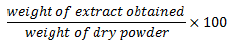
The solvents methanol, acetone and ethanol were used for extraction in absolute form and at 70% concentration. One hundred grams (100 g) of the plant powder was weighed and placed in a stoppered vessel, and 1000 ml of the desired solvent was added to the powder and shaken vigorously. The mixture was left to stand at room temperature for 7 days with frequent agitation. The mixture was strained, filtered through cotton wool, and through Whattman filter paper and pressed. The filtrate was concentrated in an oven at 50°C to remove excess solvent (Singh, 2008). The extract was weighed and the percentage extract yield was calculated as:
All samples extracted by maceration as pure solvents were labeled 100% ETH, 100% MTH, 100% ACTN, 100% E.A and 100% CHF for ethanol, methanol, acetone, ethyl acetate and chloroform, respectively. Those extracted with 70% alcohol were labeled 70% ETH, 70% MTH, and 70% ACTN for ethanol, methanol and acetone, respectively. The water extract was labeled H2O.
Soxhlet extraction
The extraction was done as described by Tandon and Rane (2008) but with modification. 10 g of plant powder was weighed and placed in a thimble in the extraction chamber of the assembled Soxhlet apparatus. 200 ml of solvent was placed in the distillation flask of the apparatus and heated to boiling until extraction was complete. The miscella was concentrated in an oven at 50°C. All extracts were stored in a refrigerator at 4°C for further processing. Soxhlet-extracted samples were labeled ETH SX, MTH SX, ACTN SX, E.A SX and CHF SX for ethanol, methanol, acetone, ethyl acetate and chloroform, respectively.
Phytochemical screening
Each extract was screened for phytochemicals such as flavonoids, saponins (frothing test), terpenoids (Salkowski test), alkaloids (Wagner's test), and phenols (follin reagent) as described by (EL-Kamali and Elshikh, 2015).
Antibacterial susceptibility testing
Standard isolates were obtained from the Microbiology Laboratory at the School of Biomedical Sciences, College of Health Sciences, Makerere University. These included S. aureus (ATCC 25923), K. pneumoniae (ATCC 700603), E. coli (ATCC 25922), MRSA (ATCC 43300), E. faecalis (ATCC 29212), and P. aeruginosa (ATCC 278853).
Agar well diffusion method
Stock solutions of the extracts were prepared by dissolving 0.5 g of extract in 1 ml of DMSO (99.7%). Fresh bacterial cultures (24 h old) were prepared, and the suspension’s turbidity was adjusted to that of a McFarland standard 0.5% by adding fresh colonies to 1 ml of normal saline until the turbidity matched. The standardised suspension had a concentration of approximately 1.5× 10⁸ colony-forming units (cfu) ml.
Standardised bacterial cultures were inoculated using a sterile rod on freshly prepared Mueller-Hinton agar plates to make a lawn. With the aid of a sterile cork borer, 6 mm wells were aseptically punched on the inoculated agar plates, allowing 30 mm between adjacent wells as well as between peripheral wells and the edge of the plate (Mbata et al., 2008).
Fifty microliters of the stock solution of the extract, ciprofloxacin 2 mg/ml (positive control) and DMSO (negative control) were each transferred to the wells made. The plates were incubated at 37°C for 24 h. The zones of inhibition were measured using a divider and a ruler and recorded in mm. The experiment was performed in triplicate for each sample, and the mean inhibition zone was calculated and recorded. Based on the results of agar well diffusion, 8 samples for each plant were chosen to run the MIC experiment.
Measuring MIC using the resazurin reduction method
Fresh cultures (24 h old) were prepared, and the suspension turbidity was adjusted to that of a 0.5% McFarland standard by emulsifying a colony in 1 ml of sterile normal saline. A 1:100 standardised bacterial suspension was made by adding 1 ml of the suspension matched with 0.5% McFarland standard to 99 ml of normal saline to make a 1:100 dilution. First, 0.5 g of extract was weighed and dissolved in 1 ml of DMSO to a concentration of 0.5 g/ml in sterile Bijou tubes.
Determination of MIC was performed according to Teh et al. (2017) but with modification. Fifty microliters of Mueller Hinton broth was dispensed into each well of a sterile 96-well microtiter plate. To the first row of the microtiter plate, 50 µl of each extract to be tested was placed into a separate well of a column leaving out two wells for the positive (ciprofloxacin) and negative (DMSO) controls. The mixture was mixed thoroughly and labelled. Fifty microliters of this mixture in the first row was transferred to the wells in the second row using a sterile multichannel pipette. The mixture in the second well was also mixed thoroughly, and 50 µl was transferred to the third well. This 2-fold dilution was repeated up to the eighth well. Finally, 50 µl of the mixture was removed from the eighth well and discarded. Fifty microliters of ciprofloxacin (0.5 mg/ml) and 50 µl of DMSO were placed in the 11 and 12th columns and serially diluted 2-fold. Twenty microliters of the standardised bacterial suspension was transferred to each of the wells and mixed thoroughly. The microtiter plates were covered and incubated at 37°C for 24 h.
After incubation, 20 µl of 0.015% w/v resazurin salt was added to each well, incubated at 37°C for 4 h and observed for colour change from purple to pink (Purple to pink indicates microbial reduction of the dye). To determine the MIC, the dilution of the last well showing no color change was multiplied by the original concentration of the extract, that is, lowest dilution with no color change × original concentration of the extract.
Statistical analysis
The experimental results of the inhibition zones were expressed as the means and standard deviations of 3 replicates. This analysis was done in Microsoft Excel 2013. Further analysis was done in Minitab-17 software to determine the difference in means using One-way analysis of variance at a 95% confidence interval. P values less than 0.05 were considered significantly different. A post hoc analysis where applicable was performed using Tukey pairwise comparisons.
RESULTS
Plant extraction yield
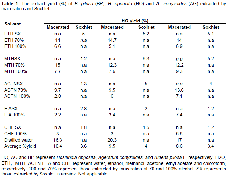
H. opposita, A. conyzoides, and B. pilosa, were extracted by Soxhlet and cold maceration using different solvents. The percentage yield for the Soxhlet and maceration extraction methods is shown in Table 1. Macerated samples showed better yield than Soxhlet samples. Water extracts had a higher yield than the rest of the solvents. Hydroalcoholic solvents (70% each of ethanol, methanol and acetone) had higher extract yields than the corresponding alcohols that were used at 100% concentration. For Soxhlet extraction, A. conyzoides had the highest yield (4%), followed by H. opposita (3.6%) and B. pilosa (3.4%). For maceration, H. opposita had the highest yield (10.4%), followed by A. conyzoides (9.5%) and B. pilosa (8.6%).
Qualitative phytochemical analysis
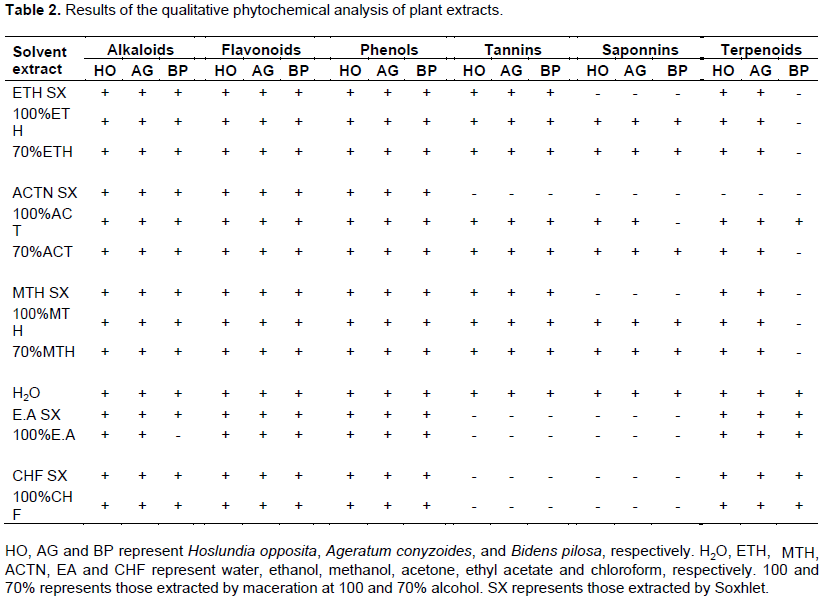
The results from the phytochemical analysis are shown in Table 2. Results showed the presence of most of the phytochemicals tested. All extracts showed the presence of alkaloids, flavonoids and phenols. Tannins and saponnins were mostly absent in ethyl acetate and chloroform extracts. Most Sohxlet extracts did not show the presence of saponnins. Terpenoids were more frequent in H. opposita and A. conyzoides extracts compared to B. pilosa.
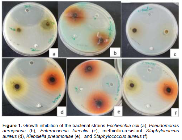
Antibacterial activity by agar well diffusion
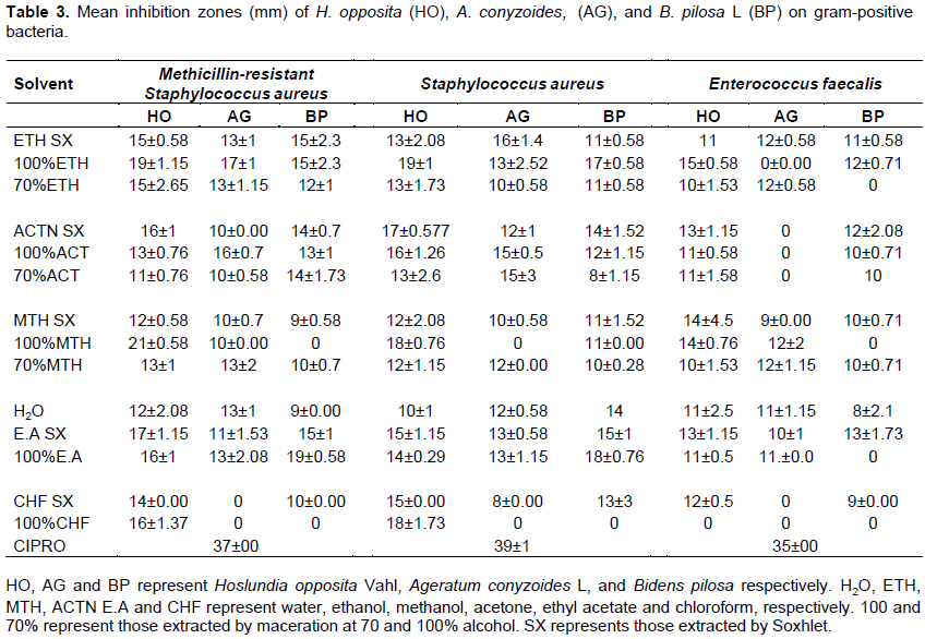
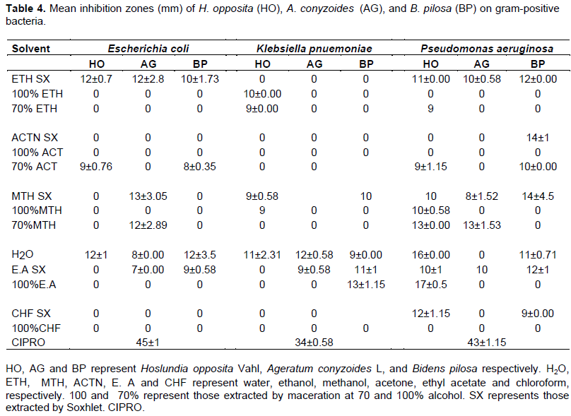
The plant extracts were screened for antibacterial activity against gram-positive and -negative bacteria using the agar well diffusion method, and the results are shown in Tables 3 and 4 as well as Figure 1.
The negative control DMSO did not inhibit the growth of any of the organisms. The positive control ciprofloxacin showed significantly higher inhibition zones above 30 mm for all organisms. The water extract of H. opposita, ethyl acetate (Soxhlet) extract of A. conyzoides as well as the water and ethyl acetate (Soxhlet) extracts of B. pilosa showed some clearance on all organisms.
MRSA was the most susceptible microorganism to most of the plant extracts. All extracts of H. opposita inhibited the growth of this organism. For B. pilosa and A. conyzoides, 12 out of 14 extracts showed clearance on MRSA. H. opposita extracts showed that the largest inhibition zone for MRSA was 21±0.58 mm by the 100% methanol extract (macerated), and the smallest was 13±1 mm by 70% acetone (macerated) and 70% methanol (macerated). For B. pilosa extracts, the largest inhibition zone was 19±0.58 mm by 100% ethyl acetate (macerated), and the smallest was 9±0.00 mm by distilled water extract. A. conyzoides showed the largest inhibition zone of 17±1 mm on MRSA by 100% ethanol (macerated) and the smallest 10±0.00 mm by 70% acetone (macerated) and 70% methanol (macerated) extracts.
The activity of the plant extracts against S. aureus was not significantly different from that against MRSA (P value=1.000). All H. opposita extracts inhibited this organism with the largest inhibition zone of 19±1.15 mm by 100% ethanol (macerated) and the smallest 10±1 mm by the distilled water extract. The largest inhibition zone for B. pilosa extracts was 18±0.76 mm by 100% ethyl acetate extract (macerated), and the smallest 8±1.15 mm by 70% acetone (macerated). For A. conyzoides, the largest inhibition zone was 16±1.4 mm by ethanol (Soxhlet), and the smallest, 8±0.00 mm by chloroform (Soxhlet).
E. faecalis was the least inhibited Gram-positive microorganism. All Hoslundia extracts except 100% chloroform (macerated) showed clearance of this bacterium, where the largest inhibition zone was 15±0.58 mm by 100% ethanol (macerated) and the smallest was 10±1.53 mm by 70% methanol (macerated). For B. pilosa, 10 out of 14 extracts showed clearance of the bacteria. The largest inhibition zone was 13±1.73 mm by ethyl acetate (Soxhlet), and the smallest was 8±2.1 mm by distilled water. Eight out of 14 extracts of Ageratum inhibited E. faecalis, where the largest inhibition zone was 12±0.71 mm by ethanol (Soxhlet), 70% ethanol (macerated), 100% methanol (macerated) and 70% methanol (macerated), while the smallest was 9±0.00 mm by methanol (Soxhlet).
P. aeruginosa was the most susceptible Gram-negative organism. Ten out of 15 extracts of H. opposita inhibited P. aeruginosa. The largest inhibition zone was 17±0.5 mm by ethyl acetate (macerated) extract, and the smallest was 9±1.15 mm by 70% ethanol (macerated) and 70% acetone (macerated). For B. pilosa, 7 out of 14 extracts showed activity on the organism. The largest inhibition zone was 14±4.5 mm by acetone (Soxhlet) and methanol (Soxhlet), and the smallest was 9±0.00 mm by the chloroform (Soxhlet) extract. For A. conyzoides, 4 out of 14 extracts inhibited the organism. The largest was 13±1.53 mm by 70% methanol (macerated), and the smallest was 8±1.52 mm by the ethyl acetate (Soxhlet) extract.
E. coli was quite resistant to most of the extracts, such that 5/14 extracts of H. opposita and B. pilosa showed some clearance of the organism, while for A. conyzoides, 4/14 extracts showed inhibition. The largest inhibition zone achieved for H. opposita was 12 mm by distilled water and ethanol (Soxhlet) extracts, and the lowest was 9±0.76 mm by 70% acetone (macerated). For B. pilosa, the largest was 12±3.5 mm by distilled water extract, and the lowest was 8±0.35 mm by 70% acetone (macerated). A. conyzoides extracts showed the largest inhibition zone of 13±0.35 mm by methanol (Soxhlet) and the smallest 7±0.00 mm by ethyl acetate (Soxhlet). K. pneumoniae was the most resistant microorganism, where 3 extracts of H. opposita, 4 extracts of B. pilosa L, and only 2 extracts of A. conyzoides showed inhibition. The largest inhibition zone obtained for H. opposita was 11±2.31 mm by distilled water extract, and the smallest was 9 mm by methanol (Soxhlet) and 70% acetone (macerated). For B. pilosa, the largest inhibition zone was 13±1.15 mm by 100% ethyl acetate (macerated), and the smallest was 9 mm by distilled water. For A. conyzoides, the largest was 12±0.58 mm by distilled water and 9±0.58 mm by ethyl acetate (Soxhlet).
Generally, the overall average inhibition zone of Gram-positive bacteria was significantly higher (P>0.001) than that of Gram-negative bacteria. The inhibition zones of MRSA and S. aureus were not significantly different (P=1.000) and these organism were the most susceptible to the plant extracts. H. opposita showed a significantly higher inhibition zone (p=0.003) than A. conyzoides. However, the overall inhibition zones for B. pilosa L and A. conyzoides, as well as H. opposita and B. pilosa, were not significantly different, with P values of 0.625 and 0.051, respectively.
The solvents that showed higher inhibition zones for all plant extracts were ethyl acetate (Soxhlet), water and ethanol (Soxhlet), with overall average inhibition zones (calculated across all three plants) of 10.8, 10.6 and 10.2 mm, respectively.
The Soxhlet-extracted samples generally showed a significantly higher average inhibition zone (p=0.03) than those extracted by maceration. However, the mean inhibition zones of extracts from the same type of solvent extracted by maceration or Soxhlet were not significantly different.
Determining the minimum inhibitory concentration (MIC)
From the first tests on antibacterial activity using agar well diffusion, 8 extracts of each plant were chosen according to their activity on a broad spectrum of bacteria by calculating the average inhibition zone obtained across the whole spectrum of bacteria tested. Their MIC was determined using the resazurin reduction method. The color change from purple to pink was observed. The change is an indication of the presence of active living bacterial cells that reduce resazurin (purple-blue) to resofurin (pink-colorless) (Elshikh et al., 2016). The MIC results are as shown in Figures 2 to 5.
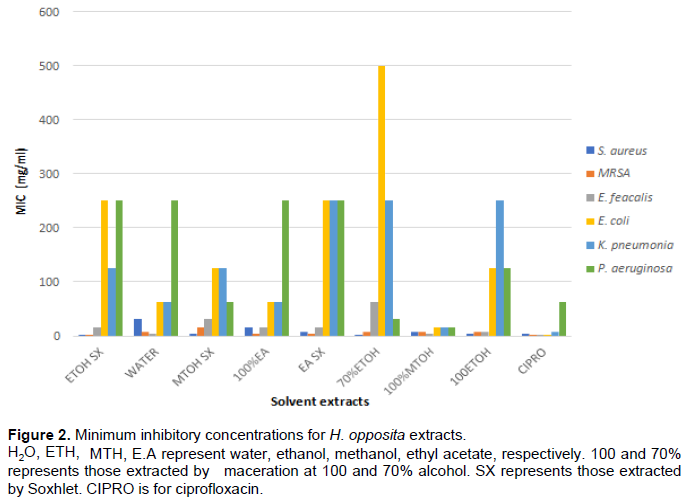
Gram-positive bacteria were greatly affected by the plant extracts, with MICs ranging from 1.95 to 62.5 mg/ml (Figures 2 to 4). S. aureus was mostly susceptible to ethanol (Soxhlet) and 70% ethanol extracts of H. opposita, with the lowest MIC of 1.95 mg/ml. This was half as much as that of ciprofloxacin (3.9 mg/ml). Methanol (Soxhlet), 100% ethanol extracts of H. opposita, and 100% ethyl acetate and ethyl acetate (Soxhlet) extracts of B. pilosa showed MICs of 3.9 mg/ml for S. aureus, similar to that of ciprofloxacin. MRSA was most affected by the ethanol (Soxhlet) extract of H. opposita, with the lowest MIC of 1.95 mg/ml, similar to that of ciprofloxacin.
E. faecalis was mostly susceptible to ethanol (Soxhlet), water, 100% ethyl acetate, and 70% ethanol extracts of A. conyzoides, with a MIC of 1.95 mg/ml, which was also similar to that of ciprofloxacin. Generally, as shown in Figure 5, S. aureus and MRSA were more susceptible than E. faecalis; however, their MICs were not sig-nificantly different (P=0.688). S. aureus and MRSA were mostly affected by H. opposita, with average MICs of 8.68 and 6.5 mg/ml, respectively, while E. faecalis was affected by A. conyzoides, with an average MIC of 11.81 mg/ml.
Gram-negative bacteria were less susceptible to the plant extracts, and their average MICs were not significantly different (0.648). E. coli was more susceptible to ethyl acetate (Soxhlet) extract of A. conyzoides, with 12.5 mg/ml MIC. K. pneumoniae and P. aeruginosa were more susceptible to the 100% methanol (macerated) extract of H. opposita, with MICs of 15.6 and 7.8 mg/ml, respectively. As shown in Figure 5, A. conyzoides extracts showed better activity on gram-negative bacteria, followed by H. opposita and B. pilosa. However, only the average MICs of A. conyzoides L and B. pilosa were significantly different (P<0.000). There was no significant difference between the average MICs of A. conyzoides and H. opposita (P=0.411) or H. opposita and B. pilosa (P=0.162). However, the average MICs of A. conyzoides were significantly lower than those of B. pilosa L. (P=0.006).
Generally, H. opposita extracts showed a higher antibacterial activity against Gram-positive bacteria, especially S. aureus and MRSA, compared to the rest of the plants, while A. conyzoides extracts showed a higher antibacterial activity against Gram-negative bacteria. Similar to the results of the inhibition zones, the Gram-positive bacteria were significantly more susceptible to all the plant extracts than the Gram-negative bacteria (P<0.000). The lowest overall average MIC (calculated across all the bacteria) for H. opposita Vahl was 11.05 mg/ml by 100% methanol (macerated) extract. For A. conyzoides L., it was 25.5 mg/ml by ethyl acetate (Soxhlet) extract, and for B. pilosa L, it was 31.2 mg/ml by the ethanol (Soxhlet) extract. Therefore, the most effective extract was the 100% methanol (macerated) extract of H. opposita.
DISCUSSION
Extraction yield and phytochemical analysis
The difference in the yield obtained using different solvents can be attributed to the polarity of the solvent. Water is more polar than the rest of the solvents, which explains the higher yield obtained by the water extracts. The higher yield of hydroalcoholic solvents compared to pure alcoholic solvents could also be attributed to polarity because the addition of water increased the polarity of the solvent. Maceration gave a better yield than Soxhlet. This was expected since the former took a longer period of extraction (7 days). When the plant is in contact with the solvent for a longer period, it allows more diffusion of active compounds from the plant powder and thus a higher yield (Paz et al., 2018). The phytochemical analysis showed presence of flavonoids, tannins, and terpenoids in both Soxhlet and macerated. These compounds are responsible for the antibacterial activity of plants.
Antibacterial activity
From the results, it was observed that the water extracts of all plants inhibited at least 3 or more bacteria, and their inhibition zones and MICs were better than some of the solvent extracts. In traditional medicine, water is the main extractant for these plants during deconcoction (Ssegawa and Kasenene, 2007). The antibacterial activity of all the water extracts against a broad spectrum of bacteria justifies their use in traditional medicine to treat wounds and other bacterial infections.
H. opposita Vahl has been used in the treatment of several diseases, and its pharmacological properties have been explored (Said, 2017). In this study, the distilled water extract of this plant showed clearance of all organisms tested. A study by Ojo and Anibijuwon (2010) also found that aqueous extracts of H. opposita Vahl were able to inhibit all the organisms tested in this study (MRSA and E. faecalis were not tested). Furthermore, the results in this study also agree with other studies that have evaluated the antibacterial activity of this plant with other solvents. For example, methanol extracts of H. opposita showed clearance of S. aureus and K. pnuemoniea (Maregesi et al., 2008).
Ethanol extracts of the same plant were found to inhibit the growth of S. aureus and E. faecalis (Atindehou et al., 2002). However, Ogbole et al. (2018) reported that methanol extracts (macerated) of H. opposita, from Nigeria did not show activity against MRSA and P. aeruginosa. Our results show that all methanol extracts (Soxhlet and macerated) of H. opposita Vahl had activity on both organisms, especially the 100% methanol (macerated). The difference in these results could be attributed to the difference in geographical location and environmental conditions, which may cause variation in secondary metabolites that are responsible for the antibacterial activity of the plant.
B. pilosa L. has been greatly explored in research for its pharmacological activities (Bartolome et al., 2013). The results obtained for extracts from this plant also correlate with other studies elsewhere. For example, in other similar studies, solvent extracts such as acetone (Adedapo et al., 2011; Shandukani et al., 2018), water (Khan et al., 2001; Adedapo et al., 2011; Silva et al., 2014; Lawal et al., 2015; ?çö z et al., 2016), ethanol (?çöz et al., 2016), methanol (Ukwubile et al., 2014), and chloroform (Owoyemi and Oladunmoye, 2017) have been found to inhibit S. aureus. Ethanol (?çöz et al., 2016) and 70% ethanol (Silva et al., 2014) extracts were found to inhibit MRSA. Methanol (Njume et al., 2016), ethanol (Khan et al., 2001; ?çöz et al., 2016) and water (Lawal et al., 2015) extracts were found to inhibit E. faecalis. Ethyl acetate, acetone and methanol extracts showed inhibition against K. pneumoniae. Ethanol (Khan et al., 2001), ethyl acetate, methanol, and acetone (Shandukani et al., 2018) extracts of B. pilosa L. have been found to have activity against E. coli. These results agree with those in our study. In contrast, ?çöz et al. (2016) and Silva et al. (2014) reported that aqueous extracts of B. pilosa L showed no activity against MRSA, while in our study, aqueous extracts showed moderate activity against this organism.
A. conyzoids L has been used to treat wounds as well as other illnesses, and its pharmacological properties have also been researched (Okunade, 2002; Singh et al., 2013). Several studies have investigated the activity of this plant, and our results are in agreement with most of them. For example, 90% methanol (Soxhlet) (Neelabh et al., 2017), 80% ethanol (Agarwal et al., 2016) and water (Jagarlamudi and Kumar, 2017) showed activity against S. aureus. Elsewhere, 80% ethanol extracts (Voukeng et al., 2016), 40% ethanol and water (Akinyemi et al., 2005) extracts of A. conyzoides L showed inhibition against MRSA. Eighty percent ethanol has been shown to inhibit various VRE strains of E. faecalis (Agarwal et al., 2016). E. coli and P. aeruginosa were fairly affected by A. conyzoides L. extracts. Previous studies have found that methanol (Garg and Grewal, 2015; Jagarlamudi and Kumar, 2016; Neelabh et al., 2017), ethyl acetate, water and hydroethanolic (Jagarlamudi and Kumar, 2016), chloroform, acetone (Garg and Grewal, 2015) and ethanol (Odeleye et al., 2014) extracts of A. conyzoides showed clearance of E. coli and P. aeruginosa. This was true for ethyl acetate, ethanol and methanol in our study but not acetone and chloroform. The contradictions in most of the studies could be attributed to the differences in extraction methods, geographical conditions, and environmental conditions. All these are reported to affect the quality and composition of the active components (Ncube et al., 2008) responsible for antibacterial activity.
Minimum inhibitory concentration
The MIC results were determined using the rezasurin reduction method. This method is recommended to quantify bacteria because it enables direct reading and the rezasurin indicator does not easily precipitate when reduction takes place (Cos et al., 2006). The color change from purple to pink that was observed is due to the presence of active living bacterial cells that reduce resazurin (purple-blue) to resofurin (pink-colorless) (Elshikh et al., 2016).
S. aureus has been reported to be a danger in surgical and burn wounds (Bowler et al., 2001). It has been globally perceived to be the number one cause of infections (Šiširak et al., 2010) and is fond of forming biofilms in chronic wounds (Fazli et al., 2009). In this study, 70% ethanol, methanol (Soxhlet), 100% methanol, and 100% ethanol extracts of H. opposita, as well as 100% ethyl acetate and ethyl acetate (Soxhlet) extract of B. pilosa, showed MICs between 1.95 and 3.9 mg/ml against this organism. This is an indication that these extracts contain compounds that are able to prevent or treat S. aureus infection in wounds. Moreover, MRSA is reported to be a resistant strain and a cause of mortality and morbidity in patients with infected surgical wounds (Šiširak et al., 2010). Our study found that MRSA, similar to S. aureus, was highly susceptible to all the plant extracts compared to other organisms. The organism was more susceptible to H. opposita, showing that compounds in these plants can be potential antibiotics against MRSA.
Vancomycin-resistant E. faecalis is the third most frequently isolated microorganism in infected surgical wounds and other types of infected wounds. It is resistant to a number of antibiotics, and is difficult to treat (Tripathi et al., 2016). In this study, E. faecalis was more susceptible to A. conyzoides extracts than other plants, where most of the extracts showed an MIC of 1.95 mg/ml. This is an indication that A. conyzoides has compounds that are likely to treat such a resistant strain.
P. aeruginosa is known worldwide for causing infection in chronic wounds and is involved in biofilm formation (Fazli et al., 2009). This organism was fairly resistant to a number of extracts. The 100% methanol (macerated) extract of H. opposite showed the most activity against this organism. Thus, compounds isolated from this extract could act as potential antibiotics against P. aeruginosa.
E. coli and K. pneumoniae were quite resistant to most of the extracts. E. coli isolates found in wounds have many virulence factors and have been reported to be resistant to drugs such as ampicillin, fluoroquinolones and tetracycline (Alharbi et al., 2019). K. pneumoniae isolates are responsible for one-third of all infections caused by Gram-negative bacteria, including wound infections (Effah et al., 2020). In this study, the 100% methanol extract (macerated) of H. opposita fairly inhibited the growth of these organisms with MIC of 7.8 mg/ml. This gives hope that further processing of compounds from this extract or making combinations with other antibiotics could lead to better results.
Generally, the activity of Gram-positive bacteria was significantly higher (P>0.001) than that of Gram-negative bacteria. This has also been reported in other similar studies (Owoyemi and Oladunmoye, 2017; Nakibuule et al., 2019). Gram-negative bacteria have a complex cell wall structure compared to that of Gram-positive bacteria. The structure consists of various proteins, polysaccharides and lipids. Additionally, the cell wall consists of an outer membrane hardly separated from it by a periplasmic space. The latter contains periplasm, which has been reported to contain bacterial enzymes responsible for destroying antibacterial agents before they can distort the cell membrane (Annan and Dickson, 2008).
The results from the agar well diffusion method did not necessarily correlate with the MIC results of some extracts, especially A. conyzoides. This difference could be attributed to the polarity of the bioactive compounds contained in the plants. It is stated that nonpolar com-pounds may not easily diffuse through agar (Eloff, 2019). Additionally, the difference in volatility, solubility and diffusion properties of the media could affect the results (Cos et al., 2006). This could explain the high activity A. conyzoides in regard to the MIC results, especially on E. faecalis and on all Gram-negative organisms, compared to the results of the inhibition zones.
From this study, it is not clear whether Soxhlet extraction gives better antibacterial activity compared to maceration because the extracts from the same solvent and plant but different extraction methods did not show significantly different antibacterial activity. In other studies, Paz et al. (2018) reported no significant difference in the antibacterial activity of 70% ethanol extracts of Hamelia patens extracted by maceration, percolation and Soxhlet extraction. However, Sankeshwari et al. (2018) reported that the antibacterial activity of cold macerated extracts of liquorice root were significantly different from those obtained by Soxhlet.
Therefore, it could be that the antibacterial activity may depend more on the type of solvent and type of plant rather than the extraction method. But, this may require further investigation.
It was also shown that the type of solvent greatly affected the antibacterial activity of the plant, as seen by the different values of the inhibition zone and MIC obtained by different solvent extracts. This could be because the quantity and composition of active com-pounds depend on the nature, polarity and concentration of the solvent (Ncube et al., 2008). In this study, ethanol, ethyl acetate, methanol and water were better solvents since most of their extracts had activity on a broad spectrum of bacteria for all three plants. Acetone seemed to be a better solvent for B. pilosa than for the other two plants. The best extract was the 100% methanol (macerated) extract of H. opposita.
The antibacterial activity of these plants can be attributed to the presence of active compounds such as phenols, flavonoids, terpenoids, sponnins, and tannins shown in Table 2. Their presence in these plants has already been investigated (Ajanaku et al., 2021; Jagarlamudi and Kumar, 2017; Odeleye et al., 2014; Okach et al., 2013; Owoyemi and Oladunmoye, 2017), and our results have confirmed the same. Their mode of action on microbes is linked to destruction of the microbial cell membrane, binding directly to the cell wall, interfering with the cell membrane and altering its integrity. They also synthesize microbial nucleic acids and inhibit multidrug resistant (MDR) pumps and microbial enzymes (Reichling, 2010). Flavonoids derive their antimicrobial activity by forming complexes with bacterial membranes and with soluble and extracellular proteins (Alihosseini, 2016). It is no wonder that they are synthesized by plants to counteract infection by microorganisms (Ncube et al., 2008). Their antimicrobial action is derived from their ability to form complexes with proteins via covalent bonding, hydrophobic effects and hydrogen bonding (Alihosseini, 2016). Most of the cholesterol-free gram-negative bacteria have their outer membrane covered by lipopolysaccharide (LPS); thus, it is claimed that saponins increase cell wall permeability by interacting with LPS (Arabski et al., 2012).
Therefore, the study confirms that these plants can be used in the further development of antibiotics for wound infections.
CONCLUSION
The objective of this study was to determine and compare the antibacterial activities of H. opposita, B. pilosa, and A. conyzoides against some common wound pathogens. We found that the type of solvent greatly affected the antibacterial activity of the plant. Ethanol, ethyl acetate, methanol and water were better solvents than acetone and chloroform. Gram-positive bacteria were more susceptible to the plant extracts than gram-negative bacteria. MRSA and S. aureus were the most affected organisms, while K. pneumoniae and E. coli were the least affected.
The antibacterial activity of these plants can be attributed to the presence of secondary metabolites shown in the phytochemical analysis. H. opposita showed better antibacterial activity against Gram-positive bacteria while A. conyzoides showed higher antibacterial activity against all Gram-negative bacteria than other plants. The best extracts from each plant were the 100% methanol (macerated) extract of H. opposita, followed by the ethyl acetate (Soxhlet) extract of A. conyzoides and the ethanol (Soxhlet) extract of B. pilosa. The overall most effective extract was the 100% methanol (macerated) extract of H. opposite. Since all the plant extracts had very good activity in one way or another, a combination of compounds isolated from these plants or with other antibiotics could produce syner-gistic action against a number of organisms. Antibiotic resistance is a great concern in the medical field. The research findings will guide the further development of pharmaceuticals from these plants as one step to combat the issue.
CONFLICT OF INTERESTS
The authors have not declared any conflict of interests.
ACKNOWLEDGMENTS
The authors are grateful for the funding from World Bank through Makerere University Africa Centre of Excellence in Materials Product Development and Nanotechnology.
REFERENCES
|
Adedapo A, Jimoh F, Afolayan A (2011). Comparison of the nutritive value and biological activities of the acetone, methanol and water extracts of the leaves of Bidens pilosa and Chenopodium album. Acta Poloniae Pharmaceutica- Drug research 68(1):83-92. |
|
|
Agarwal P, Agarwal N, Gupta R, Gupta M, Sharma B (2016). Antibacterial activity of plants extracts against methicillin-resistant Staphylococcus aureus and vancomycin-resistant Enterococcus faecalis. Journal of Microbial and Biochemical Technology 8(5):404-407. |
|
|
Ajanaku C, Echeme J, Mordi R, Bolade O, Okoye S, Jonathan H, Ejilude O (2021). In-vitro antibacterial, phytochemical, antimycobacterial activities and GC-MS analyses of Bidens pilosa leaf extract. Journal of Microbiology, Biotechnology and Food Sciences 721-725. |
|
|
Akinyemi KO, Oladapo O, Okwara CE, Ibe CC, Fasure KA (2005). Screening of crude extracts of six medicinal plants used in South-West Nigerian unorthodox medicine for anti-methicillin resistant Staphylococcus aureus activity. BMC Complementary and Alternative Medicine 5(1):1-7. |
|
|
Al-Azzawi SNA, Abdullah RM (2018). Study of the resistance of P. aeruginosa isolated from wounds and burns for some disinfects and antiseptic from some baghdad hospitals. Journal of Pharmaceutical Sciences and Research 10(6):1481-1484. |
|
|
Alharbi NS, Khaled JM, Kadaikunnan S, Alobaidi AS, Sharafaddin AH, Alyahya SA, Almanaa TN,Alsughayier MA, Shehu MR (2019). Prevalence of Escherichia coli strains resistance to antibiotics in wound infections and raw milk. Saudi Journal of Biological Sciences 26(7):1557-1562. |
|
|
Alihosseini F (2016). Plant-based compounds for antimicrobial textiles. In Antimicrobial Textiles (pp.155-195). Elsevier. |
|
|
Andreu V, Mendoza G, Arruebo M, Irusta S (2015). Smart dressings based on nanostructured fibers containing natural origin antimicrobial, anti-inflammatory, and regenerative compounds. Materials 8(8):5154-5193. |
|
|
Annan K, Dickson R (2008). Evaluation of Wound Healing Actions of Hoslundia Opposita Vahl, Anthocleista Nobilis G. Don. and Balanites Aegyptiaca L. Journal of Science and Technology (Ghana) 28(2):26-35. |
|
|
Arabski M, W?gierek-Ciuk A, Czerwonka G, Lankoff A, Kaca W (2012). Effects of saponins against clinical E. coli strains and eukaryotic cell line. Journal of Biomedicine and Biotechnology 2012. |
|
|
Atindehou K K, Kone M, Terreaux C, Traore D, Hostettmann K, Dosso M (2002). Evaluation of the antimicrobial potential of medicinal plants from the Ivory Coast. Phytotherapy Research: An International Journal Devoted to Pharmacological and Toxicological Evaluation of Natural Product Derivatives 16(5):497-502. |
|
|
Bartolome AP, Villaseñor IM, Yang WC (2013). Bidens pilosa L.(Asteraceae): botanical properties, traditional uses, phytochemistry, and pharmacology. Evidence-Based Complementary and Alternative Medicine 2013. |
|
|
Bowler P, Duerden B, Armstrong DG (2001). Wound microbiology and associated approaches to wound management. Clinical Microbiology Reviews 14(2):244-269. |
|
|
Chah K, Eze C, Emuelosi C, Esimone C (2006). Antibacterial and wound healing properties of methanolic extracts of some Nigerian medicinal plants. Journal of Ethnopharmacology 104(1-2):164-167. |
|
|
Chien SC, Young, PH, Hsu YJ, Chen CH, Tien YJ, Shiu SY, Li TH, Yang CW, Marimuthu P, Tsai LFL, Yang, WC. (2009). Anti-diabetic properties of three common Bidens pilosa variants in Taiwan. Phytochemistry 70(10):1246-1254. |
|
|
Chong KKL, Tay WH, Janela B, Yong AMH, Liew TH, Madden L, Keogh D, Barkham TMS, Ginhoux F, Becker DL (2017). Enterococcus faecalis modulates immune activation and slows healing during wound infection. The Journal of Infectious Diseases 216(12):1644-1654. |
|
|
Cortés-Rojas DF, Chagas-Paula DA, Da Costa FB, Souza CRF, Oliveira WP (2013). Bioactive compounds in Bidens pilosa L. populations: a key step in the standardization of phytopharmaceutical preparations. Revista Brasileira de Farmacognosia 23(1):28-35. |
|
|
Cos P, Vlietinck AJ, Berghe DV, Maes L (2006). Anti-infective potential of natural products: How to develop a stronger in vitro 'proof-of-concept'. Journal of Ethnopharmacology 106(3):290-302. |
|
|
Dowsett C (2004). The use of silver-based dressings in wound care. Nursing Standard 19(7): 56-60. Effah CY, Sun T, Liu S, Wu Y (2020). Klebsiella pneumoniae: An increasing threat to public health. Annals of Clinical Microbiology and Antimicrobials 19(1):1-9. |
|
|
EL-Kamali HH, Elshikh AA (2015). Preliminary Phytochemical Screening of 27 Plants Species Use in Ethnoveterinary in Khartoum State, Sudan. Advances in Life Sciences 5(2):48-52. |
|
|
Eloff JN (2019). Avoiding pitfalls in determining antimicrobial activity of plant extracts and publishing the results. BMC Complementary and Alternative Medicine 19(1):1-8. |
|
|
Elshikh M, Ahmed S, Funston S, Dunlop P, McGaw M, Marchant R, Banat IM (2016). Resazurin-based 96-well plate microdilution method for the determination of minimum inhibitory concentration of biosurfactants. Biotechnology Letters 38(6):1015-1019. |
|
|
Fazli M, Bjarnsholt T, Kirketerp-Møller K, Jørgensen B, Andersen AS, Krogfelt KA, Givskov M, Tolker- Nielsen T (2009). Nonrandom distribution of Pseudomonas aeruginosa and Staphylococcus aureus in chronic wounds. Journal of Clinical Microbiology 47(12):4084-4089. |
|
|
Garg P, Grewal A (2015). In vitro antibacterial activity of Ageratum conyzoides L. (Asteraceae). World Journal of Pharmacy and Pharmacy Sciences 4(7):893-897. |
|
|
Gounani Z, Karaman D?, Venu AP, Cheng F, Rosenholm JM (2020). Coculture of P. aeruginosa and S. aureus on cell derived matrix-An in vitro model of biofilms in infected wounds. Journal of Microbiological Methods 175:105994. |
|
|
Hassan KA, Deogratius O, Nyafuono JF, Francis O, Engeu OP (2011). Wound healing potential of the ethanolic extracts of Bidens pilosa and Ocimum suave. African Journal of Pharmacy and Pharmacology 5(2):132-136. |
|
|
Hsu YJ, Lee TH, Chang CLT, Huang YT, Yang WC (2009). Anti-hyperglycemic effects and mechanism of Bidens pilosa water extract. Journal of ethnopharmacology, 122(2):379-383. |
|
|
?çöz, ÜG., Eryilmaz M., Yazgan AN, Yilmaz BS, Altun ML (2016). Antimicrobial Activity of Some Bidens Species. Hacettepe University Journal of the Faculty of Pharmacy (2):129-134. |
|
|
Jagarlamudi A, Kumar V (2016). Ageratum conyzoides linn., and wound healing properties. Journal of Drug Delivery and Therapeutics 6(2):89-94. |
|
|
Jagarlamudi A, Kumar V (2017). evaluation of phytochemical constituents and antibacterial properties of Ageratum conyzoides linn., against the most common skin infection causing agents. Journal of Drug Delivery and Therapeutics 7(3):131-135. |
|
|
Kakki D, Konwar, B, Bayan, H, Prasad H, Ravindran R (2016). Wound healing with bidens pilosa and cassia tora in rabbits. Indian Veterinary Journal 93(03):27-29. |
|
|
Khan MR, Kihara M, Omoloso AD (2001, 2001/08/01/). Anti-microbial activity of Bidens pilosa, Bischofia javanica, Elmerillia papuana and Sigesbekia orientalis. Fitoterapia 72(6):662-665. |
|
|
Kiguba R, Ononge S, Karamagi C, Bird SM (2016). Herbal medicine use and linked suspected adverse drug reactions in a prospective cohort of Ugandan inpatients. BMC Complementary and Alternative Medicine 16(1):145. |
|
|
Kumadoh D, Archer MA, Yeboah GN, Kyene MO, Boakye-Yiadom M, Adi-Dako O, Osei-Asare C, Adase E, Appiah AA, Mintah SO (2021). A review on anti-peptic ulcer activities of medicinal plants used in the formulation of Enterica, Dyspepsia and NPK 500 capsules. Heliyon 7(12):e08465. |
|
|
Lawal OA, Amisu KO, Akinyemi SK, Sanni AA, Simelane MB, Mosa RA, Opoku AR (2015). In vitro antibacterial activity of aqueous extracts of Bidens pilosa L.(Asteraceae) from Nigeria. Microbiology Research Journal International, pp. 525-531. |
|
|
Lestari MA, Sari AIP, Amanah A (2018). Potential accelerating effect of Ageratum conyzoides l. leaves extract on fibroblasts density of incision wound of male white mice (Mus musculus). Proceedings of the International Conference on Applied Science and Health (3):82-89 |
|
|
Lubega A, Joel B, Justina Lucy N (2017). Incidence and etiology of surgical site infections among emergency postoperative patients in mbarara regional referral hospital, South Western Uganda. Surgery Research and Practice. |
|
|
Maher T, Ahmad Raus R, Daddiouaissa D, Ahmad F, Adzhar NS, Latif ES, Abdulhafiz F, Mohammed A (2021). Medicinal plants with anti- leukemic effects: A review. Molecules 26(9):2741. |
|
|
Manzuoerh R, Farahpour MR, Oryan A, Sonboli A (2019). Effectiveness of topical administration of Anethum graveolens essential oil on MRSA-infected wounds. Biomedicine and Pharmacotherapy 109:1650-1658. |
|
|
Maregesi SM, Pieters L, Ngassapa OD, Apers S, Vingerhoets, R, Cos P, Berghe D AV, Vlietinck AJ (2008). Screening of some Tanzanian medicinal plants from Bunda district for antibacterial, antifungal and antiviral activities. Journal of Ethnopharmacology 119(1):58-66. |
|
|
Maver T, Maver U, Stana Kleinschek K, Smrke DM, Kreft S (2015). A review of herbal medicines in wound healing. International Journal of Dermatology 54(7):740-751. |
|
|
Mbata T, Debiao L, Saikia A (2008). Antibacterial activity of the crude extract of Chinese green tea (Camellia sinensis) on Listeria monocytogenes. African Journal of Biotechnology 7(10). |
|
|
Mujovo SF, Hussein AA, Meyer JM, Fourie B, Muthivhi T, Lall N. (2008). Bioactive compounds from Lippia javanica and Hoslundia opposita. Natural Product Research 22(12):1047-1054. |
|
|
Nakibuule MK, Ntulume I, Mwandah DC, Tibyangye J, Bashir A, Odoki M, Okoche D, Maniga JN, Emmanue E, Kwizera E. (2019). Anti-bacterial Activity of Crude Flavonoid Fraction from Bidens pilosa Leaves against Selected Chronic Wound Bacterial Pathogens. Journal of Complementary and Alternative Medical Research pp. 1-13. |
|
|
Ncube N, Afolayan A, Okoh A (2008). Assessment techniques of antimicrobial properties of natural compounds of plant origin: current methods and future trends. African Journal of Biotechnology 7(12). |
|
|
Neelabh C, Nahid A, Kumar N (2017). Study on methanolic extract of Ageratum conyzoides for its ability to act as an antioxidant and to suppress the microbial growth. The Pharma Innovation 6(11, Part C):170. |
|
|
Njume C, Gqaza BM, Rozani C, Goduka NI (2016). Studies on bioactivity and secondary metabolites of crude extracts of Bidens pilosa L.(Asteraceae): A medicinal plant used in the Transkei region of South Africa. Pakistan Journal of Pharmaceutical Sciences 29(3). |
|
|
Odeleye O, Oluyege J, Aregbesola O, Odeleye P (2014). Evaluation of preliminary phytochemical and antibacterial activity of Ageratum conyzoides (L.) on some clinical bacterial isolates. International Journal of Engineering Science 3(6):1-5. |
|
|
Ogbole O, Segun P, Fasinu P (2018). Antimicrobial and antiprotozoal activities of twenty-four Nigerian medicinal plant extracts. South African Journal of Botany 117:240-246. |
|
|
Ojo O, Anibijuwon I (2010). Studies on extracts of three medicinal plants of south-western Nigeria: Hoslundia opposita, Lantana camara and Cymbopogon citratus. Advances in Natural and Applied Sciences 4(1):93-99. |
|
|
Okach D, Nyunja A, Opande G (2013). Phytochemical screening of some wild plants from Lamiaceae and their role in traditional medicine in Uriri District-Kenya. International Journal of Herbal Medicine 1(5):135-143. |
|
|
Okunade AL (2002). Ageratum conyzoides L.(asteraceae). Fitoterapia 73(1):1-16. |
|
|
Oladejo O, Imosemi I, Osuagwu F, Oyedele O, Oluwadara O, Ekpo O, Aiku A, Adewoyin O, Akang E (2003). A comparative study of the wound healing properties of honey and Ageratum conyzoides. African Journal of Medicine and Medical Sciences 32(2):193-196. |
|
|
Owoyemi OO, Oladunmoye MK. (2017). Phytochemical screening and antibacterial activities of Bidens pilosa L. and Tridax procumbens L. on skin pathogens. International Journal of Modern Biolology and Medicine 8(1):24-46. |
|
|
Paz JEW, Contreras CR, Munguía AR, Aguilar CN, Inungaray MLC. (2018). Phenolic content and antibacterial activity of extracts of Hamelia patens obtained by different extraction methods. Brazilian Journal of Microbiology 49:656-661. |
|
|
Reichling J (2010). Plant?Microbe Interactions and Secondary Metabolites with Antibacterial, Antifungal and Antiviral Properties. In Wink M (Ed.), Functions and Biotechnology of Plant Secondary Metabolites 39:214-347. |
|
|
Said SA (2017). Antimalarial effect and other properties of Hoslundia opposita-a review. Global Journal of Pharmacy and Pharmaceutical Sciences (4):1-5. |
|
|
Sankeshwari RM, Ankola AV, Bhat K, Hullatti K (2018). Soxhlet versus cold maceration: Which method gives better antimicrobial activity to licorice extract against Streptococcus mutans? Journal of the Scientific Society 45(2):67. |
|
|
Seni J, Najjuka CF, Kateete DP, Makobore P, Joloba ML, Kajumbula H, Kapesa A, Bwanga F (2013). Antimicrobial resistance in hospitalized surgical patients: a silently emerging public health concern in Uganda. BMC Research Notes 6(1):1-7. |
|
|
Shah A, Amini-Nik S (2017). The role of phytochemicals in the inflammatory phase of wound healing. International Journal of Molecular Sciences 18(5):1068. |
|
|
Shandukani PD, Tshidino, SC, Masoko P, Moganedi KM (2018). Antibacterial activity and in situ efficacy of Bidens pilosa Linn and Dichrostachys cinerea Wight et Arn extracts against common diarrhoea-causing waterborne bacteria. BMC Complementary and Alternative Medicine 18(1):1-10. |
|
|
Shen Y, Sun Z, Shi P, Wang G, Wu Y, Li S, Zheng Y, Huang L, Lin L, Lin X (2018). Anticancer effect of petroleum ether extract from Bidens pilosa L and its constituent's analysis by GC-MS. Journal of Ethnopharmacology 217:126-133. |
|
|
Silva JJ, Cerdeira CD, Chavasco JM, Cintra ABP, Silva CBP, Mendonça AN, Ishikawa T, Boriollo MFG, Chavasco JK (2014). In vitro screening antibacterial activity of Bidens pilosa Linne and Annona crassiflora Mart. against oxacillin resistant Staphylococcus aureus (ORSA) from the aerial environment at the dental clinic. Revista do Instituto de Medicina Tropical de São Paulo 56:333-340. |
|
|
Singh J (2008). Maceration, percolation and infusion techniques for the extraction of medicinal and aromatic plants. Extraction Technologies for Medicinal and Aromatic Plants 67:32-35. |
|
|
Singh SB, Devi WR, Marina A, Devi WI, Swapana N, Singh CB (2013). Ethnobotany, phytochemistry and pharmacology of Ageratum conyzoides Linn (Asteraceae). Journal of Medicinal Plants Research 7(8):371-385. |
|
|
Šiširak M, Zvizdi? A, Huki? M (2010). Methicillin-resistant Staphylococcus aureus (MRSA) as a cause of nosocomial wound infections. Bosnian Journal of Basic Medical Sciences 10(1):32. |
|
|
Sood A, Granick MS, Tomaselli NL (2014). Wound dressings and comparative effectiveness data. Advances in Wound Care 3(8):511-529. |
|
|
Ssegawa P, Kasenene JM. (2007). Medicinal plant diversity and uses in the Sango bay area, Southern Uganda. Journal of ethnopharmacology 113(3):521-540. |
|
|
Teh CH, Nazni WA, Norazah A, Lee HL (2017). Determination of antibacterial activity and minimum inhibitory concentration of larval extract of fly via resazurin-based turbidometric assay. BMC Microbiology 17(1):1-8. |
|
|
Tandon S, Rane S (2008). Decoction and Hot Continuous Extraction Techniques In: S. S. Handa, S. Khanuja PS, Longo G, Rakesh DD (Eds.), Extraction technologies of medicinal and aromatic plants pp. 93-106. |
|
|
Tjitraresmi A, Moektiwardoyo M, Susilawati Y, Shiono Y (2020). Antimalarial Activity of Lamiaceae Family Plants. Systematic Reviews in Pharmacy 11(7):324-333. |
|
|
Tripathi A, Shukla S, Singh A, Prasad K (2016). Prevalence, outcome and risk factor associated with vancomycin-resistant Enterococcus faecalis and Enterococcus faecium at a Tertiary Care Hospital in Northern India. Indian Journal of Medical Microbiology 34(1):38-45. |
|
|
Ugboko HU, Nwinyi OC, Oranusi SU, Fatoki TH, Omonhinmin CA (2020). Antimicrobial importance of medicinal plants in Nigeria. The Scientific World Journal 2020. |
|
|
Ukwubile CA, Agu MO, Adamu SZ, Adamu A, Kagaru DC, Matthew OJ (2014). Pharmacognostic, toxicity, antibacterial and antihaemonchosis evaluations of bidens pilosa l.(asteraceae). International Journal of Experimental Pharmarcology 4(3):162-167. |
|
|
Voukeng IK, Beng VP, Kuete V (2016). Antibacterial activity of six medicinal Cameroonian plants against Gram-positive and Gram-negative multidrug resistant phenotypes. BMC Complementary and Alternative Medicine 16(1):1-9. |
|
|
Wahyuddin M, Nurdaonah, N, Ferawati, F (2020). Activity of Bidens Pilosa Herb Infussion as Antiinflammatory. ad-Dawaa' Journal of Pharmaceutical Sciences 3(1). |
|
|
Yadav N, Ganie SA, Singh B, Chhillar AK, Yadav SS (2019). Phytochemical constituents and ethnopharmacological properties of Ageratum conyzoides L. Phytotherapy Research 33(9):2163-2178. |
|
|
Yang WC (2014). Botanical, Pharmacological, Phytochemical, and Toxicological Aspects of the Antidiabetic Plant Bidens pilosa L Evidence-Based Complementary and Alternative Medicine (2014). |
|
Copyright © 2024 Author(s) retain the copyright of this article.
This article is published under the terms of the Creative Commons Attribution License 4.0


