Full Length Research Paper
ABSTRACT
Camellia hakodae Ninh (CHN) is an ornamental plant of Vietnam, whose extracts is used as a potential phytotherapeutic beverage due to its good improvement in the immune system, reduction of inflammation and assisting the treatment of some chronic diseases. CHN production may be useful for health during this coronavirus epidemic period. This study aimed to evaluate the safety, acute and sub-chronic toxicity of aqueous extracts of CHN leaf in mice. In the acute toxicity study, CHN leaf at doses from 24.0 to 120.0 g/kg/day was given to 5 groups of Swiss mice by oral administration (equivalent in humans is 0.2 to 10 g/kg/day). Mice were observed for general behavioral changes, adverse effects and mortality up to 7 days post-treatment to find the highest dose that did not kill mice (the dose of death 0% (LD0)), the lowest dose that completely killed the mice (the dose of death 100% (LD100)) and the intermediate doses (LD50). In sub-chronic toxicity studies, CHN was given orally to 2 groups of Wistar rats at doses of 2.8 and 14 g CHN/kg/day (equivalent in humans is 0.4 and 2 g/kg) for 28 days taking CHN daily. After 28 days of taking CHN, mice were operated to observe the whole organ for histopathology. The microscopic structure of the liver, spleen, and kidney of at least 30% of mice in each group was randomly checked. The acute toxicity study in all the doses used did not cause any significant change as no LD50 was found. In addition, the sub-chronic toxicity study did not show any treatment-related abnormalities with regard to hematological and biochemical parameters. These results demonstrated that the CHN leaf aqueous extract is safe and does not appear to exert toxicity.
Key words: Camellia hakodae Ninh, golden tea, acute and sub-chronic administration, Vietnamese tea.
INTRODUCTION
Golden Camellia (Golden tea) is the important genus in the Theaceae family that had been used as high-ranking drink tea due to its extremely good health benefits with many antioxidant components such as selens, catechin, quercetin and polyphenols (Wei et al., 2015; Van et al., 2018). Thus, it can strengthen the immune system, anti-inflammatory, improve the body's resistance, strengthen vascular walls, reduce blood pressure in the hypertension patients, control dyslipidemia, reduce weight in obese patients and can prevent stroke, inhibit the cancer cells, and reduce skin pigmentation (Manh et al., 2019; Huang et al., 2013). Golden tea have been used to treat sore throats, dysentery, hypertension, diarrhea, high blood pressure, fallopian tubes, irregular menstruation and prevent cancer (Mukhtar and Ahmad, 2000; He et al., 2018). The golden tea is quite useful for health in this coronavirus epidemic period for patients with severe condition of chronic disease or weak resistance.
There are about 52 species of golden Camellia with about 43 species, especially Camellia hakodae Ninh (CHN) found only in Vietnam; it was found for the first time at Tam Dao National Park. This species grows in the valleys of evergreen forests at 400 to 500 m altitude. Its flowers are yellow, with diameters ranging from 6 to 8 cm. CHN is a small tree (3 to 4 m high), leaves stalked (8 to 15 mm long), and glabrous. Flowers are yellow, 6 to 8 cm in diameter, terminal or axillary. Petals 16 to 17, nearly rounded to elliptic, 2.0 to 5.3 cm long, 2.3 to 3.5 cm wide. Capsule nearly globose, 5 to 6 cm in diameter, 4 to 4.5 cm high, 3 to 4 seeds in each loculus, and pericarp 4.5 to 6.5 mm thick; And seeds about 2.2 cm long, pubescent (Ninh and Hakoda, 2009). This is an endemic species of Vietnam and is one of the most favorite trees being used for drinking tea (Ninh and Hakoda, 2009). However, adverse and untoward fatal effects of this tea have not been researched. This study investigates the toxicological profile of the water extract of CHN leaf after acute and sub-chronic administration in mice according to the Organization for Economic Cooperation and Development (OECD) guideline (OECD, 1981, 1987, 2004).
MATERIALS AND METHODS
General materials
The automatic biochemical testing machine used was a Sysmex Chemix 180, while the hematology testing machine used was an XE2100 Sysmex firm, analytical balance 10-4, model CP224S (Sartorius - Germany).
Collection and identification of the plant material
The fresh C. hakodae leaves were collected from Tam Dao National Park, Vietnam, in June 2019. The plant was identified and authenticated taxonomically by Dr. Nguyen The Cuong at the Institute of Ecology and Biological Resources (IEBR), Vietnam Academy of Science and Technology (VAST), Hanoi, Vietnam. A voucher specimen (NHH 001-19) was deposited and stored at IEBR, VAST, Vietnam.
Extraction of the plant materials
Fresh leaves of C. hakodae were collected, washed and dried using a freeze dryer. The dried leaves were then cut into small pieces and restrained in boiling water, according to the traditional method. The tea infusion was warmed with hot water, and the leaves (20 g) was added. The hot water was filtered and poured into the thermos; the tea was rinsed by adding water quickly to flood the tea, and then immediately removed. Boiling water (approximately 850 ml) was poured into the tea infusion. After extracting the tea 4 times, the total volume was approximately 4 L. After filtration, it was concentrated under high vacuum conditions, and stored under refrigerated conditions, until the dosage was calculated, using the weight of the dry tea leaves in g/kg of weight. Based on an expected dose in humans of 20 g/person/day, or 0.4 g/kg/day, the dose used in rats (conversion factor of 7) was set to 2.8 g/kg/day, whereas the dose used in white mice (conversion factor of 12) was 4.8 g/kg/day.
Animals
The animals used for the acute toxicity study were 40 adult Swiss mice, irrespective of breed, with body weights at the start of the experiment ranging from 18 to 22 g. The animals used for semi-long-term toxicity study were 24 adult Wistar rats, regardless of breed, with body weights at the start of the experiment ranging from 160 to 180 g. The animals had free access to food and water and were maintained under standard laboratory conditions, which included a 12-hour light-dark cycle and temperature of 28 to 30°C. Animals were acclimatized for one week prior to the study. The experimental protocol was approved by the Vietnam Military Medical University, Hanoi, Vietnam (Permission number IACUC-074/19 issued on August 08, 2019). If the CHN expected dose on humans was 20 g/person/day, equivalent 0.4 g/kg/day, the converted dosage on Swiss mice was 4.8 g/kg/day (conversion factor of 12) and Wistar rats was 2.8 g/kg/day (conversion factor of 7).
Acute toxicity study and the determination of the dose required for 50% death (LD50)
The acute toxicity study and the determination of LD50 values for reagents in Swiss mice were performed based on the procedures described by Litchfield and Wilcoxon (1949) and OECD guidance (OECD, 1987, 2004). Before conducting the experiment, the mice were fasted overnight. Mice were randomly divided into 5 groups, each containing 8 mice. The CHN samples were mixed in distilled water at different concentrations, so that the volume for each dose was 0.2 mL/10 g body weight. CHN samples were administered 3 times during a 24-h period, with each dose at least 2 h apart. Mice were treated with increasing doses to identify the highest dose that could be administered without causing death [0% Death Dose (LD0)] and the lowest dose that killed all mice [100% Death Dose (LD100)]. The general conditions of the mice were monitored for signs of intoxication (vomiting, convulsions, and agitation), and the number of dead mice in each group was recorded for 72 h after the last drug administration. A linear graph was generated to determine the LD50 dose. The overall conditions of the mice was continuously monitored (activity, eating, and excretion) in each group for 7 days after the last drug administration. Following animal death, if any, the conditions of the organs were immediately examined to determine the cause of death (Carol, 1995; Dybing et al., 2002).
Sub-chronic toxicity studies
The sub-chronic toxicity studies were performed according to the regulations of the Vietnam Ministry of Health and the guidelines of the OECD (OECD, 1981) on evaluating the safety and efficacy of traditional medicine. A total of 24 Wistar rats were randomly divided into 3 groups, each containing 8 rats. Rats were given CHN samples or distilled water (control group) daily, 2 times a day (morning and afternoon), at a volume of 10 ml/kg body weight. The dose administered to treatment group 1 was 2.8 g/kg/day, whereas the dose administered to treatment group 2 was 14 g/kg/day. Before and during treatments, the rats were evaluated for general condition, body weight, and hematopoietic functions, based on erythrocyte numbers, average red blood cell volume, hemoglobin content, hematocrit content, white blood cell counts, and platelet counts. In addition, liver function was assessed by quantifying the following enzymes and metabolites in the blood: alanine aminotransferase (ALT), aspartate aminotransferase (AST), total bilirubin, albumin, and total cholesterol; renal function was also evaluated by quantifying serum creatinine levels. All monitored parameters were evaluated before CHN administration as well as 14 and 28 days after administration. To perform histopathological evaluations, 28 days after CHN administration, whole organs were excised from the rats for observation. The microscopic structures of the liver, spleen, and kidney from at least 30% of animals in each group were randomly checked (Carol, 1995; Dybing et al., 2002).
Statistical analysis
Data were expressed as the mean ± standard deviations (SD). Statistical significance was assessed by the two-tailed unpaired Student’s t-test and P values less than 0.05 were considered statistically significant.
RESULTS
Acute toxicity study and the determination of the dose required for 50% death (LD50)
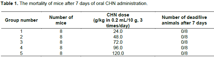
Thrice each day, Swiss albino mice were treated with CHN at different doses, ranging from 24.0 to 120.0 g/kg body weight, in a volume of 0.2 mL/10 g.
The results in Table 1 show that the tested concentration range did not produce any clinical signs of toxicity or cause death in mice. In all the research groups, there was no record of death of mice. In our observation, mice did not display any abnormal symptoms within 72 h after CHN administration or during the 7 days after administration. Even at the highest dose of 120 g/kg/day, the mice remained alive and did not display any unusual symptoms. These results demonstrated that CHN did not cause acute toxicity through oral administration. Because no mice died during this experiment, the LD50 value for orally administered CHN in mice could not be determined. Even at the highest dose administered (120 g/kg mice body weight), which represents 25-fold the expected effective dose, no acute toxicity was observed. Interestingly, in this study, food intake and water consumption were found to have no effects on the administration of CHN. Additionally, the oral administration of CHN did not appear to promote appetite suppression or other damaging effects. Thus, these results indicate that the interruption of carbohydrate, protein, or fat metabolism was unlikely, in all experimental groups.
Sub-chronic toxicity study results
Influence of CHN on rat general condition and body weight of rats during long-term administration
All Wistar rats used in this study were monitored daily for general conditions, including motor activity, eating behavior, fur, skin, and mucous membrane conditions, as well as secretions. Rats in the control group and in the CHN oral administration groups displayed normal activities, smooth fur, normal mucosal skin, normal eating behaviors, and the production of molded feces.
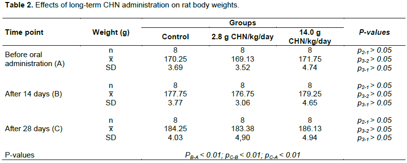
When comparing body weights of mice before and after oral CHN administration, the body weights in all three study groups increased significantly. In detail, from 14 to 28 days of oral administration, the body weights of the two groups 2.8 g CHN/kg/day and 14.0 g CHN/kg/day were increasing from 176.75 to 183.38 g, and from 179.25 to 186.13 g, respectively (Table 2), and the change was statistically significant (p < 0.01). At all examined time points, no significant difference was observed for the weights of the rats in the two administration groups compared with the weight of the rats in the control group (p > 0.05). Thus, CHN appeared to have no effects on rat body weight in this study, even over a wide concentration range.
Effects of CHN on hematological parameters
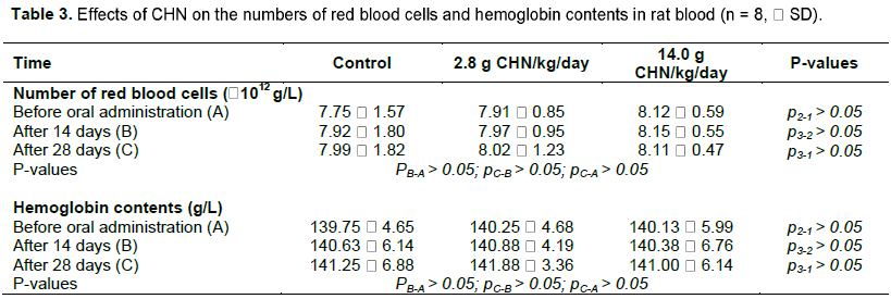
As shown in Table 3, the numbers of red blood cells and hemoglobin contents did not show significant changes following CHN administration (p > 0.05). In the group of 2.8 g CHN/kg/day, the number of red blood cells was increased from 7.97 to 8.021012 g/L in 14 to 28 days period after administration. However, in the high dose, the number of red blood cells were somewhat decreased from 8.15 to 8.111012 g/L (p > 0.05). Interestingly, the hemoglobin content ratios in both administration doses did not change so much as from 140.38  6.76 to 141.00  6.14 g/L (p > 0.05). Thus, CHN, even when administered at different doses and for different durations, did not cause any changes of red blood cell numbers or hemoglobin contents in rat blood (Table 3).
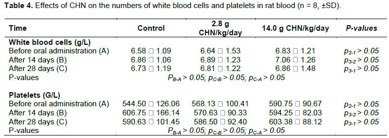
As shown in Table 4, the numbers of white blood cells and platelets in rat blood did not change significantly following CHN administration (p > 0.05). In fact, from 14 to 28 days durations, the number of white blood cells were somewhat changed from 6.89  1.23 to 6.81  1.22 g/L in the 2.8 g CHN/kg/day group and decreased from 7.06 ï‚±ï€ 1.26 to 6.86  1.48 g/L; these changes were acceptable due to the similar levels between treated mices and normal mices in control group. The levels of platelets were also recognized but not so much of changing differences were observed (p > 0.05). Thus, it was possible to note that CHN with different administration doses and for different durations did not cause any changes in white blood cell or platelet counts in rat blood.
Evaluating liver cell damage
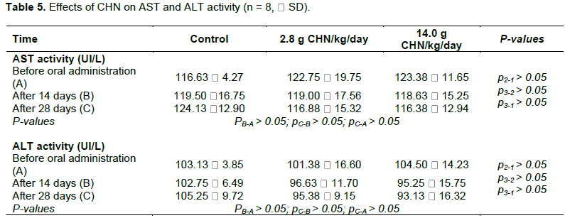
AST (or SGOT) and ALT (or SGPT) are liver enzyme indicators that can reflect liver damage. These indicators often increase, depending on the type of disease, and if treatment does not result in improvements, these indicators may drop unexpectedly, due to liver cell death (Wedemeyer et al., 2010). In this experiment, the AST and ALT enzyme activities did not change significantly following CHN administration; the enzyme levels were recorded approximately from 116.88  15.32 to 119.00 ± 17.56 UI/L in the dose of 2.8 g CHN/kg/day and from 118.63 ± 15.25 to 116.38 ± 12.94 UI/L in the dose of 2.8 g CHN/kg/day (p > 0.05) after 28 days administration (Table 5). Thus, even at different doses and for different durations, CHN administration did not significantly affect AST or ALT enzyme activity in this study, indicating that these tea leaves did not cause damage to rat liver cells.
Assessing the effects of CHN on liver function
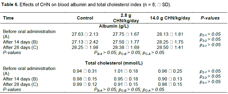
Albumin is one of the most important protein constituents of serum, accounting for 58–74% of the total serum protein content. Approximately 40% of albumin is found in plasma, whereas the remaining 60% is found in extracellular fluid. Albumin in blood has many important functions, such as maintaining colloidal osmotic pressure, stopping water from leaking out of blood vessels, providing amino acids for peripheral protein synthesis, as well as linking and transporting certain substances produced during metabolisms, such as fatty acids, bilirubin, steroid hormones, and other active substances, throughout the body. The liver is the only organ in the body that produces albumin; thus, the albumin index can reflect the functional status of the liver (Farrugia, 2010). Cholesterol is a bile component that is found in red blood cells, cell membranes, and muscles. The liver is the major organ that synthesizes cholesterol and is the only organ that esterifies cholesterol. In our experiment, both the albumin and total cholesterol indices for rat blood were evaluated before and after CHN administration. The results in Table 6 showed that these parameters were not significantly changed by CHN administration, as the number of albumin level were from 27.50 ± 1.77 to 28.50 ± 1.41 g/L in both administration doses in 28 days; meanwhile, the total cholesterol were calculated from 0.86 ± 0.15 to 0.95 ± 0.18 mmol/L (p > 0.05). Based on these results, it is worthy to note that CHN administration did not produce any significant changes in total cholesterol or albumin levels, regardless of the CHN dose used in treated animals compared with control animals.
Assessing the effects of CHN on kidney function
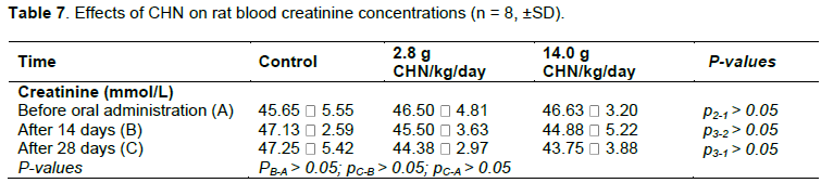
Blood creatinine is a catabolic product of creatine phosphate. Creatinine is exogenously derived from food and endogenously generated by the liver, kidney, and pancreas. Thus, quantitative creatinine testing in the blood can indicate many health problems, including impaired kidney function (Baum et al., 1975). In our experiment, creatinine levels were evaluated in rat blood both before and after oral CHN administration. As shown in Table 7, blood creatinine concentrations were 45.65 ± 5.55 mmol/L the day before oral CHN administration and significantly changed to 47.25 ± 5.42 mmol/L after 28 days (p> 0.05) in the control group. In the other 2 groups, the creatinine concentrations did not change significantly following CHN administration (p > 0.05). Thus, yellow tea leaves did not affect creatine concentrations in the blood, regardless of the dose used or the duration of treatment.
Histopathological results
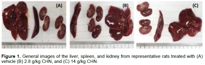
The liver, spleen, and kidney were observed with the naked eye and under a microscope, at 25× magnification (Carol, 1995; Dybing et al., 2002). The colors and morphologies of the liver, spleen, and kidney in groups treated with 2.8 g/kg CHN and 14 g/kg CHN did not differ from those of the control group (Figure 1).
The histopathological samples were examined at the pathological morphology level. The histopathology results for the liver, spleen, and kidney of treated rats showed that the oral administration of yellow flower tea leaves, at 2.8 and 14 g/kg/24 h continuously for 28 days, did not cause liver, kidney, or spleen damage in rats. The results in Figure 2 show that the structures of the hepatic lobe remained clear and conspicuous, without necrosis phenomena or hepatocellular degeneration, were observed in rats treated with vehicle (A), 2.8 g/kg CHN (B), and 14 g/kg CHN (C) after 28 days.
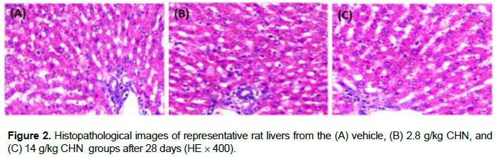
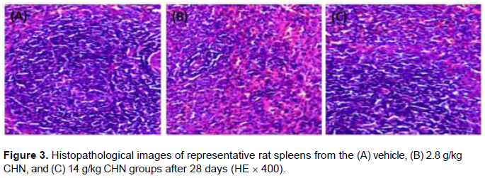
Figure 3 showed histopathological images of representative rat spleens from the vehicle (A), 2.8 g/kg CHN (B), and 14 g/kg CHN (C) groups after 28 days of oral administration. The spleen remained intact, demonstrating a white marrow, with large cell numbers concentrated in the large lymphoid follicles. Thickened penile arteries could be observed in the white marrow, and the red marrow, characterized by cyst sinuses, contains many red blood cells and some macrophages in normal growth stages.
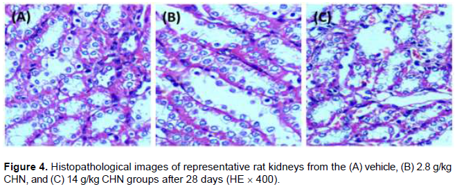
The completely undamaged kidneys of rats from all three groups remained clear and preserved (Figure 4). The medulla and the cortex remained unblemished, and the glomeruli are evenly distributed, with no inflammatory infection. Arteries and small arterioles have normal structures.
DISCUSSION
The yellow tea, Camellia sp., is a shrub and small tree belonging to the family Theaceae (Xu et al., 2018). Approximately 52 species of yellow Camellia have been described in southern China and Vietnam, of which nearly 40 species are naturally distributed in Vietnam (Manh et al., 2019; Huang et al., 2013). These plants are shade-tolerant species that grow well only in the shade. In general, the yellow Camellia is distributed at an altitude of 100–1,000 m above sea level, particularly at altitudes ranging from 300–700 m. Therefore, these plants are often found in valleys and near streams. In some cases, the plants are also found in dry soil, although they grow badly and are small in size under these conditions. In Vietnam, the yellow Camellia is often used to make tea because of its beneficial properties. Although the dried flowers are used more often than the leaves, both fresh and dried flowers and leaves can be used to make a drink similar to green tea (C. sinensis). However, dry products are preferred because they can be stored for long periods. Recently, some cosmetics have been made from yellow Camellia flowers, such as yellow silk oil, organic yellow Camellia oil, and face cream. Camellia species contain many active substances, such as polysaccharides, polyphenols, saponins, and flavonoids. Clinical research has shown that yellow Camellia products can inhibit transplant cancer, lower blood pressure, blood lipids, and cholesterol levels, and prevent atherosclerosis. Research has demonstrated that Camellia extracts possess antioxidant, with anionic superoxide and hydroxyl radical scavenging activities (Manh et al., 2019; Huang et al., 2013). Yellow Camellias, such as C. nitidissima, have been used to treat sore throats, diarrhea, high blood pressure, irregular menstruation and to prevent cancer (Mukhtar and Ahmad, 2000). Studies on C. euphlebia, a yellow Camellia grown extensively in Vietnam showed that the leaves can be used to treat dysentery, hypertension, diarrhea, fallopian tubes, and irregular menstruation (He et al., 2018). The primary natural products found in yellow Camellia include α-spinasteryl-D-glucopyranoside, stigmasta glucopyranosides, aromadendrin, catechin, phlorizin glucopyranosides, dodecanoic acid, 3β-acetoxy-20-lupanol, and 3β,6α,13β-trihydroxyolean-7-one. Water extract from yellow Camellia had been shown to have anti-anxiety, antidepressant, and anti-bacterial activities (Xu et al., 2018; Nguyen et al., 2019). Due to its high value to human health, the commercial value of yellow Camellia is much higher than that of green tea. Therefore, planting Camellia can be a potential avenue for poverty reduction among the ethnic minorities that reside in mountainous areas. In Vietnam, the recent harvests of flowers from natural forests have not been readily available because many flowering plants have been dug up and transplanted into gardens. The future development of yellow Camellia should focus on developing the best varieties, in addition to harvesting those plants that have been transplanted from the natural forest. While the natural population of yellow Camellia remains abundant in China4, very few natural populations remain in Vietnam, especially C. hakodae. Because C. hakodae is an endemic yellow tea in Vietnam, appropriate and sustainable management strategies for this golden tea should be considered. In addition, toxicological studies should be evaluated. In our study, the toxicological evaluation revealed that for both acute and sub-chronic toxicity tests, C. hakodae extract (CHN) did not produce any toxic effect in mice or rats. During the acute toxicity study, no morbidity or mortality was observed. The LD50 value of the extract was not identified, but is likely higher than 120 g/kg, suggesting that the extract is essentially non-toxic and safe for the oral administration of the tested doses. During the sub-chronic toxicology test, no deaths or treatment-related symptoms were observed in any group. The histopathological inspection of the liver, kidney, and spleen samples from both CHN- and vehicle-treated rats showed normal organ architecture, suggesting no microscopic changes or morphological disturbances were caused by the oral administration of CHN, at both low to high doses. Interestingly, no significant differences in food and water intake, weight gain, biochemical, or hematological parameters were observed between the control and treated groups during the oral administration period.
CONCLUSION
This study has revealed that aqueous extracts of C. hakodae Ninh leaves were relatively safe because treatment using this extract did not cause death or any abnormal changes during either the acute or sub-chronic toxicity studies. Further studies will be required to investigate the potential medical significance of this tea.
CONFLICT OF INTERESTS
The authors have not declared any conflict of interests.
ACKNOWLEDGMENTS
The research groups thank Associate Prof. MD. Dau Xuan Canh and the Vietnam University of Traditional Medicine for the help and good condition provided for this project.
REFERENCES
|
Baum N, Dichoso CC, Carlton CE (1975). Blood urea nitrogen and serum creatinine. Physiology and interpretations. Urology 5:583-588. |
|
|
Carol SA (1995). Acute, subchronic and chronic toxicology. In: Michael JD, Mannfred AH editors. CRC Handbook of Toxicology, CRC Press Inc.: Boca Raton, pp. 51-104. |
|
|
Dybing E, Doe J, Groten J, Kleiner J, O Brien J, Renwich AG, Schlatter J, Steinberg P, Tritscher A, Walker R, Younes M (2002). Hazard characterization of chemicals in food and diet: Dose-response, mechanism, and extrapolation issues. Food Chemical and Toxicology 42:237-282. |
|
|
Farrugia A (2010). Albumin usage in clinical medicine: Tradition or therapeutic? Transfusion Medicine Reviews 24:53-63. |
|
|
He DY, Sai X, Wang N, Li X, Wang L, Xu Y (2018). Camellia euphlebia exerts its antidepressant-like effect via modulation of the hypothalamic-pituitary-adrenal axis and brain monoaminergic systems. Metabolic Brain Disease 33:301-312. |
|
|
Huang LD, Li ZH, Lu F, Qin MQ, Luo YY (2013). Three decades of breeding Golden Camellia varieties in Nanning Golden Camellia Park. International Camellia Journal 45:101-104. |
|
|
Litchfield JTJr, Wilcoxon E (1949). A simplified method of evaluating dose-effect experiments. Journal of Pharmacology and Experimental Therapeutics 96:99-113. |
|
|
Manh TD, Thang NT, Son HT, Thuyet DV, Trung PD, Tuan NV, Duc DT, Linh MT, Lam VT, Thinh NH, Phuong NTT, Do TV (2019). Golden Camellias: A review. Archives of Current Research International 16: 1-8. |
|
|
Mukhtar H, Ahmad N (2000). Tea polyphenols: prevention of cancer and optimizing health. American Journal of Clinical Nutrition 71:1698S-1702S. |
|
|
Nguyen TT, Tran VH, Pham GD, Ninh T, Hung NT, Hoang VD (2019). A new sexangularetin derivative from Camellia hakodae. Natural Product Communications 14:1-4. |
|
|
Ninh T, Hakoda N (2009). Cac loai Tra cua Vuon Quoc gia Tam Dao, Vietnam National University Press, Hanoi, pp. 1-121. |
|
|
OECD (1981). Test No: 411, Subchronic dermal toxicity: 90-day study. Section 2. 411. Paris: OECD Publishing, pp. 5-9. |
|
|
OECD (1987). Test No: 402, Acute dermal toxicity. Section 2. 402. Paris: OECD Publishing, pp. 1-3. |
|
|
OECD (2004). Test No: 434, Acute dermal toxicity - fixed-dose procedure Section 20 to 34. 434. Paris: OECD Publishing, pp. 4-5. |
|
|
Van NTH, Inh CT, Bach PC, Tuyen TT, Thanh NT, Quynh NH, Long PQ (2018). Flavonoid glycoside isolated from Camellia chrysantha. Vietnam Journal of Chemistry 56:335-340. |
|
|
Wedemeyer H, Hofmann WP, Lueth S, Malinski P, Thimme R, Tacke F, Wiegand J (2010). ALT screening for chronic liver diseases: Scrutinizing the evidence. Zeitschrift für Gastroenterologie 48:46-55. |
|
|
Wei JB, Li X, Song H, Liang YH, Pan YZ, Ruan JX, Qin X, Chen YX, Nong CL, Su ZH (2015). Characterization and determination of antioxidant components in the leaves of Camellia chrysantha (Hu) Tuyama based on composition activity relationship approach. Journal of Food and Drugs Analysis 23:40-48. |
|
|
Xu J, Wang M, Zhao J, Wang HD, Tang Q, Khan IA (2018). Yellow tea (Camellia sinensis L.), a promising Chinese tea: Processing, chemical constituents and health benefits. Food Research International 107:567-577. |
|
Copyright © 2024 Author(s) retain the copyright of this article.
This article is published under the terms of the Creative Commons Attribution License 4.0


