Full Length Research Paper
ABSTRACT
The highly selective serotonin reuptake inhibitors are successful in the treatment of depression mood and anxiety disorders. The objective of this study is to investigate the effect of fluexotine and buspirone when given orally in three dose levels (5, 10 and 15 mg/ kg) not only for eight weeks on gastric lesions and oxidative markers in normal rats' mucosal integrity, but also for four weeks on gastric mucosal lesions in rats treated with indomethacin- induced ulcers. The results of this study showed that in normal rats, the administration of fluoxetine induced gastric lesions while buspirone caused no lesions. The indomethacin administration resulted in the development of gastric mucosal lesions. Furthermore, co-administration of fluoxetine and indomethacin enhanced the development of gastric mucosal lesions that were coupled with disturbance in antioxidant status. Buspirone, in contrast, significantly decreased the development of gastric mucosal lesions in rats treated with indomethacin. In conclusion, fluoxetine caused the development of gastric mucosal lesions and aggravate the effect of indomethacin to induce ulcer, however, buspirone had a protective effect that may be attributed to its antioxidant properties.
Key words: Fluoxetine, buspirone, peptic ulcer, indomethacin-induced ulcer, oxidative stress.
INTRODUCTION
Serotonin is a neurotransmitter that plays an integral role in mediating a number of physiological processes; a wide distribution in the brain and gut (van Praag, 1980; Hanson and Hurley, .2014). Several studies (Kuhn et al., 1980; Rickels and Schweizer, 1990; Ahmed and Simmons, 2013) said that selective serotonin reuptake inhibitors (SSRIs) are very successful in the treatment of psychological depression (Dworkin et al., 2007). Interestingly, although administration of an SSRI causes an immediate increase in synaptic release of 5-HT, antidepressant effects are not experienced until approximately 3 to 4 weeks of chronic administration in humans (Rickels and Schweizer, 1990; Stahl, 1988) and 2 weeks in rat models of affective disorder (Gambarana et al., 2001).
Most serotonin is found in the gut being produced by the enterochromaffin cells in the gastric and intestinal mucosa (Ener et al., 2003). Gut serotonin is involved in the control of smooth muscle tone and motility (Mazda et al., 2004; Fink et al., 2006). Studies in humans have suggested an association between the intake of SSRIs and increased episodes of gastric bleeding in patients with depressive disorders (De Abajo et al., 2006). As serotonin promotes platelet aggregation, and it is thought that SSRIs limit the uptake of blood serotonin by platelets consequently to serotonin promoting platelet aggregation (Meijer et al., 2004). The present work aimed to investigate the effect of long term treatment with drugs influencing serotonergic neurotransmission on gastric mucosal integrity in rats.
In this experimental study, the effect of fluoxetine and buspirone were investigated in normal rats' stomach. In addition, the effect of both drugs on oxidant and antioxidant parameters in rats' stomach tissue was evaluated, as a first approach to investigate the mechanism behind their effect on the stomach.
MATERIALS AND METHODS
Animals
Adult albino wistar rats, weighting 120 to 130 g of body weight, were obtained from the animal house colony in National Research Center (Giza, Egypt). The animals were housed in a conditioned room at 25 ± 2ºC, standard diet and tap water source were supplied ad libitum. Approval for this study was obtained from the Ethics Committee of The National Research Center-Egypt and in accordance with the recommendations of the proper care and use of laboratory animals.
Drugs
Fluexotine was purchased from lilly (England), buspirone from Squibb (Egypt) and indomethacin from Khahira Pharmaceutical and Chemical IND Company (Egypt). The doses of the drugs were selected from published literature according to Abdel-Salam et al. (2003), Chial et al. (2003) and Ilahi et al. (2006), respectively.
Studies in the intact rat
Rats were randomly allocated into 7 groups, each consisting of 16 rats. Daily administration orally of the test drugs in three dose levels was carried out for eight weeks. The animals were treated according to the following scheme: Group 1: Received saline and served as a negative control. Groups 2, 3 and 4 received fluexotine 5, 10 and 15 mg/kg, respectively. Finally groups 5, 6 and 7 received buspirone 5, 10 and 15mg/kg, respectively.
Studies in combination with indomethacin on normal rats
Eleven groups, each consisting of 8 rats, daily administration orally of the test drugs in three dose levels was carried out orally for four weeks, along with indomethacin 1 mg / Kg / 24 h (Ilahi et al., 2006). Animals were treated according to the following scheme: Group 1: Received saline and served as the control. Group 2: Received indomethacin and served as the control +ve. Groups 3, 4 and 5 received fluexotine 5, 10 and 15 mg/kg, respectively and indomethacin. Groups 6, 7 and 8 received buspirone 5, 10 and 15 mg/kg, respectively and indomethacin.
At the end of the first experiment, eight animals of each group were sacrificed by decapitation. After which the brains were quickly isolated, the brains were frozen stored at -80°C, until analysis of serotonin brain levels.
At the end of two experiments, animals were sacrificed by cervical dislocation then the abdominal cavity was opened and the stomach was removed. The stomach was opened along the greater curvature and pinned on a plastic board. The mucosa was examined for mucosal necrotic lesions, red streaks and red erosions (Mózsik et al., 1982). Immediately after gross lesion examination, the stomach was placed over an ice-cold surface. The glandular mucosa was cut, weighed then homogenized in ice-cold saline to obtain a 10 % (W/V) homogenate by using glass homogenizer. This was performed to determine the level of glutathione, lipid peroxides and nitric oxide.
The stomach mucosa was examined for mucosal necrotic lesions, red streaks and red erosions (Mózsik et al., 1982). Total lesion number was counted and the lesion severity was determined based on the following scores:
0 = no ulcer
1 = lesion size ≤ than 1 mm.
2 = lesion of size 1-2 mm.
3 = lesion of size 2-3 mm.
4 = lesion of size 3-4 mm.
5 = lesion of size > 4 mm.
Determination of oxidative stress
Lipid peroxides were determined according to the method described by Mihara and Uchiyama (1978) and expressed as nmol/g wet tissue. Lipid peroxidation products were estimated by the determination of the level of TBARS that were measured as malondialdehyde (MDA). The latter is the decomposition product of the process of lipid peroxidation and is used as an indicator of this process. The principle of the assay depends on the colorimetric determination of a pink pigment product, resulting from the reaction of TBARS with thiobarbituric acid (TBA) in an acidic medium, at high temperature. Reduced glutathione (GSH) content was determined in the stomach homogenates according to the method of Beutler et al. (1963) and expressed as mg/g wet tissue. The method depends on the fact that both protein and non-protein thiol (SH-) groups (mainly GSH) react with Ellman’s reagent [5,5' -dithiobis (2- nitrobenzoic acid)] to form a stable yellow color of 5-mercapto -2- nitrobenzoic acid, which can be measured colorimetrically at 412 nm. Stomach NO metabolites were determined according to the method described by Miranda et al. (2001) and expressed as μM/g wet tissue. The assay determines the total NOx content based on the reduction of any nitrate to nitrite by vanadium, followed by the detection of total nitrite (intrinsic + nitrite obtained from reduction of nitrate) by Griess reagent. The Griess reaction leads to the formation of a chromophore from the diazotization of sulfanilamide by acidic nitrite, followed by coupling with bicyclic amines such as N-(1-naphthyl) ethylenediamine. The chromophoric azo derivative can be measured colorimetrically at 540 nm.
Estimation of serotonin
Estimation of serotonin in the midbrain region using rapid and precise liquid chromatography with mass spectrometric detection (LC/MS) method for the identification and quantification of serotonin from rat brain tissue without any pre-analysis adjustment of the sample such as pre-concentration or derivatization has been developed (Cao et al., 2006).
Histopathological examination
The dissected stomachs of different groups were washed with saline and fixed in 10% formalin for histopathological assessment. The prepared sections were stained with Haematoxylin and Eosin for assessing histopathological changes.
Statistical analysis
Values were expressed as means ± S.E.; the results of the ulcer number and severity were analyzed using Kruskal-Wallis non-parametric one way analysis of variance (ANOVA), followed by Mann Whitney multiple comparison test. The results of the remaining experiments were analyzed using one way ANOVA followed by least significant difference (LSD) multiple comparison test. P<0.05 was accepted as being significant in all types of statistical tests. Statistical analysis of results, were done using software
RESULTS
Effect on ulcer index
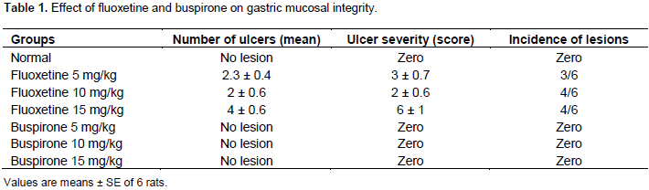
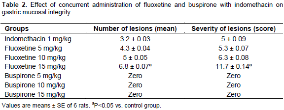
In normal rats, after eight weeks of drugs administration, fluoxetine caused lesions compared with buspirone (Table 1). The concurrent administration of fluoxetine and indomethacin for four weeks enhanced the development of gastric mucosal lesions (Table 2).
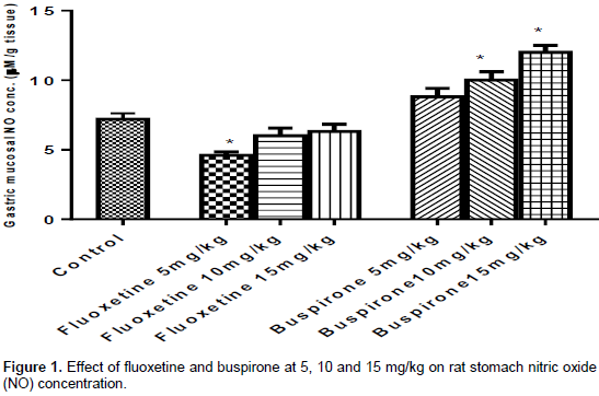
After eight weeks the administration of fluoxetine reduced gastric NOx content while buspirone elevated gastric NOx (Figure 1). The concurrent administration of fluoxetine and buspirone with indomethacin for four weeks results in gastrointestinal mucosal damage decreasing NO cause by indomethacin. Moreover, gastric lesions caused by NSAIDs can be aggravated by vascular ischemia, which can explain why fluoxetine enhanced lesions with indomethacin co-administration through their decrease in nitric oxide.in the same manner gastric NOx content was reduced by fluoxetine (Figure 2).
Effect on gastric NOx content
Effect of fluoxetine and buspirone on gastric NOx in normal rats
The effect of fluoxetine on NO concentration, in normal rats that received fluoxetine at 5 mg/kg, showed a significant decrease by 35% compared to the control (saline treated) group. The other two doses of fluoxetine 10 and 15 mg/kg resulted in an insignificant reduction in NO concentration compared to the normal group.
The rats, that received buspirone at 5 mg/kg showed an insignificant increase in NO concentration by 23% compared to the normal group. In rats that were administered buspirone at 10 and 15 mg/kg, NO concentration was significantly increased compared to normal group by 41 and 55%, respectively (Figure 1).
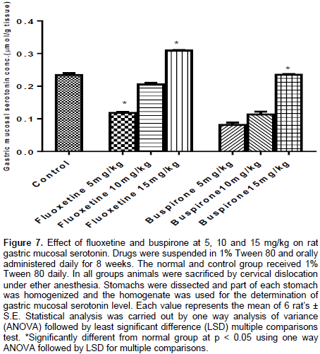
Drugs were suspended in 1% Tween 80 and orally administered daily for 8 weeks. The normal and control group received 1% Tween 80 daily. In all groups, animals were sacrificed by cervical dislocation under ether anesthesia. Stomachs were dissected and part of each stomach was homogenized and the homogenate was used for the determination of gastric total nitrate/nitrite (NOx) content. Each value represents the mean of 6 rats ± S.E. Statistical analysis was carried out by one way analysis of variance (ANOVA) followed by least significant difference (LSD) multiple comparison test.
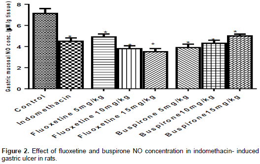
Effect of fluoxetine and buspirone NO concentration in indomethacin induced gastric ulcer in rats
Indomethacin significantly decreased NO content by 36% compared to the control group. Fluoxetine at 5, 10 and 15 mg/kg was given to indomethacin treated rats resulting in significant decrease in NO concentration compared to the control group by 31, 46 and 50% respectively. In the same manner, NO concentration showed insignificant decreased compared to the indomethacin treated group. Buspirone was given at 5, 10 and 15 mg/kg to indomethacin treated rats resulting in a significant decrease in NO concentration compared to the control group by 45, 40 and 30%, respectively. However, no significant effects were seen after treating with buspirone in NO concentration compared to indomethacin treated group.
Drugs were suspended in 1% Tween 80 and orally administered daily for four weeks with 1% indomethacin. The normal received 1% Tween 80 daily and the control group received 1% indomethacin. In all groups, animals were sacrificed by cervical dislocation under ether anesthesia. Stomachs were dissected and part of each stomach was homogenized and the homogenate was used for the determination of gastric total nitrate/nitrite (NOx) content. Each value represents the mean of 6 rats ± S.E. Statistical analysis was carried out by one way analysis of variance (ANOVA) followed by least significant difference (LSD) multiple comparison test.
Effect on gastric GSH content
Effect of fluoxetine and buspirone gastric on glutathione (GSH) content in normal rats
Normal rats were given fluoxetine; GSH content showed an insignificant change compared to the control group. Rats that received buspirone at 5 mg/kg showed an insignificant increase in GSH content compared to the control group. In rats treated with buspirone at 10 and 15 mg/kg, GSH content was a significantly increased compared to the control group by 27 and 36% respectively (Figure 3).
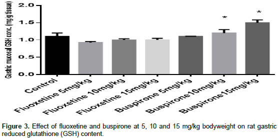
Drugs were suspended in 1% Tween 80 and orally administered daily for 8 weeks. The normal and control group received 1% Tween 80 daily. In all groups, animals were sacrificed by cervical dislocation under ether anesthesia. Stomachs were dissected and part of each stomach was homogenized and the homogenate was used for the determination of gastric total reduced glutathione content. Each value represents the mean of 6 rat's ± S.E. Statistical analysis was carried out by one way analysis of variance (ANOVA) followed by least significant difference (LSD) multiple comparison test.
Effect of fluoxetine and buspirone GSH content in indomethacin- induced gastric ulcer in rats
Indomethacin was given at 1 mg/kg for four weeks, resulting in an insignificant increase in GSH content compared to the control group. Fluoxetine administration to indomethacin treated rats at 5 mg/kg caused an insignificant decrease in GSH content compared to both the control and the indomethacin treated group. Fluoxetine at 10 and15 mg/kg administrations to indomethacin treated rats showed a significant increase in GSH content compared to the control group by 38 and 84%, respectively. Fluoxetine at 15 mg/kg caused a significant increase in GSH content by 72% compared to the indomethacin treated group.
Buspirone at 5, 10 and 15 mg/kg was given to indomethacin treated rats resulted in a significant increase in GSH content not only compared to the control group by 29, 57and 84%, but also to the indomethacin treated group by 27, 54 and 72%, respectively (Figure 4).

Drugs were suspended in 1% Tween 80 and orally administered daily for four weeks with 1% indomethacin. The normal group received 1% Tween 80 daily and the control group received 1% indomethacin. In all groups animals were sacrificed by cervical dislocation under ether anesthesia. Stomachs were dissected and part of each stomach was homogenized and the homogenate was used for the determination of gastric reduced glutathione content. Each value represents the mean of 6 rats ± S.E. Statistical analysis was carried out by one way analysis of variance (ANOVA) followed by least significant difference (LSD) multiple comparison test.
Effect on gastric MDA content
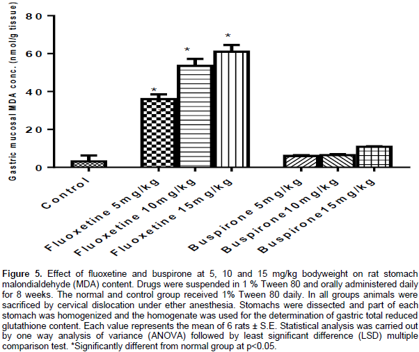
Effect of fluoxetine and buspirone on normal rat stomach malondialdehyde (MDA) content
The fluoxetine group resulted in a significant increase in MDA content compared to the control group. Buspirone at 5, 10 and 15 mg/kg, MDA content showed an insignificant change compared to the control group (Figure 5).
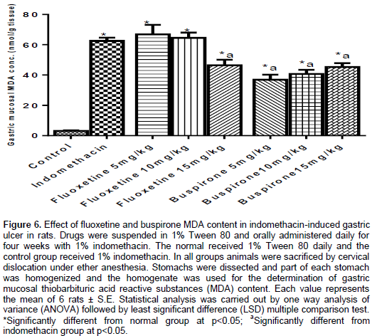
Effect of fluoxetine and buspirone MDA content in indomethacin induced gastric ulcer in rats
Indomethacin given at 1 mg/kg for four weeks resulted in significant increase in MDA content compared to the control group. Fluoxetine at 5 and 10 mg/kg given to indomethacin treated rats, resulted in a significant increase in the MDA content compared to the control group. This elevation was insignificant compared to the indomethacin treated group. However, fluoxetine at 15 mg/kg given to indomethacin treated rats, resulted in a significant increase in the MDA content compared to both the control and the indomethacin treated groups.
Buspirone was given to indomethacin treated rats, resulted in a significant decrease in gastric mucosal the MDA content compared to the indomethacin treated group (Figure 6).
Effect on serotonin level
Effect of fluoxetine and buspirone on rat gastric mucosal serotonin
In normal rats, that were given fluoxetine at 5 and 10 mg/kg, the gastric mucosal serotonin level was decreased compared to the control group by 50 and 12%, respectively. Fluoxetine at 15 mg/kg increased gastric mucosal serotonin level by 32% compared to the control group. Buspirone at 5 and 10 mg/kg decreased the gastric mucosal serotonin level compared to the control group. Buspirone at 15 mg/kg showed no effect on the gastric mucosal serotonin level compared to the control group (Figure 7).
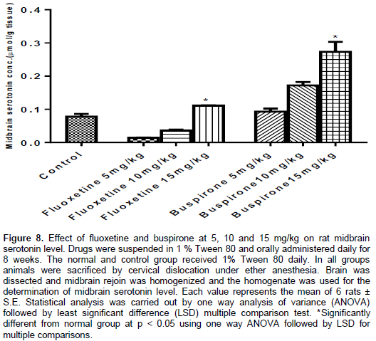
Effect of fluoxetine and buspirone on rat midbrain serotonin level
Fluoxetine at 5 and 10 mg/kg, decreased the brain serotonin level compared to the control group by 82 and 52% respectively. Fluoxetine at 15 mg/kg increased the brain serotonin level by 42% compared to the control group. Buspirone at 5, 10 and 15 mg/kg, increased the brain serotonin level compared to the control group (Figure 8).
Histopathological changes
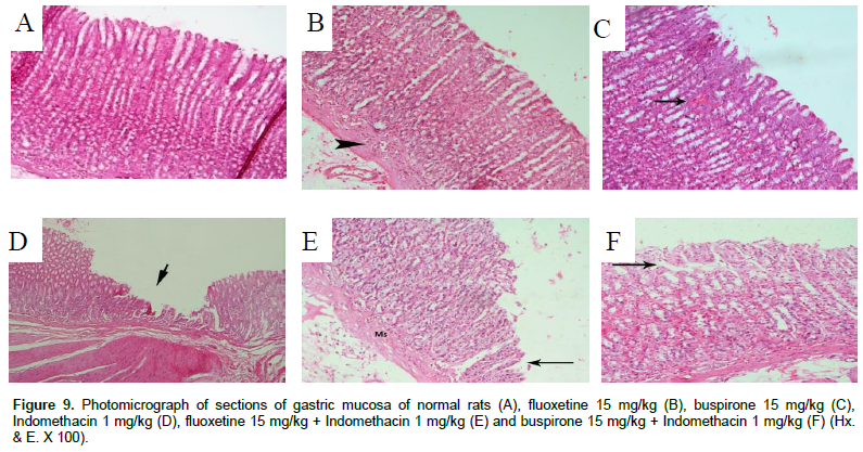
Examination of the gastric mucosa of normal rats showed normal stomach structure (Figure 9A). Gastric mucosa of normal rats treated with fluoxetine at 15 mg/kg showed slight cellular infiltration in the muscularis mucosa layer. The rest of the tissue appears normal (Figure 9B). Gastric mucosa of normal rats treated with buspirone at 10 mg/kg showed quite normal gastric mucosal structure (Figure 9C).
Gastric mucosa of rats subjected to indomethacin showed a wide area of damage in the upper 2/3 of the gastric mucosa. Only the bases of the fundic glands are still present, while numerous sorts of cells were separated. (Figure 9D). Gastric mucosa of rats subjected to indomethacin and fluoxetine at 15 mg/kg showing marked thickening of muscularis mucosa (Ms). The gastric mucosa shows partial detachment of the upper third or half of the gastric glands (Figure 9E). Gastric mucosa of rats subjected to indomethacin and buspirone at 15 mg/kg showed mild gaps in between the gastric glands occupied by connective (fibrous) tissue, with a mild deformity of the normal structure of gastric mucosa (Figure 9F).
DISCUSSION
The gastric side effects of SSRI drugs have been reported (Lewis et al., 2008). However, numerous serotonin 1A receptor (5HT1-A) agonists, as buspirone, developed as anxiolytics, appeared with antisecretory and gastroprotective effects in rats (Glavin et al., 1995), by decreasing stomach and intestinal distension (Tack, 1999). Fluoxetine was found to cause a few number of ulcers with moderate severity in a dose-dependent manner. On the other hand, buspirone was found to be safe on the gastric mucosal. This study investigated fluoxetine and buspirone effect on GSH, NO and MDA activities in normal stomach tissue of rats, as a first approach to investigate the mechanism behind their effect on the stomach.
In the current study, all doses of fluoxetine and buspirone increased gastric NO levels significantly when compared to the normal group.
The present investigation revealed that eight weeks administrations of fluoxetine resulted in a decreased production of gastric NO content of the gastric mucosa in a dose dependent-manner with a marked decrease in low doses. By contrast, administrations of buspirone resulted in an increased gastric NO content in a dose dependent-manner; however, both markedly increased gastric mucosal lipid peroxides.
The increase in gastric GSH content was reported in the current results. Buspirone showed marked increase in the gastric GSH content; in the same way fluoxetine showed a slight increase in the gastric GSH content. This lesser increase in reduced glutathione was observed at the higher dose of fluexotine which markedly enhanced lipid peroxidation in gastric content.
The findings that lipid peroxidation was markedly increased by fluoxetine, was correlated with its ability to cause gastric lesions. Alternatively, buspirone insignificantly increased gastric lipid peroxidation was visually previewed compared to control group in all experiments. Conversely, the increase of GSH by buspirone, may explain the effect of buspirone on gastric mucosa compared to fluoxetine.
SSRIs increase the 5-HT concentration in tissue, as a result of the inhibition of 5-HT reuptake at nerve endings and platelets. An increased serotonin level in the stomach plays a role in the aggravation of gastric lesions due to the vasoconstrictor effect of serotonin as shown by Cho et al. (1989); that the blood flow was decreased by 5-HT in a dose dependent-manner. On other hand, Takeuchi et al. (2011) found that aggravating effect of SSRIs to induce lesions were mimicked by exogenous 5-HT, symptomatic of the effect of endogenous 5-HT in this action. Ohta et al. (1997) confirmed the current results by serum serotonin concentration, an index of mast cell degranulation, increased with the formation of gastric mucosal lesions, and this increased serotonin level was attenuated with lesion progression and recovery.
A conseguential increase in the serotonin level in the midbrain may be due to the enhancement of serotonin synthesis secondary to an increased hydroxylation of tryptophan by tryptophan hydroxylase; the rate-limiting enzyme of the serotonin biosynthetic pathway. Serotonin additionally represents the aggressive ulcerogenic amines, which stimulate ulcer formation by raising total acidity and decreasing the volume of gastric secretion (Ibrahim et al., 1996).
CONCLUSION
The administration of fluoxetine caused the development of gastric mucosal lesions. This effect may be attributed to oxidative stress and an imbalance in gastric acid, peptic activity, mucin, GSH and NO, as well as increase in MDA contents. Buspirone appeared to be devoid of a deleterious effect on the gastric mucosa through antioxidant properties and by improving of the immunity in the gastric mucosa.
CONFLICT OF INTERESTS
The authors have not declared any conflict of interests.
ACKNOWLEDGMENT
This study was supported by the National Research Center in Cairo, Egypt (Number 7/2/1).
REFERENCES
|
Abdel-Salam OM, Nofal SM, El-Shenawy SM (2003). Evaluation of the anti-inflammatory and anti-nociceptive effects of different antidepressants in the rat. Pharmacol. Res. 48(2):157-165. |
|
|
Ahmed A, Simmons Z (2013). Pseudobulbar affect: prevalence and management. Ther. Clin. Risk Manag. 9:483-489. |
|
|
Beutler E, Duron O, Kelly BM (1963). Improved method for the determination of blood glutathione. J. Lab. Clin. Med. 61:882-888. |
|
|
Cao J, Murch SJ, O'Brien R, Saxena PK (2006). Rapid method for accurate analysis of melatonin, serotonin and auxin in plant samples using liquid chromatography-tandem mass spectrometry. J. Chromatogr. A 1134(1-2):333-337. |
|
|
Chial HJ, Camilleri M, Ferber I, Delgado-Aros S, Burton D, McKinzie S, Zinsmeister AR (2003). Effects of venlafaxine, buspirone, and placebo on colonic sensorimotor functions in healthy humans. Clin. Gastroenterol. Hepatol. 1(3):211-218. |
|
|
Cho CH, Pang SF, Chen BW, Pfeiffer CJ (1989). Modulating action of melatonin on serotonin-induced aggravation of ethanol ulceration and changes of mucosal blood flow in rat stomachs. J. Pineal Res. 6(1):89-97. |
|
|
De Abajo FJ, Montero D, Garcı’a Rodrı’guez LA, Madurga M (2006). Antidepressants and Risk of Upper Gastrointestinal Bleeding. Basic & Clin. Pharmacol. Toxicol. 98:304-310. |
|
|
Dworkin RH, O'connor AB, Backonja M, Farrar JT, Finnerup NB, Jensen TS, Kalso EA, Loeser JD, Miaskowski C, Nurmikko TJ, Portenoy RK (2007). Pharmacologic management of neuropathic pain: evidence-based recommendations. Pain 132(3):237-251. |
|
|
Ener RA, Meglathery SB, Van Decker WA, Gallagher RM (2003). Serotonin syndrome and other serotonergic disorders. Pain Med. 4(1): 63-74. |
|
|
Fink C, Tatar M, Failing K, Hospes R, Kressin M, Klisch K (2006). Serotonin containing cells in the gastrointestinal tract of newborn foals and adult horses. Anat. Histol. Embryol. 35(1):23-27. |
|
|
Gambarana C, Scheggi S, Tagliamonte A, Tolu P, De Montis MG (2001). Animal models for the study of antidepressant activity. Brain Res. Protoc. 7(1):11-20. |
|
|
Glavin GB, Alvarez I, Colombo M, Farré AJ (1995). Effects of a novel 5-HT1A receptor agonist, E4424, on gastric adherent mucus levels following restraint stress in rats. Dig. Dis. Sci. 40(11):2317-2320. |
|
|
Hanson JL, Hurley LM (2014). Context-dependent fluctuation of serotonin in the auditory midbrain: the influence of sex, reproductive state and experience. J. Exp. Biol. 217(4):526-535. |
|
|
Ibrahim YI, Khalifa MMA, Abdel-Hakim SM (1996). Biochemical Changes in brain catecholamine and serotonin during gastric ulcer induced by cold restraint stress in male albino rats. El-Menya Med. Bull. 7(1):84-95. |
|
|
Ilahi M, Khan J, Inayat Q, Abidi TS (2006). Histological changes in parts of foregut of rat after indomethacin administration. J Ayub Med Coll Abbottabad 18(3):29-34. |
|
|
Kuhn MD, Wolf WA, Lovenberg W (1980). Review of the role of central serotonergic neuronal system in blood pressure regulation. Hypertension 2: 243-255. |
|
|
Lewis JD, Strom BL, Localio AR, Metz DC, Farrar JT, Weinrieb RM, Nessel L, Brensinger C, Kimmel SE (2008). Moderate and high affinity serotonin reuptake inhibitors increase the risk of upper gastrointestinal toxicity. Pharmacoepidemiol Drug Saf. 17:328-335 |
|
|
Mazda T, Yamamoto H, Fujimura M, Fujimiya M (2004). Gastric distension-induced release of 5-HT stimulates c-fos expression in specific brain nuclei via 5-HT3 receptors in conscious rats. Am. J. Physiol. Gastrointest Liver Physiol. 287(1):G228-G235. |
|
|
Meijer WE, Heerdink ER, Nolen WA, Herings RM, Leufkens HG, Egberts AC (2004). Association of risk of abnormal bleeding with degree of serotonin reuptake inhibition by antidepressants. Arch. Intern. Med. 164:2367-2370. |
|
|
Mihara M, Uchiyama M (1978). Determination of malondialdehyde precursor in tissues by thiobarbituric acid test. Anal. Biochem. 86:271-278. |
|
|
Miranda KM, Espey ME, Wink DA (2001). A Rapid, simple spectrophotometric method for simultaneous detection of nitrate and nitrite. Biol. Chem. 5: 62-71. |
|
|
Mózsik G, Móron F, Jávor T (1982). Cellular mechanisms of the development of gastric mucosal damage and of gastrocytoprotection induced by prostacyclin in rats. A pharmacological study. Prostagland. Leukot. Med. 9:71-84. |
|
|
Ohta Y, Kobayashi T, Nishida K, Ishiguro I (1997). Relationship between changes of active oxygen metabolism and blood flow and formation, progression, and recovery of lesions is gastric mucosa of rats with a single treatment of compound 48/80, a mast cell degranulator. Dig. Dis. Sci. 42(6):1221-1232. |
|
|
Rickels K, Schweizer E (1990). Clinical overview of the serotonin reuptake inhibitors. J Clin. Psychiatry 51:9-12. |
|
|
Stahl SM (1988). Mechanism of action of serotonin selective reuptake inhibitors. Serotonin receptors and pathways mediate therapeutic effects and side effects. J. Affect. Disord. 51:215-235. |
|
|
Tack JA (1999). A placebo-controlled trial of buspirone, a fundusrelaxing drug in functional dyspepsia: effect an symptoms and gastric sensory and motor function. Gastroenterology 116:A325. |
|
|
Takeuchi K, Tanaka A, Nukui K, Kojo A, Gyenge M, Amagase K (2011). Aggravation by Paroxetine, A Selective Serotonin Re-Uptake Inhibitor, of Antral Lesions Generated by Nonsteroidal Anti-inflammatory Drugs in Rats. J. Pharmacol. Exp. Ther. 338(3):850-859. |
|
|
Van Praag HM (1980). Central monoamine metabolism in depressions. I. Serotonin and related compounds. Compr. Psychiatry 21(1):30-43. |
|
Copyright © 2024 Author(s) retain the copyright of this article.
This article is published under the terms of the Creative Commons Attribution License 4.0


