Full Length Research Paper

ABSTRACT
In Malawi, fish is regarded as a cheapest source of animal proteins and other macronutrients. Recent epizootic ulcerative syndrome (EUS) outbreaks reported in countries sharing Chobe-Zambezi river system like Zambia, Democratic Republic of Congo, Botswana, Namibia, Zimbabwe and Republic of South Africa, have posed a major threat to fish production. Malawi’s biggest and important Shire River connects to Zambezi River and there is sharing of waters with Zambia during floods in some areas in north western Malawi. Active surveillance in Malawi was, therefore, conducted in four high risk areas to establish the presence or absence of EUS. Fish were inspected for EUS-like lesions by a trained surveillance team. No fish was found with EUS-like lesions. However, one Barbus paludinosus from Vwaza Marsh had a reddish and swollen caudal peduncle which after doing laboratory tissue squash did not show any evidence of fungal hyphae to suspect EUS but numerous inflammatory cells were seen.
Key words: Barbus paludinosus, marsh, EUS, Aphanomyces invadans, sampling sites.
INTRODUCTION
EUS is a seasonal epizootic condition caused by a fungal pathogen called Aphanomyces invadans listed in OIE (Office International des Epizooties) Aquatic Code as a notifiable pathogen (Yadav et al., 2014; Huchzermeyer et al., 2017). A. invadans is detected by doing histological analysis of infected fish samples to demonstrate presence of mycotic granulomas (Iberahim et al., 2018; Kar, 2016; OIE, 2018). Diagnosis of EUS and understanding its epizootiology is very important to attempt prevention or control (OIE, 2018). Recent outbreaks of emerging trans-boundary aquatic animal diseases notably EUS infection in the Zambezi river basin have posed potential threat to fish production from capture fisheries and aquaculture thereby affecting those that rely on fishing for income (Nsonga et al., 2013, Walakira, 2017; Huchzermeyer et al., 2017). In addition, EUS outbreaks threaten food security for subsistence fishers, fish farmers and subsequently people’s physical health, as fish is an important source of animal protein (Iberahim et al., 2018; Afzali et al., 2015). In case of an outbreak in Malawi, EUS will reduce the contribution of fish production to the economic growth even further and this will affect the majority of people’s livelihoods (Murphy et al., 2013; Kar, 2016). The fisheries sector significantly contributes to economic growth of Malawi as it adds 4% to the Gross Domestic Product (GDP). The incursion of EUS in Malawi will further add to the existing challenges of HIV/AIDS, Malaria, Tuberculosis, Cholera, Newcastle Disease, Foot and Mouth Disease that have already put a strain on the country’s social, health, and economic systems (GOM, 2016a; GOM, 2016b).
MATERIALS AND METHODS
Study area
The study was carried out in Vwaza marsh (11°8'7.908''S, 33° 38'57.084''E) in the northern region and in the southern region at Ndindi marsh (17° 5' 30.912'' S, 35° 14' 6.504'' E), Elephant marsh (16°31'45.012''S, 35°5'32.784''E) as well as Lower Shire River (16°34'50.628''S, 35°7'46.668''E) shown in Figure 1. Sampling site visited at Vwaza marsh was about 618.24 km away from Elephant marsh. The distance between the sites sampled at Elephant marsh and Shire River was 6.99 km whereas Ndindi marsh and Shire River was 57.41 km apart.

Fish sampling
A purposive sampling was used where the selection parameter was fish manifesting EUS-like lesions (Cameron, 2002; Ilker et al., 2016). A total of 1320 fish were investigated during the surveillance period, 330 in each sampling site.
Sample collection, preparation and processing
The fish were mainly caught using a multifilament seine net measuring 80 m long, 3 m deep with a mesh size of 12 mm. Other samples were purchased from fishers upon landing. Each sampled fish was given a code, identified using a fish guide as described by Skelton (2001), measured (total length and body weight), sexed, inspected for EUS-like lesions and recorded on a data sheet. At the Central Veterinary Laboratory, the preserved fish sample was processed and analyzed using direct detection method as described by OIE (2018). To detect A. invadans in the sample, the affected area of the fish was removed using a sharp scalpel blade number 11 with an overall length of 140 mm and then muscular tissue was cut at the edge of the affected area. The cut out tissue was then placed on a cutting board to partition into thin slices. The sliced tissue was placed between two glass slides of 1mm thick and compressed with minimum energy using fingers. One of the glass slides was removed and replaced with a cover slip to cover the tissue. This was viewed under a light microscope to find the non-septate hyphae structure of A. invadans (12 to 25 µm in diameter). Water samples were collected using a plastic van dorn sampler of model Kahlsico Calif USA 92022 No. 135 three times to minimize sampling errors and poured in clean 150 ml plastic bottles to measure water temperature and pH using HACH portable meter model HQ40d. Equipment blank samples of deionized water certified to be free of organic and inorganic compounds were passed through the water sampling equipment to verify initial cleanliness.
Data analysis
The data were analyzed using Microsoft Excel 2016 where mean water temperature and pH values were compared by ANOVA at 5% significance level. Post-hoc t-test was used to confirm where significant differences occurred between the groups. Simple binomial analysis method available in EpiTools for epidemiology and research was applied to demonstrate population freedom from disease in a larger population.
Quality assurance
As recommended by Cameron (2002), EUS Surveillance was conducted by a well-trained sampling team where clear standard operating procedures were applied. To prevent cross-contamination, fishing gear used in one sampling site was not used again in another. Mock sampling was conducted to test the methodology and validate sampling equipment in order to enhance credibility and reliability of results.
RESULTS
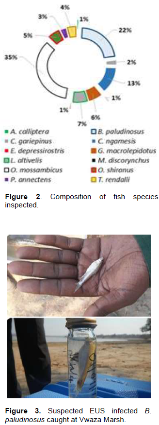
The fish species inspected were: Barbus paludinosus, Oreochromis mossambicus, Clarias gariepinus, Clarias ngamesis, Protopterus annectensbrieni, Eutrophius depressirostris, Oreochromis shiranus, Tilapia rendalli, Labeo altivelis, Gnathonemus macrolepidotus, Marcusenius discorhynchus and Astototilapia calliptera.
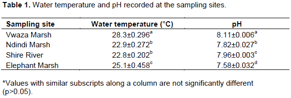
However, all the fish species are reported to be susceptible to EUS (McHugh et al., 2014; OIE, 2018). Fish samples were dominated by O. mossambicus (35%) followed by B. paludinosus (22%) and C. ngamesis (13%). The rest of the samples contributed less than 8% (Figure 2). In all the four sampling sites 0% of fish were found with mycotic granulomas suggestive of EUS. The chance of the population having EUS-diseased fish was smaller (probability of not having disease = 1) at 95% confidence level. One fish sample of B. paludinosus collected at Vwaza Marsh had reddish and swollen caudle peduncle (Figure 3). Appearance of the waters in all the sampling sites was turbid.
DISCUSSION
This study established that no fish was found to be infected with A. invadans. Muscle tissue squash laboratory procedure carried out on B. paludinosus revealed that the fish had a traumatic lesion as evidenced by the presence of inflammatory cells and this did not demonstrate presence of fungal hyphae or granulomas suggestive of EUS (McHugh et al., 2014; Paladini et al. 2017). Turbidity of the waters in all the four sampling sites was due to frequent mixing of bottom mud with water. At Vwaza marsh water turbidity was caused by constant movements of hippopotamuses as this sampling site is one of their habitat for cooling off. At Shire River, Elephant and Ndindi marshes the turbidity was due to winter cropping and active fishing. Communities clear bushy areas along the shores to create gardens to grow crops due to the presence of fertile alluvial soils. Also, frequent canoeing by fishers and or farmers going to various maize gardens while using long paddling sticks reaching the bottom sediments contributed a lot to the turbidity. EUS usually occurs when water temperatures are low between 18°C and 25°C (OIE, 2015; Gomo et al., 2016; FAO, 2017) and the Current study recorded a temperature range of 22.8°C to 28.5°C (Table 1). This shows that fish population in Ndindi marsh and Shire River was prone to the risk of EUS infection with Elephant marsh slightly within the normal range of fungal sporulation. Water temperature recorded at Vwaza marsh was higher rendering A. invadans substantially inactive (Huchzermeyer et al., 2017). Mean values of water temperature showed no significant differences between Ndindi and Shire River (p=0.78).
In terms of pH, the mean values were significantly different (p<0.05) with a mean value of 7.86±0.059. This pH value does not agree to the findings reported by Thapa and Pal (2015), and OIE (2018) who stated that EUS outbreaks occur in acidic waters because the fungus is able to grow and multiply. Despite the fact that water from EUS-infected Zambezi River mixes with those from Malawi’s Shire River and that there is continual downstream and upstream movement of fish, combination of water temperature and pH in the sampling sites could not initiate sporulation of A. invadans to influence severity of EUS lesions.
CONCLUSION
Malawi has never reported cases of EUS infection and this study has been the first of its kind. Based on the findings of this active surveillance, there is no evidence of EUS presence in Malawi. Previous studies reported by Nsonga et al (2013), McHugh et al (2014), Huchzermeyer et al (2017) and FAO (2017) have already demonstrated that a combination of environmental factors such as low water temperature and pH compromise normal fish skin defences. This enables A. invadans to attach to the skin and invade underlying tissues. Therefore, more studies are required targeting clustering period when the water pH and temperature are most suitable for A. invadans sporulation in order to conclude that Malawi is free from EUS and that proper measures and strategies can be put in place to prevent introduction of the disease from other infected areas through various risk factors.
CONFLICT OF INTERESTS
The authors have not declared any conflict of interests.
ACKNOWLEDGMENTS
This active EUS surveillance was funded by FAO through GCP/SFS/001/MUL (Strengthening Controls of Food Safety, Plant and Animal Pests and Diseases for Agricultural Productivity and Trade in Southern Africa Project). We are grateful to Dr Melba Reantaso for making technical and administrative arrangements needed for the surveillance in Malawi. Dr Nihad Fejzic, international consultant, for guidance on data collection and analysis. Director of National parks and wildlife for the approval to sample Vwaza Marsh and for providing security during fish sampling inside Vwaza wildlife Reserve. Profound appreciations go to Directors in the Departments of Fisheries, and Animal Health and Livestock Development for encouragements and inspirations and all the Staff members who supported the surveillance work in Malawi.
REFERENCES
|
Afzali SF, Hassan MD, Sharifpour I, Afsharnasab M, Shankar S (2015). Experimental Infection of Aphanomyces invadans and susceptibility in seven species of Tropical fish. Veterinary World 8(9):1038-1044 |
|
|
Cameron A (2002). Survey Toolbox for Aquatic Animal Diseases. A Practical Manual and Software Package. Australian Centre for International Agricultural Research, Canberra, P. 375. |
|
|
Skelton P (2001). A Complete Guide to Freshwater Fishes of Southern Africa. Struik Publishers, Singapore. |
|
|
Murphy E, Erickson K, Chima J (2013). USAID Office of Food for Peace: Food Security Country Framework for Malawi. FHI360/FANTA, Washington, DC. |
|
|
Nsonga A, Mfitilodze W, Samui KL, Sikawa D (2013). Epidemiology of Epizootic Ulcerative Syndrome in the Zambezi River System: A case study for Zambia. HVM Bioflux 5(1):1-8. |
|
|
McHugh KJ, Christison KW, Weyl OLF, Smit NJ (2014). Histological confirmation of epizootic ulcerative syndrome in two cyprinid species from Lake Liambezi, Zambezi Region, Namibia. African Zoology 49(2):311-316. |
|
|
Yadav MK, Pradhan PK, Sood N, Chaudhary DK, Verma DK, Debnath C, Jena JK (2014). Innate immune response of Indian major carp, Labeo rohita infected with oomycete pathogen Aphanomyces invadans. Fish and Shellfish Immunology 39(2):524-531. |
|
|
OIE (2015). Infection with Aphanomyces invadans (Epizootic Ulcerative Syndrome). Chapter 10.2. In: Aquatic Animal Health Code 10(2):1-6. |
|
|
OIE (2018). Manual of Diagnostic Tests for Aquatic Animals. |
|
|
Thapa GB, Pal J (2015). Role of physicochemical factors of pond water on the outbreak of epizootic ulcerative syndrome and histopathology of affected fishes from eastern Nepal. International Journal of Fisheries and Aquatic Studies 2:1-8. |
|
|
Ilker E, Sulaiman AM, Rukayya SA (2016). Comparison of Convenience Sampling and Purposive Sampling. American Journal of Theoretical and Applied Statistics 5(1):1-4. |
|
|
Government of Malawi (GOM) (2016a). National Agriculture Policy. Lilongwe. |
|
|
Government of Malawi (GOM) (2016b). National Fisheries and Aquaculture Policy. 2nd Edition, Lilongwe. |
|
|
Gomo C, Hanyire T, Makaya PV, Sibanda S (2016). Outbreak of epizootic ulcerative Syndrome (EUS) in Seranochromis robustus Fish species in Darwendale dam, Zimbabwe. African Journal of Fisheries Science 4(1):204-205. |
|
|
Kar D (2016). Pathology of Epizootic Ulcerative Syndrome. Epizootic Ulcerative Fish Disease Syndrome, pp. 83-93. |
|
|
FAO (2017). Report of the international emergency fish disease investigation mission on a suspected outbreak of epizootic ulcerative syndrome (EUS) in the Democratic Republic of the Congo, 13-19 March 2015. |
|
|
Huchzermeyer CF, Huchzermeyer KDA, Christison KW, Macey BM, Colly PA, Hang'ombe BM, Songe MM (2017). First record of epizootic ulcerative syndrome from the Upper Congo catchment: An outbreak in the Bangweulu swamps, Zambia. Journal of Fish Diseases 41(1):87-94. |
|
|
Kim A, Nguyen TL, Kim D (2017). Modern Methods of Diagnosis. In: Austin B, Newaj-Fyzul A (Eds). Diagnosis and Control of Diseases of Fish and Shellfish. pp. 109-146 |
|
|
Paladini G, Longshaw M, Gustinelli A, Shinn AP (2017). Parasitic Diseases in Aquaculture: Their Biology, Diagnosis and Control. In: Austin B, Newaj-Fyzul A (Eds). Diagnosis and Control of Diseases of Fish and Shellfish, pp. 37-108. |
|
|
Walakira J (2017). Mapping study of aquatic animal diseases-Southern Africa. AU-IBAR. |
|
|
Iberahim NA, Trusch F, Van West P (2018). Aphanomyces invadans, the causal agent of Epizootic Ulcerative Syndrome, is a global threat to wild and farmed fish. Fungal Biology Reviews 32(3):118-130. |
|
Copyright © 2024 Author(s) retain the copyright of this article.
This article is published under the terms of the Creative Commons Attribution License 4.0


