Full Length Research Paper

ABSTRACT
The present work evaluated the inhibitor potential of acetylcholinesterase, the preliminary toxicity and determination of active components of different leaf and seed extracts of Parkia platycephala. All extracts were obtained through hot extraction in a closed system (Soxhlet). To obtain the leaf and seed hexanic, methanolic, and ethanolic extracts (LHE, LME, LEE, and SHE, SME, and SEE), sequential extraction was performed on the same plant sample using hexane, methanol, and hydroethanol solution (70%). Preliminary Phytochemical analysis and the characterization by gas chromatography coupled to the mass spectrometer (GC-MS) was performed. The content of phenols total flavonoids and the antioxidant potential was then quantified.The preliminary toxicity against Artemia salina was also evaluated and the potential for acetylcholinesterase inhibition was determined. The presence of tannins, flavonoids, saponins, phytosterols/triterpenoids and alkaloids were detected in phytochemical analysis. The leaf extracts showed antioxidant potential, LEM (IC50 = 30.19 ± 0.75 μg/ml) and LEE (IC50 = 40.62 ± 0.65 μg/ml). The analysis by GC-MS indicated a diversity of volatile compounds, evidencing urs-12-ene (triterpenoid) and 1,2,3-benzenetriol (phenol) in the leaf extracts, and linoelaidic acid (fatty acid), (Z)-9-octadecenamide, tricycle [20.8.0.0 (7.16)] triacontane,1(22),7(16)-diepoxy-, (Z)-7-hexadecenal (fatty aldehyde) in the seed extracts. The preliminary toxicity analysis demonstrated that the use of P. platyceplaha leave and seeds for medicinal purposes is relatively safe. All the extracts inhibited acetylcholinesterase, compared to the physostigmine control, with IC50 values in the range of 9.85 to 15.68 mg/ml. Thus, these data support the use of P. platycephala as a potential therapy for Alzheimer's disease.
Key words: Phytochemistry, antioxidant activity, toxicity, fava-de-bolota, Alzheimer.
INTRODUCTION
The origin of Alzheimer's disease (AD), despite various indications, is still fueled by the cholinergic hypothesis, which relates the symptoms of dementia to the decrease in the level of acetylcholine, a neurotransmitter and neuromodulator (Kuppusamy et al., 2017; Sharma, 2019). The availability of acetylcholine is regulated by the enzyme acetylcholinesterase (AChE), that is, the inhibition of this enzyme increases the concentration of acetylcholine in synapses, improving communication between cells (Decker and Duncan, 2020; Mota et al., 2012).
Among the main therapeutic approaches for AD, a compound isolated from the species Galanthus spp. known as galantamine, has the ability to inhbit acetylcholine production (Heinrich, 2010). The interest in plant species (herbs) with medicinal action against Alzheimer's (Soheili et al., 2021) stems from the numerous side effects associated with the synthetic drugs currently available for AChE inhibition (iAChE), including peripheral toxicity, hepatotoxicity, and gastrointestinal tract disorders (Shaikh et al., 2021).
Despite the advancement of scientific research into new actives against AD, data confirming the iAChE effect of Brazilian plants is still insignificant (less than 1%) when compared to national biodiversity (Santos et al., 2018). Aside from the low national rate, regional studies of the Cerrado Biome discovered that only qualitative iAChE tests were conducted, highlighting the need for a more detailed investigation of the species in the region.
Parkia platycephala is popularly known in Brazil as "fava-de-bolota", "faveira", "faveira-preta", "visgueiro", "fava-de-boi" or "sabi" (Chaves et al., 2020). It has an extensive regional representation as the state of Tocantins' symbol tree and also peculiar characteristics resulting from its location on the border between the Cerrado and the Brazilian Caatinga (Lorenzi, 2020; Tocantins, 2012). Despite the fact that its extracts and isolated actives, primarily from pods, are well-known in the fields of nutrition and pharmacology (gastrointestinal infections in animals), little is known about their therapeutic potential (Araújo et al., 2019; Cavada et al., 2020; Santos et al., 2018). There are reports of the presence of phenolic compounds, flavonoids, flavones, phytosteroids, condensed tannins, and saponins in this species. Such compounds are of great interest in the health field (Oliveira et al., 2017). Among its therapeutic uses, the antinociceptive and anti-inflammatory effects were observed in mice, after intravenous application with purified lectin, obtained from the seed of P. platycephala (Bari et al., 2016). In addition, gastroprotective and anthelmintic effects have been reported in studies with lectin against multiresistant strains of Staphylococcus aureus and Escherichia coli following its association with gentamicin (Silva et al., 2019). The ethanolic extract and the hexane, acetatic and methanolic fractions of the leaf of P. platycephala inhibited trichostrongylid egg hatching (Figueiredo et al., 2020). Although scarce, there are studies with other therapeutic indications for extracts of the specie P. platycephala, such as the preliminary analysis of the iAChE, in which the seed ethanol extracts from plant species of the Brazilian semiarid region were evaluated, indicating anti-cholinesterase activity (71.5%) of this species (Farias et al, 2013). With a view to identifying new iAChEs of natural origin, this research aimed to identify the phytochemical composition, and evaluate the preliminary toxicity of the P. platycephala leaf and seed extracts, as well as the potential acetylcholinesterase inhibitory activity of these extracts.
MATERIALS AND METHODS
Plant material
The samples of the leaves and seeds of P. platycephala were collected within the Federal University of Tocantins – Campus of Palmas, between the coordinates 10°10’55”S and 48°21'45” W. Anfterwards, were registered and incorporated into Tocantins Herbarium (HTO) collection, located at the Federal University of Tocantins (UFT) in Porto Nacional, under the number HTO 12007. The project is registered with SIGEN under the number A06B860.
Preparation of extracts
The plant materials (leaf and seed) were dried in an oven at 60°C for 48 h, powdered with the aid of the Willye knife mill (Fortinox STAR FT 50) with granulometry below Mesh 20 and stored in a contamination-free glass container, preserving it from moisture. Sequential hot extractions were performed in a closed system, using a Soxhlet apparatus. The extraction with hexane was performed to degrease the sample, for a period of 5 h of reflux, obtaining the leaf hexanic extract (LHE) and the seed hexanic extract (SHE). After completing the extraction time, before starting the extraction with the subsequent solvent, the plant sample was dried at room temperature for 12 h. Then, the extraction process was repeated for each solvent (methanol and 70% ethanol), obtaining the leaf and seed methanol extracts (LME, SME) and the leaf and seed ethanol extracts (LEE, SEE). The solvent from all extracts was removed using a rotary evaporator (FISATOM brand) at -600 mmHg at 45°C. Then the extracts were frozen at -70°C, lyophilized in LIOTOP bench freeze dryer L101 and stored in airtight flasks for further analysis.
Phytochemical prospecting
The qualitative characterization of the main classes of secondary metabolites was performed through analysis based on precipitation and/or staining reactions, with specific reagents for flavonoids, tannins, phytosterols, terpenoids, quinones, saponins and alkaloids (Matos, 2009; Saraiva et al., 2018; Simões et al., 2017).
Characterization by gas chromatography mass spectrometry (GC-MS)
The extracts were analyzed by GC-MS using a Shimadzu® chromatograph model QP2020 Ultra equipped with a ZB-5HT column (30 m long ×0.25 mm internal diameter x 0.25 µm film thickness). It was performed under the following conditions: heating at 50°C for 1 min, until reaching 320°C in 35 min. Injection temperature: 320°C; Interface temperature: 320°C; carrier gas (Helium): 1 ml/min; The electron energy was 70 eV and the temperature of the ion source was 320°C; scan mode. 1 µl of each extract was injected, in which the constituents were identified by comparison with the mass spectra of the NIST 14 library.
Determination of the content of total phenolic compounds
The quantification of the total phenolic content was performed using the Folin–Ciocalteu method (Amorim et al., 2012), with modifications, using gallic acid as standard. Samples (0.2 ml) of methanol solutions (1 mg/ml, p/v) from extracts (LHE, LME, LEE, SHE, SME, SEE) or from the standard (4 to 100 μg/ml, p/v) were mixed with an aqueous solution of the Folin-Ciocalteu reagent (0.5 mL to 10%, v/v), sodium carbonate (1 ml to 7.5%, p/v) and 8.3 ml of distilled water, carefully stirred and kept for 30 min protected from light. The absorbance was measured at 760 nm using a UV-VIS HACH DR 5000 spectrophotometer. The total phenol content was determined by interpolating the absorbances of the samples against a calibration curve constructed with the different concentrations of the gallic acid standard (y = 0.006x + 0.0449, adjusted R2 = 0.997), expressed as milligrams equivalent of gallic acid per gram of lyophilized extract (mg EAG/g). All experiments were performed in triplicate.
Determination of total flavonoid content
The quantification of total flavonoids was performed according to Peixoto-Sobrinho (2008), with modifications (Soares et al., 2017). By mixing 0.5 ml of methanol solution (1mg/ ml, p/v) of the extracts (LHE, LME, LEE, SHE, SME, SEE) or the standard (10 to 400 μg/ml, p/v) with an aqueous solution of acetic acid (0.5 ml to 60%, v/v), methanolic solution of pyridine (2 ml to 20%, v/v), aluminum chloride (1 ml to 5%, p/v) and distilled water (6 ml). The blank was constructed by joining all the components of the reaction and the extract or standard, replacing the aluminum chloride with water. The reaction complex and the blank were carefully shaken and kept for 30 minutes protected from light, and the absorbances were measured at 420 nm in a spectrophotometer. Total flavonoid contents were determined by interpolating the absorbances of the samples (discounting the blank absorbance) against a calibration curve constructed with the different concentrations of the standard rutin (y = 0.0022x – 0.0124, adjusted R2 = 0.999), expressed in equivalent milligrams of rutin per gram of lyophilized extract (mg ER/g). All experiments were performed in triplicate.
Determination of antioxidant activity
The antioxidant capacity was measured by the elimination of the stable free radical 2,2-diphenyl-1-picrylhydrazyl (DPPH) according to Peixoto-Sobrinho et al. (2011), using the rutin pattern as a positive control. In triplicate, 0.5 ml of different concentrations of extracts or standards (10 – 200 μg/ml, p/v) were added to a methanolic solution of DPPH (3 ml to 40 μg/ml, p/v). The blank was constructed by replacing DPPH with methanol in the reaction medium. The reaction complex and the blank were shaken and kept for 30 min protected from light, and the absorbances were measured at 517 nm in a spectrophotometer calibrated with methanol. The absorbance of the DPPH solution at 40 μg/ml was also measured and used as a negative control. The free radical scavenging activity or antioxidant activity (AA) was expressed as the percentage of inhibition determined by the equation:
where AA (%) is the percentage of antioxidant activity; Ac, the absorbance of the negative control; Aa, the absorbance of the sample; Ab, the absorbance of the blank. The IC50 (μg/ml) was obtained using the calibration curves obtained by plotting the different concentrations in relation to AA%.
Determination of preliminary toxic activity
The analysis of the preliminary toxicity was performed using the Artemia salina method according to the methodology of Lacerda et al. (2011) with adaptations. Initially, 1 L of 3% saline solution was prepared, with synthetic sea salt, pH adjusted between 8 to 9 (with 1 M sodium carbonate), for incubation of A. salina eggs (0.1 g), which were exposed to artificial light (60W incandescent light bulb) for 24 h to hatch.
To perform the bioassay, ten nauplii were placed in triplicate in test tubes containing, separately, 5 mL of solution of the extracts (LHE, LME, LEE, SHE, SME, SEE) diluted in saline solution of DMSO (1%) in concentrations of 4, 200, 1000 and 4000 μg/ml. A control group was prepared containing only DMSO solution (1%) diluted in saline solution (3%). After a period of 24 h, the number of static nauplii was counted and the percentage of mortality was determined. The mortality percentages of each concentration were used for the linear regression calculations, from which it was possible to determine the IC50 of each extract. The classification of the extract followed the criteria established by NGUTA et al. (2011). Both organic extracts and aqueous extracts with IC50 values < 100 µg/ml have high toxicity, 100 ≤ IC50 ≤ 500 µg/ml have moderate toxicity and IC50 > 1000 µg/ml are considered non-toxic.
Determination of anticholinesterase activity
The inhibiton of the acetylcholinesterase enzyme (AChE) was measured in 96-well flat-bottom plates using an Elisa BIOTEK reader, model ELX 800, software “Gen5 V2.04.11”, based on the methodology described by Ellman et al. (1961). The following solutions were used per well: 25 µl of acetylthiocholine iodide (15 mM), 125 µl of 5.5'-dithiobis-[2-nitrobenzoic] in the Tris/HCl solution (50 nM, pH = 8, with 0.1 M of NaCl and 0.02 M of MgCl2.6H2O. (3 mM, DTNB or Ellman's reagent)), 50 µl of the Tris/HCl solution (50 nM, pH = 8, with 0.1% bovine serum albumin (BSA)), 25 µl of the extract sample dissolved in DMSO (1%) and diluted 10 times in Tris/HCl solution (50 mM, pH = 8) to obtain a final concentration of 0.2 mg/mL (Rhee et al. 2001; Trevisan and Macedo, 2003).
The absorbance was measured at 405 nm for 30 seconds. Then, 25 µl of the enzyme acetylcholinesterase (0.25 U/ml) was added and the absorbance was measured once per minute for a total of 25 min of incubation of the enzyme. As a negative standard, all solutions were used except for the sample. The dilutions of the samples and the positive standards used in the quantitative evaluations in microplate, starting from a stock solution with a concentration of 2 mg/ml were: 200, 100, 50, 25, 12.5, 6.25, 3.12, 1.56 and 0.78 µg/ml. All samples were analyzed in triplicate. The values ??referring to the natural colorings of the extracts were extinguished from the analysis. The percentage of inhibition of acetylcholinesterase was calculated by comparing the reaction rates (substrate hydrolysis) of the samples in relation to the blank (considered total AChE activity, 100%). The standard used as a positive control was physostigmine (eserine).
Statistical analysis
The content of total phenols, flavonoids, antioxidant activity and anticholinesterase activity are presented in mean ± standard deviation (SD) of three determinations. The data were analyzed using the SISVAR version 5.6 program. The IC50 was determined using the GRAPHPAD PRISM program (v.8.0). Analyses of variance (ANOVA) was used to compare the mean values ??obtained in the analysis and the p < 0.05 values ??were considered statistically significant by the Tukey test.
RESULTS AND DISCUSSION
Phytochemical screening
The results obtained for the phytochemical screening of extracts of P. platycephala are shown in Table 1. The qualitative phytochemical screening of leaf and seed extracts of P. platycephala revealed the presence of flavonoids, tannins, phytosterols/triterpenoids, saponins and alkaloids. There are records of the presence of flavonoids, alkaloids, phenols, tannins, saponins, glycosides, terpenoids, anthraquinones and sterols in the leaf, bark, seed and pod of the genus Parkia (P.) (Chanu et al., 2018; Chhikara et al., 2018; Sá-Santos et al., 2018).

It is noticed that extraction with nonpolar solvent (LHE, SHE) was proved to be more efficient in the phytochemical screening of the phytosterol/triterpene metabolites, due to the fact that these compounds are fat soluble (Cheok et al., 2014). The flavonoid and tannin metabolites had a more pronounced presence in the extracts obtained after degreasing (LME, LEE, SME, SEE), that is, with polar solvents. Thus, sequential extraction optimizes the obtainment of secondary metabolites. Extraction with nonpolar solvent eliminates the competition between some metabolites, which have no selectivity when only polar extraction occurs (Soares et al., 2017).
Analysis by GC-MS
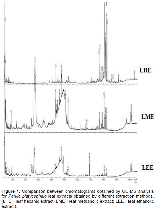
Figures 1 and 2 show the chromatograms obtained by GC-MS from the extracts of the leaves (LHE, LME, LEE, Figure 1) and seeds (SHE, SME, SEE, Figure 2) of P. platycephala. Figures 1 and 2 show the results obtained by GC-MS. It is verified in each extract a variability of compounds, influenced by the polarity of the solvents. Table 2 lists the dominant compounds of LHE, LME, LEE, SHE, SME and SEE.

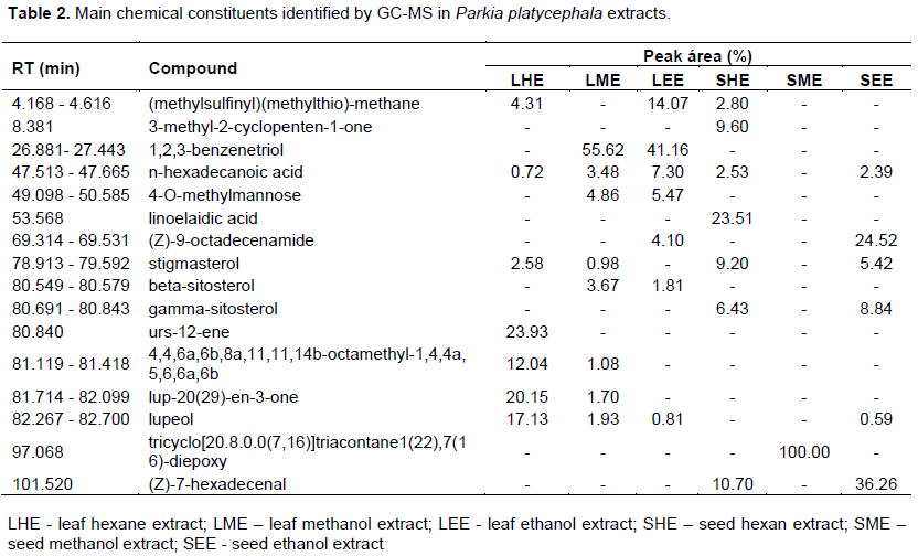
The compounds (methylsulfinyl)(methylthio)-methane, n-hexadecanoic acid (palmitic acid), (Z)-9-octadecenamide (oleamide) and stigmasterol were detected both in the leaf and seed of P. platycephala. It is worth noting that of these, 9-octadecenamide-(Z) is present only in ethanol extracts, mostly in the SEE (24%). The 1,2,3-benzenetriol compound (pyrogallol) was the majority in the LME and LEE, showing greater affinity for extraction with polar solvents (above 40%). Other compounds exclusive to polar leaf extraction are 4-O-methylmannose and beta-sitosterol.
In the hexane extracts, there were some compounds with highly non-polar characteristics, urs-12-ene (23.93%), 4, 4, 6a, 6b, 8a, 11, 11,14b-octamethyl-1,4,4a, 5, 6.6a, 6b (12.04%), lup-20(29) -en-3-one (20.15%) and lupeol (17.13%) in LHE and 2-cyclopenten-1-one, 3-methyl- (9.60%) and linoelaidic acid (23.51%) in SHE. It should be noted that the compounds 3-methyl-2-cyclopenten-1-one, linoelaidic acid, tricyclo[ 20.8.0.0(7.16)]triacontane,1(22),7(16)-diepoxy and (Z)-7-hexadecenal were detected only in the extracts of the seed of P. platycephala. There are reports of the presence of the compounds 1,2,3-benzenetriol and 4-O-methyl-mannose in extracts of the bark and root of P. biglobosa sequentially extracted with hexane, ethyl acetate, ethanol and water (Ibrahim et al., 2013). Gnansounou et al. (2019) detected stigmasterol and sitosterol derivatives in P. biglobosa leaf and bark, while Shahidah et al. (2019) confirmed (Z)-9-octadecenamide-in seeds. Lupeol was detected in various parts of the species P. biglobosa, P. bicolor and P. speciosa (Saleh et al., 2021). Palmitic acid (hexadecanoic acid), which is also found in the genus Parkia, was identified in this study (Olowokere et al., 2018; Sangodare et al., 2017).
As well as urs-12-ene detected in LHE, there are reports of the presence of ursolic acid in the leaves of the species P. javanica (Dinda et al., 2009), both derived from ursane. Derivatives of pentacyclic triterpenoid compounds are highly attractive due to the promising expectations given the variability of biological properties of this class (Luchnikova et al., 2020). The main compounds in Table 2 are biologically active. According to Beulah et al. (2018) pyrogallol has an antimicrobial, anti-inflammatory, antioxidant, analgesic, insecticide, carcinogenic and cytotoxic effect. Palmitic acid, in addition to antioxidant and hypocholesterolmeic activity, has nematicide, pesticide, lubricant, antiandrogenic, hemolytic and 5-alfareductase inhibitor effects (Beulah et al., 2018). Not least, (Z)-7-hexadecenal has antiviral activity (Devakumar et al., 2017), and (Z)-9-octadecenamide has high anti-inflammatory power (Ano et al., 2015).
Pentacyclic triterpenoids (urs-12-ene, lup-20(29)-en-3-one, lupeol) have anti-cancer activity (SHAN et al., 2016), chemopreventive (Prasad et al., 2007), anti-inflammatory (Feng et al., 2018), anti-HIV (Smith et al., 2007) and antimicrobials (Duric et al., 2013). The compound tricyclo[20.8.0.0(7.16)]triacontane,1(22),7(16)-diepoxy, despite being mentioned in some studies of chemical characterization of plant extracts (Mohiuddin et al., 2018), is not related to specific studies of bioactive compounds. The compounds methane (methylsulfinyl) (methylthio), 3-methyl-2-cyclopenten-1-one, 4,4, 6a, 6b, 8a, 11, 11, 14b-octamethyl-1, 4, 4a, 5, 6, 6a, 6b, lup-20(29)-en-3-one, tricycle [20.8.0.0 (7,16)] triacontane, 1(22), 7(16)-diepoxy- and (Z)-7-hexadecenal appear for the first time in the profile of volatile compounds of a species of the genus Parkia.
Content of phenolic, flavonoids and evaluation of antioxidant activity
The analysis of the contents of total phenols, total flavonoids and the antioxidant activity of P. platycephala leaf and seed extracts are shown in Table 3. In the leaf extracts, it was detected contents of total phenols superior to the seed extracts. Regarding phenols, it was also noticed that there was no significant difference between LME (85.11 ± 0.25 mg GAE/g) and LEE (84.17 ± 0.33 mg GAE/g) and that these values 84.57 ± 1.06 mg GAE/g are approximate to those reported in the literature for the ethanol extract of P. biglobosa leaf, obtained after sequential extraction (hexane, ethyl acetate, and ethanol) (Ibrahim et al., 2013). However, for the flavonoid content, this equivalence is not maintained, with the LEE (25.64 ± 0.91 mg RE/g) being 100% higher than the LME (11.85 ± 0.69 mg RE/g).
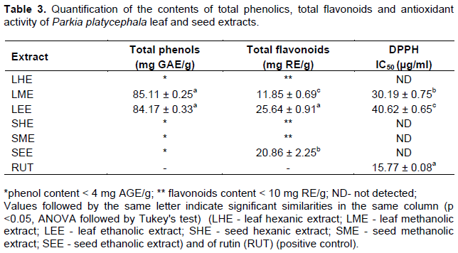
The total phenol content of the seed extracts was less than 4.00 mg GAE/g. Ghasemzadeh et al. (2018) quantified the content of total phenols and flavonoids for the seed extracts of the species P. speciosa and despite the superiority of the total phenol content (14.9 ± 2.03 mg GAE/g) in relation to the seed extracts of the species P. platycephala, the levels of flavonoids (12.4 ± 3.51 mg RE/g) of P. speciosa were lower than the SEE (20.86 ± 2.25 mg RE/g) of P. platycephala.
P. platycephala is described as a species with a high percentage of phenolic compounds, which are normally directly associated with the antioxidant potential (Chun et al., 2005; Figueiredo et al., 2020). The analysis of the antioxidant activity of the extracts, according to the classification adopted by Melo et al. (2010), indicates that the LME and LEE of P. platycephala have high potential, since they do not exceed the limit of three times the coefficient of the positive control, in this case, the rutin (IC50 = 15.77 ± 0.08 µg/ml). In addition, the antioxidant potential of LME (IC50 = 30.19 ± 0.75 µg/ml) is statistically superior to that of LEE (IC50 = 40.62 ± 0.6 µg/ml). Dluya et al. (2017) detected an IC50 = 45.72 µg/ml for the radical scavenging activity (DPPH) of methanol extracts of the species P. biglobosa, which when compared to the antioxidant potential of the LME (IC50 = 30.19 ± 0.75 µg/mL) indicates superiority of the methanol extract of P. platycephala. SHE, SME, SEE and LHE did not reach a 50% dose-response in this test, indicating that their antioxidant activity was low. Farias et al. (2013) confirm the difficulty of quantifying the antioxidant potential of the seed extract of this species. For hexane extracts (LHE, SHE), the low antioxidant activity is justified due to the low affinity of phenolic compounds for nonpolar solvents.
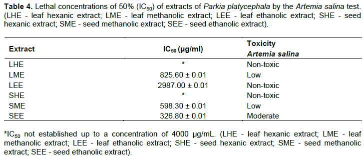
Toxicity
Table 4 defines the IC50 values and toxicity ratios for each P. platycephala extract. LHE and SHE were found to be non-toxic, with IC50 > 4000 µg/ml and LEE with IC50 = 2987.00 ± 0.01 µg/ml. It is noteworthy that the sequential extraction in the seed enabled the isolation of some compounds, which caused the SEE to have moderate toxicity, that is, 100 < IC50 < 500 µg/ml. Figueiredo (2014) reported the non-toxicity of P. platycephala leaf extracts (IC50 > 1000 µg/ml).
In in vivo tests on mice, Fernandes et al. (2010) stated that the leaf ethanolic extracts of P. platycephala did not present acute toxicity (1000 mg/kg) and neither cytotoxicity in erythrocytes of rats (100 µg/ml).
Saleh et al. (2021) confirm these results, stating that the acute toxicity in fish is within the range of 500-5000 mg/kg of body weight, and in rats the IC50 > 5000 mg/kg (Builders, 2012), suggesting that the seed extracts of the genus Parkia are not potentially dangerous as they present moderate or low toxicity.
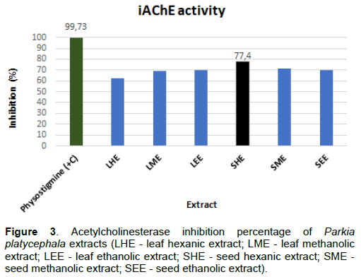
Anticholinesterase activity
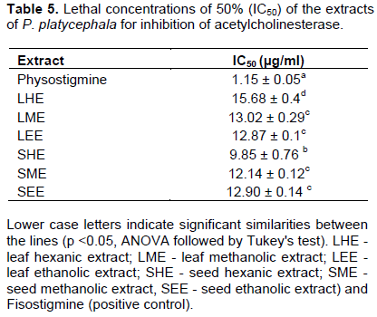
With the extracts chemically characterized and their toxicities established, we can strengthen the potential of the biological activities of P. platycephala and emphasize the inhibitory evaluation of acetylcholinesterase as shown in Figure 3. It is reported that only extracts with iAChE values ??equal to or greater than 50% have a potential for successful fractionation and isolation of the active ingredients capable of inhibiting the enzyme acetylcholinesterase (Trevisan and Macedo, 2003). In this case, as shown in Figure 3, all extracts analyzed in this study showed this characteristic of percentage inhibition, with emphasis on the SHE that obtained the highest percentage of inhibition (77.40%). Previous studies with the seed ethanolic extract of P. platycephala and aqueous from P. biglobosa, the latter defatted, confirm iAChE > 50% (Ademiluyi, 2018; Farias et al., 2013). Table 5 shows all the lethal concentrations of 50% of the extracts of P. platycephala.
According to Table 5, using physostigmine (IC50 = 1.15 ± 0.05 µg/ml) as a positive control, the seed hexanic extract of P. platycephala showed the best iAChE activity, (IC50 = 9.85 ± 0.76 µg/ml). It is noteworthy that the LME, LEE, SEE, SME had significantly similar iAChE activity. These data are the first quantitative reports of the species P. platycephala of the potential for inhibition of this enzyme. In this study, the inhibition of acetylcholinesterase from extracts of P. platycephala can be related to the pyrogallol compound, which, according to Sarikaya (2015), exhibits potent iAChE activity (IC50 = 10.2 μM), and to palmitic acid with moderate inhibitory power of AChE (IC50 = 33.9 µM or 8.69 µg/mL) (Fang et al., 2010).
Ayaz et al. (2017) detected in in vitro and in vivo studies that the beta-sitosterol compound has a strong iAChE (IC50 = 55 μg/ml) with double efficiency in the tests, inhibiting the enzyme acetylcholinesterase and eliminating free radicals. The presence of beta-sitosterol prevents oxidative stress induced by glucose oxidase and lipid peroxidation, presenting a high potential against neurodegenerative diseases such as AD (Shi et al., 2013). Inhibition of acetylcholinesterase has also been observed in studies using stigmasterol (Gade et al., 2017).
Recent studies have verified the iAChE effect of a linoleic acid isomer, demonstrating a threefold reduction in acetylcholine enzyme activity (Cigliano et al., 2019). In this context, tests with a nanoplatform with this same isomer suggested a delivery system actively directed at the central nervous system, with a sophisticated potential to reduce the symptoms of AD (Agwa et al., 2020). The compound oleamide is a chemopreventive agent against AD, has protective activity against scopolamine-induced memory loss, and also regulates microglial activity (Ano et al., 2015; HEO et al., 2003).
In SME, the iAChE effect may be related to the presence of the triacontane derivative. Dalai et al. (2014) reported inhibition of AChE, IC50 = 45.88 ± 1.94 µg/ml, after tests with cinnamon oil, which contained the triacontane compound in its composition. It is also evident that flavonoids, present in the LME, LEE, SME and SEE, have neuro and nephroprotective activities (Athira et al., 2016; Xicota et al., 2017). The iAChE potential of the flavonoids is justified due to the presence and position of the hydroxyl group (OH) in ring A and ring B, and also due to the unsaturation of ring C (Khan et al., 2018). As seen in this study, Morais et al. (2013) stated that there is no direct relationship between antioxidant activity and acetylcholinesterase inhibition, but there is a dependence on these activities due to their structural patterns (Karmakar et al., 2019).
According to Santos et al. (2018), studies relating data from plant species and iAChE, have shown relevant limitations in not associating the antioxidant and toxic effects of the extracts. This research brings concern about minimizing this limitation and confirms that extracts with high iAChE power, IC50 < 20 µg/ml (Santos et al., 2018) do not need to present high toxicity (Huq et al., 2014; Nwidu et al., 2017). Thus, it is thought that it is important to continue research with the extracts evaluated, since they have a remarkable potential for inhibiting acetylcholinesterase and there are few studies on this species.
CONCLUSION
This study provides the first evidence of the inhibition of acetylcholinesterase from the leaf and seed of P. platycephala. Sequential extraction with increasing polarity helped in optimizing the extraction of volatile chemicals and it was efficient in terms of toxicity and acetylcholinesterase inhibition. These data indicate the potential therapeutic use of this plant in medical conditions related to Alzheimer's disease.
CONFLICT OF INTERESTS
The authors have not declared any conflict of interests.
ACKNOWLEDGEMENTS
The authors are grateful to the Dean of Research (PROPESQ) and the Federal University of Tocantins for the financial incentive received. This publication received financial support from Edital nº 38/2020 of the Pro-Rectory for Research (PROPESQ) by the Federal University of Tocantins.
REFERENCES
|
Ademiluyi AO (2018). Local condiments from fermented tropical legume seeds modulate activities of critical enzymes relevant to cardiovascular diseases and endothelial function. Food Science & Nutrition 6(3):602-608. |
|
|
Agwa MM, Abdelmonsif DA, Khattab SN, Sabra S (2020) Self- assembled lactoferrin-conjugated linoleic acid micelles as an orally active targeted nanoplatform for Alzheimer's disease. International Journal of Biological Macromolecules 162:246-261. |
|
|
Amorim ELC, Castro VTNA, Melo JG, Corrêa AJA, Peixoto-Sobrinho TJS (2012). Standard operating procedures (SOP) for the spectrophotometric determination of phenolic compounds contained in plant samples. Latest research into quality control 1:47-66. |
|
|
Ano Y, Ozawa M, Kutsukake T, Sugiyama S, Uchida K, Yoshida A, Nakayama H (2015). Preventive effects of a fermented dairy product against Alzheimer's disease and identification of a novel oleamide with enhanced microglial phagocytosis and anti-inflammatory activity. Plos One 10(3):e0118512. |
|
|
Araújo MJ, Miranda HH, Marques CAT, Batista L, Carvalho FJV, Jácome DLS, Edvan RL, Silva TPD, Bezerra LR, Lima AGVO, Oliveira RL (2019). Effect of replacing ground corn with Parkia platycephala pod meal on the performance of lactating Anglo-Nubian goats. Animal Feed Science and Technology 258:114313. |
|
|
Athira K, Madhana RM, Lahkar M (2016). Flavonoids, the emerging dietary supplement against cisplatin-induced nephrotoxicity. Chemic-Biological Interactions 248:18-20. |
|
|
Ayaz M, Junaid M, Ullah F, Subhan F, Sadiq A, Ali G, Ovais M, Shahid M, Ahmad A, Wadood A, El-Shazly M, Ahmad N, Ahmad S (2017). Anti-Alzheimer's Studies on β-Sitosterol Isolated from Polygonum hydropiper L. Frontiers in pharmacology 8:697. |
|
|
Bari A U, Santiago MQ, Osterne VJS, Pinto-Junior VR, Pereira LP, Silva-Filho JC, Debray H, Rocha BAM, Delatorre P, Teixeira CS, Neto CC, Assreuy AMS, Nascimento KS, Cavada BS (2016). Lectins from Parkia biglobosa and Parkia platycephala: A comparative study of structure and biological effects. International Journal of Biological Macromolecules 92:194-201. |
|
|
Beulah GG, Soris PT, Mohan VR (2018). GC-MS Determination of Bioactive Compounds of Dendrophthoe falcata (L.F) Ettingsh: An Epiphytic Plant. International Journal of Health Sciences & Research, 8(11). |
|
|
Builders M (2012). Toxicity studies of the extracts of Parkia biglobosa Stem Bark in Rats. British Journal of Pharmaceutical Research 2(1):1-16. |
|
|
Cavada BS, Bari AU, Pinto-Junior VR, Lossio CF, Silva MTL, Souza LAG, Oliveira MV, Souza-Filho CHD, Correia SEG, Vital APMS, Lima LD, Osterne VJS, Nascimento KS (2020). Purification and partial characterization of a new lectin from Parkia panurensis Benth. ex H.C. Hopkins seeds (Leguminosae family; Mimosoideae subfamily) and evaluation of its biological effects. International Journal of Biological Macromolecules 145:845-855. |
|
|
Chanu KV, Leishangthem GD, Srivastava SK, Thakuria D, Kataria M, Telang, AG (2018). Phytochemical analysis and evaluation of anticancer activity of Parkia javanica seeds. The Pharma Innovation Journal 7(5):305-311. |
|
|
Chaves SR, Santos RR, Silva ALG (2020). Reproductive biology of Parkia platycephala Benth (leguminosae, caesalpinioideae, clado mimosoide) / Biologia reprodutiva de Parkia platycephala Benth (leguminosae, caesalpinioideae. Brazilian Journal of Development, 6(10):79442-79458 |
|
|
Cheok CY, Salman HAK, Sulaiman R (2014). Extraction and quantification of saponins: A review. Food Research International, 59(1):16-40. |
|
|
Chhikara N, Devi HR, Jaglan S, Sharma P, Gupta P, Panghal A (2018). Bioactive compounds, food applications and health benefits of Parkia speciosa (stinky beans): A review. Agriculture & Food Security, 7(1):1-9. |
|
|
Chun SS, Vattem DA, Lin YT, Shetty K (2005). Phenolic antioxidants from cloral oregano (Origanum vulgare) with antimicrobial activity against Helicobacter pylori. Process Biochemistry 40(2):809-816. |
|
|
Cigliano L, Spagnuolo MS, Boscaino F, Ferrandino I, Monaco A, Capriello T, Bergamo P (2019). Dietary supplementation with fish oil or conjugated linoleic acid relieves depression markers in mice by modulation of the Nrf2 pathway. Molecular nutrition & food research, 63(21):1900243. |
|
|
Costa BA, De Oliveira JM, Sales PA, Lira SRDS, Silva SMDS, Costa LM, Muratori M, Costa AP (2013). Systemic and reproductive toxicity induced by Parkia platycephala ethanolic extract in female Wistar rats. Brazilian Journal of Pharmacognosy 23(6):920-926. |
|
|
Dalai MK, Bhadra S, Chaudhary SK, Chanda J, Bandyopadhyay A, Mukherjee PK (2014). Anticholinesterase activity of Cinnamomum zeylanicum L. leaf extract. CELLMED 4(2):11-1. |
|
|
Decker AL, Duncan K (2020). Acetylcholine and the complex interdependence of memory and attention. Current Opinion in Behavioral Sciences 32(1):21-28. |
|
|
Devakumar J, Keerthana V, Sudha SS (2017). Identification Of Bioactive Compounds By Gas Chromatography-Mass Spectrometry Analysis Of Syzygium Jambos (L.) Collected From Western Ghats Region Coimbatore, Tamil Nadu. Asian Journal of Pharmaceutical and Clinical Research 10(1):364-369. |
|
|
Dinda B, Mohanta BC, Debnath S, Ghosh B, Arima S, Sato N, Harigaya Y (2009). Iridoid glucosides from leaves and stem barks of Parkia javanica. The Journal of Asian Natural Products Research 11(36):229-235. |
|
|
Dluya T, Daniel D, Yusuf U (2017). In vitro Antioxidant Activity and Phytochemical Evaluation of Five Medicinal Plants Extract. The Pharmaceutical and Chemical Journal 4(5):73-82. |
|
|
Duric K, Kovac-Besovic E, Niksic H, Sofic E (2013). Antibacterial activity of methanolic extracts, decoction and isolated triterpene products from different parts of birch, Betula pendula, Roth. Journal of Plant Studies 2(2):61-70. |
|
|
Ellman GL, Courtney KD, Andres V, Featherstone RM (1961) A new and rapid colorimetric determination of acetylcholinesterase activity. Biochemical Pharmacology 7(2):88-90. |
|
|
Fang Z, Jeong SY, Jung HA, Choi JS, Min BS, Woo MH (2010). Anticholinesterase and Antioxidant Constituents from Gloiopeltis furcata. Chemical and Pharmaceutical Bulletin 58(9):1236-1239. |
|
|
Farias DF, Souza TM, Viana MP, Soares BM, Cunha AP, Vasconcelos IM, Ricardo NMPS, Ferreira PMP, Melo VMM, Carvalho AFU (2013). Antibacterial, Antioxidant, and Anticholinesterase Activities of Plant Seed Extracts from Brazilian Semiarid Region. BioMed Research International 2013:510736. |
|
|
Feng J, YI X, Huang W, Wang Y, HE X (2018). Novel triterpenoids and glycosides from durian exert pronounced anti-inflammatory activities. Food Food Chemistry 241:215-221. |
|
|
Fernandes HB, Silva FV, Passos FFB, Bezerra RDS, Chaves MH, Oliveira FA, Oliveira RCM (2010). Gastroprotective effect of the ethanolic extract of Parkia platycephala Benth. leaves against acute gastric lesion models in rodents. Biological Research 43(1):451-57. |
|
|
Figueiredo BNS (2014). Análise fitoquímica, potencial anti-helmíntico e ensaio toxicológico em artemia salina de plantas presentes no ecótono amazônia e cerrado: Leucaena leucocephala, Parkia platycephala e Senna alata. [Dissertação]. Araguaína: Universidade Federal do Tocantins. |
|
|
Figueiredo BNS, Sato MO, Moura LTS, Mariano SMB, Alvim TC, Soares IM, Kawai S, Ascêncio SD, Santos HD, Paiva JA, Sato M, Maruo VM (2020). Preliminary Report on the Effect of Savanna Plants Leucaena leucocephala, Parkia platycephala and Senna alata against Eggs and Immature Stages of Trichostrongylid Nematodes In Vitro. Pathogens 9(12):986. |
|
|
Gade S, Rajamanikyam M, Vadlapudi V, Nukala KM, Aluvala R, Giddigari CS, Karanam NJ, Barua NC, Pandey R, Upadhyayula VSV, Sripadi P, Amanchy R, Upadhyayula SM (2017). Acetylcholinesterase inhibitory activity of stigmasterol & hexacosanol is responsible for larvicidal and repellent properties of Chromolaena odorata. Biochimica et Biophysica Acta General Subjects 1861(3):541-550. |
|
|
Ghasemzadeh A, Jaafar HZE, Bukhori MFM, Rahmat MH, Rahmat A (2018). Assessment and comparison of phytochemical constituents and biological activities of bitter bean (Parkia speciosa Hassk.) collected from different locations in Malaysia. Chemistry Central Journal 12:12. |
|
|
Gnansounou S, Iskandar S, Abou L, Robin M, Giorgio C, Dahouenon E, Piccerelle P, Sonhounhlou D (2019). GC-MS screening and evaluation of the anti-inflammatory and antioxidant activities of ethanolic leaves and stem barks extracts from Dialium guineense Willd, Parkia biglobosa (Jacq.) R. Br. ex Benth. and Tamarindus indica L. Journal of Pharmacognosy and Phytochemistry 8(1):295-301. |
|
|
Heinrich M (2010). Galanthamine from Galanthus and other Amaryllidaceae-chemistry and biology based on traditional use, The Alkaloids: Chemistry and Biology.Elsevier Acedamic Press pp. 157-165. |
|
|
Heo HJ, Park YJ, Suh YM, Choi SJ, Kim MJ, Cho HY, Chang YJ, Hong B, Kim HK, Kim E, Kim CJ, Kim BG, Shin DH (2003). Effects of oleamide on choline acetyltransferase and cognitive activities. Bioscience, Biotechnology and Biochemistry 67(6):1284-1291. |
|
|
Huq AKM, Jamal JA, Stanslas J (2014). Ethnobotanical, Phytochemical, Pharmacological, and Toxicological Aspects of Persicaria hydropiper (L.) Delarbre. Evidence-Based Complementary and Alternative Medicine 2014:782830. |
|
|
Ibrahim MA, Koorbanally N, Islam MS (2013). In Vitro Anti-Oxidative Activities of The Various Parts of Parkia Biglobosa And GC-MS Analysis of Extracts With High Activity. African Journal of Traditional, Complementary, and Alternative Medicines 10(5):283-291. |
|
|
Karmakar A, Ambure P, Mallick T, Sreeparna DAS, Kunal R, Begum NA (2019). Exploration of synthetic antioxidant flavonoid analogs as acetylcholinesterase inhibitors: an approach towards finding their quantitative structure-activity relationship. Medicinal Chemistry Research 28(5):723-741. |
|
|
Khan H, Marya, Amin S, Kamal MA, Patel S (2018). Flavonoids as acetylcholinesterase inhibitors: Current therapeutic standing and future prospects. Biomedicine & Pharmacotherapy 101:860-870. |
|
|
Kuppusamy A, Arumugam M, George S (2017). Combining in silico and in vitro approaches to evaluate the acetylcholinesterase inhibitory profile of some commercially available flavonoids in the management of Alzheimer's disease. International Journal of Biological Macromolecules 95:199-203. |
|
|
Lacerda AM, Modolo AK, Matias RC, Pistori H, Yano M, Roel AR, Porto KRA (2011). Screening de plantas com potencial fitotóxico. Revista Brasileira de Farmácia 92(4):352-355. |
|
|
Lorenzi H (2020). Árvores Brasileiras: manual de identificação e cultivo de plantas arbóreas nativas do Brasil. 8ed. São Paulo: Instituto Plantarum. |
|
|
Luchnikova NA, Grishko VV, Ivshina IB (2020). Biotransformation of Oleanane and Ursane Triterpenic Acids. Molecules 25(23):5526. |
|
|
Matos FJA (2009). Introdução à Fitoquímica Experimental. 3ed. Fortaleza: UFC. Fortaleza 147 Pp. |
|
|
Melo JG, Araújo TAS, Castro VTNA, Cabral DLV, Rodrigues MD, Nascimento SC, Amorim ELC, Albuquerque UP (2010). Antiproliferative activity, antioxidant capacity and tannin content in plants of semi-arid northeastern Brazil. Molecules 15(12):8534-8542. |
|
|
Mohiuddin YG, Nathar VN, Aziz WN, Gaikwad NB (2018). Investigations on important secondary metabolites from aerial parts of Artemisia absinthium L. using GC-MS. Journal of Pharmacognosy and Phytochemistry 7(1):820-827. |
|
|
Morais SM, Lima KSB, Siqueira SMC, Cavalcanti ESB, Souza MST, Menezes JESA, Trevisan MTS (2013). Correlação entre as atividades antiradical, antiacetilcolinesterase e teor de fenóis totais de extratos de plantas medicinais de farmácias vivas. Revista Brasileira de Plantas Medicinais 15(4):575-582. |
|
|
Mota WM, Barros ML, Cunha PEL, Santana MVA, Stevam CS, Leopoldo PTG, Fernandes RPM (2012). Avaliação da inibição da acetilcolinesterase por extratos de plantas medicinais. Revista Brasileira de Plantas Medicinais 14(4):624-628. |
|
|
Murray AP, Faraonia MB, Castro MJ, Alza NP, Cavallaro V (2013). Natural AChE Inhibitors from Plants and their Contribution to Alzheimer's Disease Therapy. Current Neuropharmacology 11(4):388-413. |
|
|
Nguta JM, Mbaria JM, Gakuya D, Gathumbi PK (2011). Biological screening of Kenya medicinal plants using Artemia salina L. (Artemiidae). Pharmacologyonline 2:458-78. |
|
|
Nwidu LL, Elmorsy E, Thornton J, Wijamunige B, Wijesekara A, Tarbox R, Warren A, Carter WG (2017). Anti-acetylcholinesterase activity and antioxidant properties of extracts and fractions of Carpolobia lutea. Pharmaceutical Biology 55(1):1875-1883. |
|
|
Oliveira LV, Anjos CJF, Confessor M, Vilar DA, Vilar MSA 2017. Fitoterapia como alternativa ao retardamento do alzheimer. Available at: http://www.editorarealize.com.br/artigo/visualizar/2933. |
|
|
Olowokere JA, Onen AI, Odineze MC, B'aga ID, Akoji JN (2018). Extraction and Characterization of Oil from African Locust Bean (Parkia biglobosa) Seed. Asian Journal of Applied Chemistry Research 2(2):1-11. |
|
|
Peixoto-Sobrinho TJS, Silva CHTP, Nascimento JE, Monteiro JM, Albuquerque UP, Amorim ELC (2008). Validação de metodologia espectrofotométrica para quantificação dos flavonoides de Bauhinia cheilantha (Bongard) Steudel. Revista Brasileira de Ciências Farmacêuticas 44(4):683-689. |
|
|
Peixoto-Sobrinho TJDS, Castro VTNA, Saraiva AM, Almeida DM, Tavares EA, Amorim ELC (2011). Phenolic content and antioxidant capacity of four Cnidoscolus species (Euphorbiaceae) used as ethnopharmacologicals in Caatinga, Brazil. African Journal of. Pharmacy and Pharmacology 5(20):2310-2316. |
|
|
Prasad S, Kalra N, Shukla Y (2007). Hepatoprotective effects of lupeol and mango pulp extract of carcinogen induced alteration in Swiss albino mice. Molecular Nutrition & Food Research 51(3):352-359. |
|
|
Quansah L, Mahunu GK, Tahir HE, Mariod AA (2019). Parkia biglobosa: Phytochemical Constituents, Bioactive Compounds, Traditional and Medicinal Uses. In Mariod A. (eds) Wild Fruits: Composition, Nutritional Value and Products. Springer International Publishing 1:271-284. |
|
|
Rhee IK, Van de Meent M, Ingkaninan K, Verpoorte R (2001). Screening For Acetylcholinesterase Inhibitors From Amaryllidaceae Using Silica Gel Thin-Layer Chromatography In Combination With Bioactivity Staining. Journal of Chromatography A 915(1-2):217-223. |
|
|
Saleh MSM, Jalil J, Zainalabidin S, Asmadi AY, Mustafa NH, Kamisah Y (2021). Genus Parkia: Phytochemical, Medicinal Uses, and Pharmacological Properties. International Journal of Molecular Sciences 22(2):618. |
|
|
Santos TC, Gomes TM, Pinto BAS, Camara AL, Paes AMA (2018). Naturally Occurring Acetylcholinesterase Inhibitors and Their Potential Use for Alzheimer's Disease Therapy. Frontiers in Pharmacology 9:1192. |
|
|
Sangodare RSA, Okibe PO, Mohammed M, Jajere MU, Abubakar A, Aribido OS, Bala S, Danmallam AA, Ali EO, Onuora O, Ukwueje N, Kolo MT (2017). Chemical Investigation Of Parkia biglobosa Fruit Hull Using GC-MS. American Journal of Research Communication 5(2):59-65. |
|
|
Saraiva LCF, Maia WMN, Leal FR, Maia Filho ALM, Feitosa CM (2018). Triagem fitoquímica das folhas de Moringa oleífera. Boletim Informativo Geum 9(2):12-19. |
|
|
Sá-Santos MM, da Silva FMP, da Silva JFM, Pimenta RS (2018). Phytochemistry and antibacterial activity of aqueous and hydroalcoholic extracts of three medicinal plants against food pathogens. Acta Scientiarum Biological Sciences 40:1-6. |
|
|
Sarikaya SBO (2015). Acethylcholinesterase inhibitory potential and antioxidant properties of pyrogallol, Journal of Enzyme Inhibition and Medicinal Chemistry 30(5):761-766. |
|
|
Shahidah AA, Farouq AA, Magashi MA, Sokoto AM (2019). Comparative Amino Acid and Volatile Flavor Profile of Dawadawa Produced from the Seeds of P. biglobosa, G. max and H. sabdariffa. Journal of Advances in Microbiology 14(3):1-13. |
|
|
Shaikh S, Dhavan P, Ramana MMV, Jadhav BL (2021). Design, synthesis and evaluation of new chromone-derived aminophosphonates as potential acetylcholinesterase inhibitor. Molecular Diversity 25:811-825. |
|
|
Shan J, Xuan Y, Zhang Q, Zhu C, Liu Z, Zhang S (2016). Ursolic acid synergistically enhances the therapeutic effects of oxaliplatin in colorectal cancer. Protein & Cell 7(8):571-585. |
|
|
Sharma K (2019). Cholinesterase inhibitors as Alzheimer's therapeutics (review). Molecular Medicine Reports 20(2):1479-1487. |
|
|
Shi C, Wu F, Zhu X, Xu J (2013). Incorporation of β-sitosterol into the membrane increases resistance to oxidative stress and peroxidation via estrogen receptor-mediated PI3K/GSK3β signaling lipid. Biochimica et Biophysica Acta 1830(3):2538-2544. |
|
|
Silva RRS, Silva CR, Santos VF, Barbosa CRS, Muniz DF, Santos ALE, Santos MHC, Rocha BAM, Batista KLR, Costa-Júnior LM, Coutinho HDM, Teixeira CS (2019). Parkia platycephala lectin enhances the antibiotic activity against multi-resistant bacterial strains and inhibits the development of Haemonchus contortus. Microbial Pathogenesis, 135:103629. |
|
|
Simões CMO, Schenkel EP, Mello JCP, Mentz LA, Petrovick PR (2017). Farmacognosia: do produto natural ao medicamento. Porto Alegre, RS: Artmed. |
|
|
Smith PF, Ogundele A, Forrest A, Wilton J, Salzwedel K, DOTO J, Allaway GP, Martin DE (2007). Antimicrobial agents and chemotherapy 51(10):3574-3581. |
|
|
Soares IM, Rireiro MF, Costa OJ, Sousa EE, Aguiar AA, Barbosa RS, Alvim TC, Ascêncio SD, Aguiar RWS (2017). Application of a degreasing process and sequential ultrasound-assisted extraction to obtain phenolic compounds and elucidate of the potential antioxidant of Siparuna guianensis Aublet. Journal of Medicinal Plant Research, 11(1):357-66. |
|
|
Soheili M, Karimian M, Hamidi GhA, Salami M (2021). Alzheimer's disease treatment: The share of herbal medicines. Iran Journal of Basic Medicinal Science 24(2):123-135. |
|
|
Tocantins (2012). Lei nº 2.619, de 9 de agosto de 2012. Define os Símbolos da Natureza do Estado do Tocantins, e adota outras providências. Diário Oficial do Estado do Tocantins 3691:1. |
|
|
Trevisan MTS, Macedo FVV (2003). Seleção de plantas com atividade anticolinesterase para tratamento da doença de Alzheimer. Química Nova 26(3):301-304. |
|
|
Xicota L, Rodriguez-Morato J, Dierssen M, La Torre R (2017). Potential Role of (-)-Epigallocatechin-3-Gallate (EGCG) in the Secondary Prevention of Alzheimer Disease. Current Drug Targets 18(2):174-195. |
|
Copyright © 2024 Author(s) retain the copyright of this article.
This article is published under the terms of the Creative Commons Attribution License 4.0


