Full Length Research Paper

ABSTRACT
Family Clusiaceae is spread throughout tropical as well as temperate zones, presenting many triterpenes, reported as selective antitumoral agents. The aim of this study is to isolate and identify triterpenes from Clusia studartiana C. M. Vieira & Gomes da Silva and evaluate their cytotoxicity in melanoma (SKMEL 28) and myeloid leukemia (K562) cell lines. Thus, the hexanic extract was processed by phytochemical methods to isolate and purify the pentacyclic triterpenes: 3-oxo-friedelin (1), 3-β-hydroxy-friedelin (2) and 3-oxo-olean-12-en-28-oic-acid (3), identified by spectroscopic methods. Cytotoxicity was evaluated by MTT assay, apoptosis/necrosis by Annexin V/PI, caspase activity by FLICA in flow cytometry and P-gp modulation was measured by interference in the efflux of Rhodamine 123. From the results, triterpenes 2 and 3 showed inhibitory effect in K562 cells proliferation, and only the compound 3 was able to increase percentage of AnnexinV+PI- in cells (p<0.05), with 40% increase in caspase 3/7+ cells and showed inhibition on P-gp activity.
Key words: Apoptosis, caspase, Clusiaceae, Clusia studartiana, cytotoxicity, multidrug resistance.
INTRODUCTION
The family Clusiaceae is spread in temperate and tropical areas regions with 14 genera distributed in 800 species, including trees and shrubs, hemiepiphytes, epiphytes and lianes (Stevens, 2017). The plants of this family are cultivated for various purposes, such as production of noble wood, edible fruits and to bioactive compounds for the pharmaceutical industries (Souza et al., 2013; Kotraswamy et al., 2016). In ethnomedicine, this family has been used for the treatment of numerous diseases, including inflammation, infection and cancer (Melo et al., 2014). In particular, many pentacyclic triterpenes have already been found in this family (Jamila et al., 2015; Duprat et al., 2017; Ribeiro et al., 2019).
The number of studies using natural products as chemotherapeutic agents has increased exponentially in recent years and pentacyclic triterpenes are often described with a wide range of pharmacological activities. These compounds are divided into three structural types: oleanane, ursane and friedelane (Salvador et al., 2017). In this context, oleanane, ursane and friedelane-types exhibit inhibitory activity against various intra and extracellular targets in eukaryotic cells and proteins that have participation in different processes such as cellular development, differentiation, inflammation, angiogenesis, apoptosis and metastasis (Iqbal et al., 2018; Peron et al., 2018; Ren and Kinghorn, 2019). Thus, they are considered as multitarget agents, acting through several signaling pathway, involved in cancer progression and survival. This ability makes them interesting for the development of new antineoplastics agents (Salvador et al., 2017; Ren and Kinghorn, 2019). In addition, pentacyclic triterpenes present selective cytotoxicity, capable to distinguish between tumor and non-tumor-cells, possibly associated with their ability to modulate tumor microenvironment and the immune system (Kamble et al., 2014). Therefore, these compounds have low toxicity in normal cells and high efficiency on cancer cells, reported in many in vitro and in vivo studies, including clinical trials (Gill et al., 2016; Zhang et al., 2014).
In cancer chemotherapy, the occurrence of multidrug resistance (MDR) is one of the main obstacles to the successful treatment. MDR has various molecular mechanisms such as the increase of drug efflux, activation of detoxification systems, alteration of drug targets, changes in cell cycle, activation of DNA repair and escape of apoptosis (Ye et al., 2019). In this context, P-glycoprotein (P-gp), a drug transporter more frequently related to MDR pumps substrates out of cells by an ATP-dependent mechanism, promoting decrease in the intracellular accumulation of many antitumor drugs to sub-therapeutic levels, reducing or abolishing the efficacy of the treatment. Some MDR reversal agents have been developed and participated in clinical trials. However, most of them failed due to severely adverse effects (Liu et al., 2015; Robey et al., 2018). In this context, natural products such as terpenoids, alkaloids and flavonoids are a source of promising anti-cancer compounds and also as potential modulators of MDR (Kumar and Jaitak, 2019), through its combination with chemotherapeutics agents (Mahdizadeh et al., 2016). Considering the chemical profile and antineoplastic activity already described for the Clusiaceae family, the present study aimed to evaluate the chemical composition, cytotoxicity, pro-apoptotic nature and the effects of MDR with the isolated compounds of Clusia studartiana C.M.Vieira and Gomes da Silva
MATERIALS AND METHODS
Plant material
Aerial parts of C. studartiana C.M. Vieira & Gomes da Silva (Clusiaceae) were collected at the Ecological Reserve of Macaé de Cima, Sophronites Site, Atlantic Forest, Nova Friburgo, Rio de Janeiro RJ (22°23'56.8"S 42°30'34.3"W), identified by Dr Marcos Nadruz. The specimen was deposited in the Herbarium of the Botanic Garden of Rio de Janeiro, number RB 336999. The species is registered in the Genetic Patrimony (SISGEN) under the number AB5D582.
Extraction, isolation and purification of pentacyclic triterpenes
The extract of the aerial parts (1.40 kg) of C. studartiana was prepared by dynamic maceration, at room temperature, with hexane until exhaustion. The extract was filtrated and evaporated under reduced pressure to obtain the hexanic extract (26.31 g). During concentration of the hexanic extract, an amorphous solid (10.14 g) was obtained. This solid was chromatographed on silica gel, using mixtures of solvents with increasing polarities (cyclopentane, cyclopentane/ethyl acetate, ethyl acetate and methanol). The fractions were combined according to their chromatographic profile by thin layer chromatography (TLC) and visualized with Godin detection reagent. After successive recrystallizations, it was obtained the pentacyclic triterpenes: 3-oxo-friedelin (3.29 g), 3β-hydroxy-friedelin (0.23 g), 3-oxo-olean-12-en-28-oic acid (5.78 g) pures.
The purity and identification of these compounds were determined by GC-MS and/or the structural characterization by one- and two-dimensional methods in 1H- and 13C-NMR (500MHz). The molecular formula of the acid isolated was determined through direct insertion in a Bruker microTOF–QII mass spectrometer by accurate mass and algorithmic analysis of its isotopic pattern (Bruker SmartFormula 3D True Isotopic Pattern). This acid was methylated with diazomethane (Black, 1983). For pharmacological assays, a pentacyclic triterpenes stock solution (40mM) was performed using cell culture grade DMSO (Sigma), diluted to working concentrations, in cell culture medium minutes prior treatment.
Cellular cytotoxicity assay with colorimetric method of the MTT
For this assays, human myeloid leukemia cells (K562) and human melanoma cells (SK-MEL-28) were respectively cultured in RPMI 1640 or DMEM supplemented with 10% FBS, 100 U/I penicillin, 0.1 mg/mL streptomycin and 0.05 mg/mL gentamicin and maintained in log growth phase. Twenty-four hours before treatment with samples, 100 μL of the cell suspension (5×10⁴ cells/ml) was added to 96-well plates and maintained in a 5% CO2 atmosphere at 37°C. Treatment with the pentacyclic triterpenes was performed in a single (10 μM) or multiple concentrations (0.01 to 100 µM), each in triplicate. Negative control consisted of cells incubated with vehicle (0.25% of cell culture grade DMSO in medium; SIGMA), and staurosporine (SIGMA; 1 μM) as inhibition control. After 48 h, the 3-[4,5-dimethylthiazol-2-yl]-2,5-diphenyltetrazolium bromide (MTT; SIGMA) was added and the plate maintained in CO2 incubator for 4 h. After the incubation time, the supernatant was aspirated and formazan crystals, formed by cellular activity, solubilized with isopropyl alcohol (SIGMA) (Mosmann, 1983; Spinner, 2001). Absorbance was measured at 540 nm wavelength in a microplate reader (Victor X5; Perkin Elmer USA)
Determination of apoptosis/necrosis by flow cytometry analysis (Annexin V/propidium iodide)
For apoptosis assays, 105 cells K562 cells were seeded (1 ml at 105 cells/ml) in a 24-well plate and incubated in a 5% CO2 atmosphere at 37 °C. Twenty-four hours after seeding, cells were treated in duplicate with test samples (10 and 100 μM) for 4 h. Negative control consisted of cells incubated with vehicle (0.25% of cell culture grade DMSO in medium; SIGMA), and staurosporine (Sigma; 5 μM) was used as positive control. Next, cells were collected in 4 mL tubes, centrifuged and washed with sterile phosphate buffered saline (PBS). After centrifugation, the supernatant was discarded and cells resuspended in 100 μl of binding buffer (10 mM HEPES, 140 mM NaCl, and 2.5 mM CaCl2, pH 7.4.) containing Annexin V, as recommended by the manufacturer (Annexin V-Alexa Fluor 488 – Molecular Probes). Cells were incubated at room temperature (20 min) and protected from light. Then 400 μl of binding was added and contents were transferred to flow cytometry tubes kept on ice. It was added 2 μl of a propidium iodide (PI; SIGMA) solution (250 μg/ml in binding buffer) at the time of acquisition, (within 5 min) to avoid false positive due to overexposition with PI. Data acquisition was performed with a FACScalibur flow cytometer (Becton Dickinson, San Jose, CA) using CellQuestTM software (Becton Dickinson). Forward- and side-scatters were set to exclude debris. At least 104 cells were analyzed per sample. Apoptosis status was classified by cell staining profile: early apoptosis (Annexin V+/PI-); late apoptosis (Annexin V+/PI+); necrosis (Annexin V-/PI+).
Determination of caspase activity by flow cytometry analysis (FLICA)
For this procedure, 105 cells K562 cells were seeded (1 ml at 105 cells/ml) in a 24-well plate and incubated in a 5% CO2 atmosphere at 37°C. Twenty-four hours after seeding, cells were treated in duplicate with samples (100 μM), for 6 h and centrifuged. Negative control consisted of cells incubated with vehicle (medium containing 0.25% of cell culture grade DMSO; SIGMA) or staurosporine (Sigma; 5 μM), as positive control. Supernatant was discarded and the cells resuspended in 100 μl culture medium and FLICA solution was added according to the manufacturer's instructions (Vybrant FAM Caspase-3 and -7 Assay Kit - Molecular Probes); cells were incubated for 60 min at 37°C in a CO2 protected from light. After the incubation time, cells were washed twice and resuspended in wash buffer and fixed by adding the fixing buffer. Experiments were performed twice in duplicate with a total of 104 events acquired. Data acquisition was performed with a FACScalibur flow cytometer (Becton Dickinson, San Jose, CA) and analysis using CellQuestTM software (Becton Dickinson). Forward- and side-scatters were set to exclude debris.
Monitoring assay of P-gp activity
The methodology for monitoring P-gp activity was adapted from the procedure described by Maia et al. (1996). K562-Lucena 1 (a K562 resistant cell line with multidrug resistant (MDR) characteristics) was cultured in RPMI 1640 medium at 37°C in a 5% CO2 atmosphere. In a U-bottom 96-well plate, 2×10⁵ cells/well were incubated with vehicle as negative control (medium containing 0.25% of cell culture grade DMSO; SIGMA), samples (100 μM) or verapamil (10 μM ; positive control for P-gp inhibition) in the presence of rhodamine 123 (200 ng/ml) for 45 min at 37°C. After incubation, cells were centrifuged, the supernatant discarded and cells washed by adding ice-cold PBS (200 μl). Cells were maintained on ice until data acquisition in a FacsCalibur flow cytometer. Experiments were performed twice in triplicate, a total of 104 events were acquired and Rhodamine 123 (Rh123) fluorescence monitored on FL-1 channel and analysis performed by CellQuest program.
Statistical analysis
The results were expressed as the mean, plus or minus the standard error of the mean (SEM). Statistical analysis was performed by One Way ANOVA variance analysis, followed by Newman Keuls test and considered significant when p < 0.05.
RESULTS AND DISCUSSION
Three compounds were isolated and purified by phytochemical methods from C. studartiana namely 3-oxo-friedelin (1), 3-β-hydroxy-friedelin (2), 3-oxo-olean-12-en-28-oic-acid (3). The compound methyl olean-12-en-3-oxo-28-oate (4) was obtained after derivatization of acid (3) by diazomethane (Black 1983) (Figure 1). Triterpenes were characterized by one- and two-dimensional methods in 1H- and 13C-NMR and confirmed by comparison with spectral data from the literature (Queiroga et al., 2000; Mkounga et al., 2016).

These pentacyclic triterpenes were tested on K562 and SK-MEL-28 cell lines for their ability to inhibit in vitro cell proliferation. We have adopted the strategy of testing molecules at a single concentration (10 µM) and, if an inhibition above 30% was observed, the molecule was further tested in multiple concentrations. This methodology follows the NCI strategy for drug discovery (Shoemaker, 2006). Among compounds tested at 10 µM, the compounds 2 and 3 showed inhibitory effect (41.59 % and 34.27 %, respectively) on K562 cells, while the compounds 1 and 4 showed inhibitory effect lower than 30%.All of these compounds have had no cytotoxicity on SK-MEL-28 cells (Table 1).

Compounds (10 mM) were tested for cell proliferation, as described in methodology with Staurosporine (STAU; 1 mM) as a positive control. For calculation purposes, cells cultured in medium containing 0.25% DMSO were used as 100% proliferation. Percentages of inhibition are shown as mean ± standard error of mean (SEM) of a triplicate.
Compounds 2 and 3 caused growth inhibition on K562 myeloid leukemia cells, in a dose response manner with IC50 of 8.92 µM and 26.67 µM respectively, with a maximum inhibition of 89.32% triggered by compound 2 at 100 µM (Figure 2). Due to its performance and good IC50 on K562 cell line, we have also tested compound 2 on K562 Lucena cell line proliferation (MDR phenotype). We have observed, as expected, an increase in the compound 2 IC50 from 8.92 µM on K562 cell line to 23.69 µM on K562 Lucena cell line. Cells cultured in medium containing 0.25% DMSO were used as controls for 100% proliferation, points represent mean ± standard error of mean (SEM) of six replicates from two independent experiments. Curves were analyzed using Prism 5 software for IC50 determination. Between these compounds only 3 was able to modulate P-gp activity on K562 Lucena, a K562 resistant cell line with multidrug resistant (MDR) characteristics. This acid increased retention of rhodamine 123, observed through fluorescence of labelled cells. This suggests that this terpenoid could alter MDR phenotype of tumor cells (Figure 3). Further studies are necessary to better understand this aspect of this terpenoid.


Samples were processed and the mean fluorescence intensity (MFI) was evaluated by flow cytometry. Columns represent mean + SEM of a triplicate from a representative experiment. Differences were considered significant when p<0.05 after statistical analysis by One Way ANOVA followed by Newman Keuls. * means p<0.05, when compared to cells treated with medium + means p<0.05, when compared to cells treated with medium and verapamil.
Other structure related to pentacyclic triterpenes, such as betulinic, oleanolic and pomolic acids, present inhibitory effect on K562 proliferation (Fernandes et al., 2003), suggesting that triterpenes evaluated in this study may have a mechanism of action similar of those terpenoids (Kumar et al., 2018). Li et al. (2010) have described that friedelin and other structure-related molecules inhibit HL-60 proliferation. Moreover, Huang et al. (2006) demonstrated that compound 3 presented cytotoxicity against several tumoral cells in vitro (HELF, Ketr-3, Bel7402, A549, HT-1080, PC-3M, KB and KB-V cells) and in vivo (B16-BL6), reinforcing the antitumoral component of compound 3 observed on K562 cells. Herein we have shown, for the first time, the cytotoxic activity of compound 3 on K562 leukemia cells and its ability to modulate the P-gp activity on K562 Lucena.
Samples were processed in duplicate and analyzed for Annexin V expression and PI labelling by flow cytometry. Apoptosis status classified by cell staining profile: early apoptosis (Annexin V+/PI-); late apoptosis (Annexin V+/PI+); necrosis (Annexin V-/PI+). Percentages of a representative sample from a valid experiment are shown. We have analyzed apoptosis and caspase activation to better understand the mechanisms of growth inhibition on K562 cells by compounds 2 and 3. As shown in Figure 4, compound 3, but not compound 2, was able to change annexin V expression in K562 cells in a single cell level. We have observed that compound 3 treatment was able to increase the percentage of annexin V+ cells (Figure 5B); however these cells were not stained with PI, suggesting that cells were in early apoptosis (annexin V+/PI-; Figure 5A). However, compound 2 failed to change apoptosis or necrosis after treatment. This suggests a different mechanism of action for this compound, probably a delayed caspase activation or a caspase-independent mechanism (Giampazolias et al., 2017).

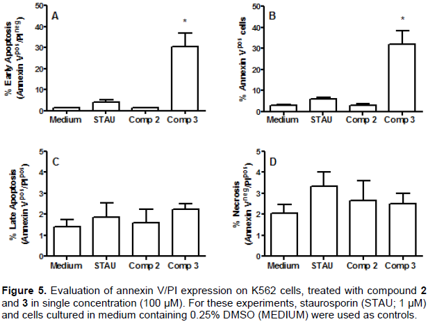
Samples were processed in duplicate and analyzed by flow cytometry and apoptosis status classified by cell staining profile: early apoptosis (Annexin V+/PI-); late apoptosis (Annexin V+/PI+); necrosis (Annexin V-/PI+). Columns represent mean + SEM, from two independent experiments. Differences were considered significant when p<0.05 after statistical analysis by One Way ANOVA variance analysis, followed by Newman Keuls. * means p<0.05, when compared to cells treated with medium.
Pentacyclic triterpene 2 failed to trigger apoptosis or necrosis in K562 cells. This data were supported by evaluation of caspase 3/7 activity as there was no increase in activity after 2 treatment, suggesting that this triterpenoid may trigger a delayed caspase activation or, more interestingly, causing cell death in a caspase-independent manner (Figure 6). On the other hand, we have observed a raise in 40% of caspase 3/7+ in 3 treated cells (from 2.03% in medium to 2.85% after treatment), when compared to cells cultured in medium alone, in agreement with proliferation and annexin V expression.

Triterpenoids such as betulinic acid, friedelin, their derivatives and structure related molecules have antitumor activities (Peron et al., 2018; Kumar et al., 2018). These molecules may trigger intrinsic apoptosis via different pathways. Moreover, betulinic acid may also trigger apoptosis in a caspase-independent fashion (Kumar et al., 2018). Herein, we have described that compound 2 have a inhibitory activity on cell proliferation (Figure 2) without changes on annexin V expression in the outer membrane (Figure 5) and caspase 3/7 activity (Figure 6). This suggests that compound 3 may activate a different pathway for apoptosis induction, independent of caspase activation, that still need to be investigated. On the other hand, similarly to observed after treatment of cancer cells with betulinic acid or friedelin-like molecules (Raghuvar et al., 2005; Subash-Babu et al., 2017; Kumar et al., 2018), compound 3 increased annexin V expression and induced a discrete increase in caspase 3/7 activity, suggesting that this compound may trigger the mitochondrial pathway. The exact mechanisms underpinning apoptosis triggered by compound 3 are still under investigation. Samples were processed and analyzed by flow cytometry, and results shown as mean + SEM of a duplicate. Differences were considered significant when p<0.05 after statistical analysis by One Way ANOVA variance analysis, followed by Newman Keuls. ** means p<0.05, when compared to cells treated with medium.
CONCLUSION
In conclusion, compound 3 caused cell growth inhibition in human myeloid leukemia cells (K562); it was able to induce apoptosis, by a mechanism that involved the activation of caspases 3/7 and promoted discrete inhibition of P-gp activity, an important aspect of MDR phenotype. Further studies are needed for a better understanding of apoptosis-induction processes triggered by this compound.
CONFLICT OF INTERESTS
The authors have not declared any conflict of interests.
ACKNOWLEDGEMENT
The authors appreciate Claudia Stuz Zubieta (Fiocruz) for technical assistance in K562 Lucena proliferation assays and Central Analítica Fernanda Coutinho/UERJ and Departamento de Métodos Analíticos/FIOCRUZ for the 1H- and 13C-NMR and ESI-MS spectra.
REFERENCES
|
Black TH (1983). The Preparation and reactions of diazomethane. Aldrichimica 16(1):3-4. |
|
|
Duprat RC, Anholeti MC, Sousa BP, Pacheco JPF, Figueiredo MR, Kaplan MAC, Santos MG, Gonzalez MS, Ratcliffe NA, Mello CB, Paiva SR, Feder D (2017). Laboratory evaluation of Clusia fluminensis extracts and their isolated compounds against Dysdercus peruvianus and Oncopeltus fasciatus. Revista Brasileira Farmacognosia 27(1):59-66. |
|
|
Fernandes J, Castilho RO, Costa MR, Wagner-Souza K, Kaplan MAC, Gattass CR (2003). Pentacyclic triterpenes from Chrysobalanaceae species: cytotoxicity on multidrug resistant and sensitive leukemia cell lines. Cancer Letters 190(2):165-169. |
|
|
Giampazolias E, Zunino B, Dhayade S, Bock F, Cloix C, Cao K, Roca A, Lopez J, Ichim G, Proïcs E, Rubio-Patiño C, Fort L, Yatim N, Woodham E, Orozco S, Taraborrelli L, Peltzer N, Lecis D, Machesky L, Walczak H, Albert ML, Milling S, Oberst A, Ricci JE, Ryan KM, Blyth K, Tait SWG (2017). Mitochondrial permeabilization engages NF-κB-dependent anti-tumor activity under caspase deficiency. Nature Cell Biology 19(9):1116-1129. |
|
|
Gill BS, Kumar S (2016). Triterpenes in cancer: significance and their influence. Molecular Biology Reports 43(9):881-896. |
|
|
Huang D, Ding Y, Li Y, Zhang W, Fang W, Chen X (2006). Anti-tumor activity of a 3-oxo derivative of oleanolic acid. Cancer Letters 233(2):289-296. |
|
|
Iqbal J, Abbasi BA, Ahmad R, Mahmood T, Kanwal S, Ali B, Khalil AT, Shah SA, Alam MM, Badshah H (2018). Ursolic acid a promising candidate in the therapeutics of breast cancer: Current status and future implications. Biomedicine and Pharmacotherapy 108:752-756. |
|
|
Jamila N, Khairuddean M, Yeong KK, Osman H, Vikneswaran M (2015). Cholinesterase inhibitory triterpenoids from the bark of Garcinia hombroniana, Journal of Enzyme Inhibition and Medicinal Chemistry 30(1):133-139. |
|
|
Kamble SM, Goyal SN, Patil CR (2014). Multifunctional pentacyclic triterpenoids as adjuvants in cancer chemotherapy: A review. RSC Advances 4(63):33370-33382. |
|
|
Kotraswamy KM, Shaikh IN, Ankalgi RF, Ankalgi SR, Shaikh IN, Bagwan UF (2016). Studies on industrially important Guttiferae and Palmae family. Journal of Pharmacognosy and Phytochemistry 5(6):194-198. |
|
|
Kumar P, Bhadauria AS, Singh AK, Saha S. (2018) Betulinic acid as apoptosis activator: Molecular mechanisms, mathematical modeling and chemical modifications. Life Science 209:24-33. |
|
|
Kumar A, Jaitak V (2019) Natural products as multidrug resistance modulators in cancer. European Journal of Medicinal Chemistry 176:268-291. |
|
|
Li YZ, Li ZL, Yin SL, Shi G, Liu MS, Jing YK, Hua HM. (2010). Triterpenoids from Calophyllum inophyllum and their growth inhibitory effects on human leukemia HL-60 cells. Fitoterapia 81(6):586-589. |
|
|
Liu DL, Li YJ, Yang DH, Wang CR, Xu J, Yao N, Zhang XQ, Chen ZS, Ye WC, Zhang DM (2015). Ganoderma lucidum derived ganoderenic acid B reverses ABCB1-mediated multidrug resistance in HepG2/ADM cells. International Journal of Oncology 46:2029-2038. |
|
|
Mahdizadeh S, Karimi G, Behravan J, Arabzadeh S, Lage H, Kalalinia F (2016). Crocin suppresses multidrug resistance in MRP overexpressing ovarian cancer cell line. DARU Journal of Pharmaceutical Science 24(1):17. |
|
|
Maia RC, Wagner K, Cabral RH, Rumjanek VM (1996). Heparin reverses Rhodamine 123 extrusion by multidrug resistant cells. Cancer Letters 106(1):101-108. |
|
|
Melo MS, Quintans JSS, Araújo AAS, Duarte MC, Bonjardim LR, Nogueira PCL, Moraes VRS, Araújo-Júnior JX, Ribeiro EAN, Quintans-Júnior LJ (2014). A systematic review for anti-inflammatory property of Clusiaceae family: a preclinical approach. Evidence-Based Complementary and Alternative Medicine, Article ID 960258, 10 pages. |
|
|
Mosmann T (1983). Rapid colorimetric assay for cellular growth and survival:application to proliferation and cytotoxicity assays. Journal of Immunology Methods 65(1-2):55-63. |
|
|
Peron G, Marzaro G, Dall'Acqua S (2018). Known triterpenes and their derivatives as scaffolds for the development of new therapeutic agents for cancer. Current Medicinal Chemistry 25(10):1259-1269. |
|
|
Queiroga CL, Silva GF, Dias PC, Possenti A, de Carvalho JE (2000). Evaluation of antiulcerogenic activity of friedelan-3-β-ol and friedelin isolated from Maytenus ilicifolia (Celastraceae). Journal of Etnopharmacology 72(3):465-468. |
|
|
Raghuvar GDV, Narkar AA, Badrinath Y, Mishra KP, Joshi DS. (2005). Betulinic acid induces apoptosis in human chronic myelogenous leukemia (CML) cell line K-562 without altering the levels of Bcr-Abl. Toxicological Letters 155(3):343-351. |
|
|
Ren Y, Kinghorn AD (2019) Natural Product Triterpenoids and Their Semi-Synthetic Derivatives with Potential Anticancer Activity. Planta Medica 85(11/12):802-814. |
|
|
Ribeiro PR, Ferraz CG, Cruz FG (2019). New steroid and other compounds from non-polar extracts of Clusia burle-marxii and their chemotaxonomic significance. Biochemical Systematics and Ecology 82:31-34. |
|
|
Robey RW, Pluchino KM, Hall MD, Fojo AT, Bates SE, Gottesman MM (2018). Revisiting the role of ABC transporters in multidrug-resistant cancer. Nature Reviews Cancer 18:452-464. |
|
|
Salvador JAR, Leal AS, Valdeira AS, Gonçalves BMF, Alho DPS, Figueiredo SAC, Silvestre SM, Mendes VIS (2017). Oleanane-, ursane-, and quinone methide friedelane-type triterpenoid derivatives: Recent advances in cancer treatment. European Journal of Medicinal Chemistry 142:95-130. |
|
|
Shoemaker RH (2006). The NCI60 human tumour cell line anticancer drug screen. Nature Reviews Cancer 6(10):813-823. |
|
|
Souza IGB, Souza VAB, Lima PSC (2013). Molecular characterization of Platonia insignis Mart. ("bacurizeiro") using inter simple sequence repeat (ISSR) markers. Molecular Biology Reports 40(5):3835-3845. |
|
|
Spinner DM. (2001). MTT growth assays in ovarian cancer. Methods Molecular Medicine 39:175-177 |
|
|
Stevens PF (2007 onwards). Angiosperm Phylogeny Website. Version 14, July 2017. |
|
|
Subash-Babu P, Li DK, Alshatwi AA. (2017). In vitro cytotoxic potential of friedelin in human MCF-7 breast cancer cell: Regulate early expression of Cdkn2a and pRb1, neutralize mdm2-p53 amalgamation and functional stabilization of p53. Experimental and Toxicological Pathology 69(8):630-636. |
|
|
Ye K, Liu K, Shen Q, Li Q, Hao J, Han F, Jiang R (2019) Reversal of Multidrug Resistance in Cancer by Multi-Functional Flavonoids Frontiers in Oncology 9:1-16. |
|
|
Zhang W, Men X, Lei P (2014). Review on anti-tumor effect of triterpene acid compounds. Journal of Cancer Research and Therapeutics 10(5):14-19. |
|
Copyright © 2024 Author(s) retain the copyright of this article.
This article is published under the terms of the Creative Commons Attribution License 4.0


