Full Length Research Paper

ABSTRACT
There are many reports which show a positive relationship between free radical damage and diseases such as cancer. Free radicals are formed when humans are exposed to ionizing radiation or during oxidation process in human beings can damage tissues and may potentially lead to development of cancers. Since radiation may be used in the management of cancer, there is need for the development of a radioprotective herbal medicine that can be used to increase the therapeutic index of radiotherapy and chemotherapy treatments by offering protection to non cancerous cells. Since most fruits contain polyphenols, they are radical scavengers which can effectively prevent cancer development due to increased reactive oxygen species. The aim of this research was to evaluate how fruit extracts of Annona stenophylla diels and Flacourtia indica may help in the prevention of cancer using UV light induced red blood cell (RBC) haemolysis and1, 1-diphenyl-2-picrylhydrazyl (DPPH) assays. Since cancer pathogenesis may involve lipid peroxidation, UV induced haemolysis and DPPH assays were used to examine the role of the fruit extracts in radioprotection and free radical quenching in order to prevent peroxidation. Phenolic compounds were extracted using aqueous methanol and the two assays were carried out using concentrations ranging from 20 to 200 µg/ml. The results were analysed using T test. The fruit extracts from the two plants showed a marked antioxidant and radioprotection efficacy against lipid peroxidation in the presence of UV light. Both assays showed a significant absorbance inhibition at (p < 0.05) implying that the fruit extracts were effective radical scavengers and radioprotectors. The results obtained suggest that the fruit extracts were good plasma membrane stabilizers and good radical quenchers. The membrane of red blood cells was stabilized by preventing UV induced peroxidation, a process in which free radicals are formed. These findings suggests that fruit extracts obtained from A. stenophylla diels and F.indica plants can be incorporated in pharmaceuticals formulations as nutraceuticals for prevention cancer and improvement of radiotherapy and chemotherapy therapeutic index.
Key words: DPPH, radioprotective, cancer, UV visble, A. stenophylla diels, F.indica, reactive oxygen species.
INTRODUCTION
Role of radiation and free radicals in cancer development
Radiation exposure to human beings may trigger mutation which may lead to development of cancer. Normal people who are exposed to radiation include professionals handling radioactive materials and patients undergoing radiotherapy or radio-diagnosis. Ionizing radiation generates free radicals in healthy cells and may induce mutations which lead to the development of cancer.
Most degenerative diseases including cancer are caused by DNA damage due to oxidative stress. The radicals O2•- and OH• that are produced during oxidative stress are chief culprits responsible for oxidative DNA damage (Fulgentius et al., 2013). Several studies have determined 8-hydroxy-2- deoxyguanosine (8-OHdG) as the target for oxidative attack on DNA. 8-hydroxy-2- deoxyguanosine (8-OHdG) was initially identified as a marker of carcinogenesis by Floyd (1990). In other studies it was found that sugar-phosphate backbone of DNA is also attacked by these free radicals (Prasad and Amit, 2017).
Need for pharmaceutical radioprotection
X rays were discovered by Roentgen in 1895 and Becquerel then discovered radioactivity in 1896. These two discoveries were considered to be a turning point in human health care since X rays were able to get inside the human body. The harmful effects of radiation were reported within months after the discovery of X rays; however, the extent of damage could not be ascertained. Studies of workers exposed to radiation were later carried out on workers such as physicians and research scientists handling radioactive material. The studies gave a clear picture of the harmful effects of radiation and the evidence was further strengthened by survivors of 1945 Japanese atomic bomb (Smith et al., 2017; Ganesh, 2007). It is now known that radiation has harmful effects to living organisms and it is reasonable to do research on pharmacological agents of plant origin to safeguard human beings against radiation exposure. Lead shielding can be used by workers handling radiation, however it may not protect against radiation exposure due to accidents, background radiation and terror attacks; therefore, the use of pharmacological medicines of plant origin either alone or to complement lead shielding to protect humans against ionizing radiation is still recommended. Free radical damage forms the basis of some chemotherapy drugs in cancer treatment such as anthracyclines (Mut-Salud et al., 2016). The side effects of chemotherapy which include hair loss and immune-suppression are well documented. These side effects which result from the barrage of free radicals that indiscriminately attack both malignant and health cells can be reduced by radioprotectors (Mut-Salud et al., 2016).
Chemical radioprotection and radical protection
The need to safeguard humans against radiation from atomic weapons was first attempted in 1949 after World War II by Patt and co-workers. Patt and co-workers wanted to use a free radical scavenger amino acid cystein which proved to be efficacious in preclinical studies. However, due to the high toxicity of this thiol compound it could not be used and this necessitated search for safe alternative agents; thereafter, it was thought that agents of plant origin can be used as nontoxic radioprotectors (Shukla and Gupta, 2010).
In view of the above, most investigators have now diverted their attention towards natural products of plant origin in the last decades. Plants have played an important role in drug discovery and development. This is interestingly true in areas of infectious diseases and cancer, where more than 75 and 60% of the drugs respectively, were derived from plants (Smith et al., 2017). A chemical protector is considered to be good if it can protect against harmful effects of ionising radiation and background irradiation. A desirable radioprotector should be cheap and should possess the following pharmacological properties; non toxic over a wide dose range, should be administered orally, quickly absorbed in the gut and act through several mechanisms. This implies that the best radioprotector should consist of a mixture of herbal extracts to increase its capacity to quench the effects of radiation through multiple mechanisms.
Basically radioprotection process involves radical scavenging; therefore, radio-protectors can also act as radical quenchers and can play a significant role in the prevention of cancer due to oxidative stress. The advantage of using agents of plant origin is that plants are used in traditional health systems that are widely accepted by human beings; however, their use as radioprotectors requires scientific investigation and validation (Mut-Salud et al., 2016).
Radio-protective potential of plants and herbs
Plants can be useful radio-protectors, notably those that have antioxidant, anti-inflammatory, antimicrobial and anti-stress properties. Radio-protection capacity is due to phenolic compounds that are produced by plants. These phenolic compounds are typically found in leaves, roots, stem, rhizomes, fruits and seeds. Phenolic compounds are normally produced in response to specific physical or physiological noises (Adeboye et al., 2014) which include exposure to UV light or infection by parasites and extreme temperatures (Soto et al., 2015). Phenol compounds are synthesized by three pathways which are the pentose, shikimate and phenylpropanoid pathways (Adeboye et al., 2014). Phenolic compounds are basically produced by nearly all plants. Phenol implies that the complex secondary metabolite contains one or several phenyl ring moieties in its structure (Lattanzio, 2013). Each of the phenyl moieties in these complex structures contains one or more hydroxyl groups (Min et al., 2015). Phenol compounds prevent effects of free radicals and radio-induced radicals by accepting the radical or donating hydrogen, thus terminating the possible propagation step which generates more radicals. Short term in vitro tests for free radical and antioxidant status and lipid peroxidation of the pharmacological agent may provide some leads with regard to radioprotective and free radical scavenging potential of the agent. The best approach to select a desirable plant candidate with radio-protective effects is to look for a plant with anti-inflammatory, antioxidant, antimicrobial, immuno-modulatory or free radical scavenging properties. Lipid peroxidation tests can be done by irradiating red blood cells with UV light, whereas the scavenging capacity can be done by determining the effects of plant extracts on DHHP absorbance.
The mechanisms of cancer prevention
Cancer prevention mechanism targets the physiological processes necessary for tumour development. Plant extracts can block insults that cause damage to cellular DNA thus preventing cancer initiation, which is achieved by quenching free radicals. Radical quenchers can prevent the formation of carcinogens from precursor substances (Gopalakrishnan and Kong, 2008).
Cancer initiation blockers also stimulate molecular pathways including Nrf2 (nuclear factor-erythroid 2p45 (NFE2)- related factor 2) to promote the synthesis of protective phase II enzymes such as UDP-glucuronyltransferases, acetyltransferases, glutathione-S-transferases (GSTs), NAD(P)H: sulphotransferases and quinone oxidoreductase (Gopalakrishnan and Kong, 2008). It has also been reported that dietary antioxidants perform their protective effects both as cancer initiation blockers and by inducing de novo expression of genes that code defensive genes (Gopalakrishnan and Kong, 2008).
These genes that are stimulated by xenobiotics in response to stress contain a common cis-element genes, called antioxidant response elements (ARE). The expression of these genes is modulated by transcription factors that include NRF (NF-E2 related factor). These transcription factors bind to specific ARE sequences to promote expression of genes (Mut-Salud et al., 2016).
The cancer promotion suppressors are agents that can activate specific cellular pathways to express signals that induce apoptosis in abnormal cells. They can also terminate malignant cell growth by repairing damaged DNA material (Mut-Salud et al., 2016). Mitogen-activated protein kinases (MAPKs) are intracellular signalling pathways responsible for regulation of cellular proliferation (Yu and Kensler, 2005: Kwon et al., 2007).
Dietary antioxidants targeting different moieties in MAPKs can silence or activate the signalling system, thereby preventing abnormal cell proliferation (Mut-Salud et al., 2016). Excessive stimulation of NF-κB leads to increased resistance to cellular apoptosis and neoplastic cellular transformation. Several phytochemicals have been found to target the NF-κB genes to suppress the expression of these genes thus preventing development of cancer (Martin-Moreno et al., 2008).
Classification of radio-protectors
Primary radio-protectors
These are compounds that donate an electron to radiation induced free radical present in the system (for example, lipid radical) forming a new radical more stable than the initial one. Primary antioxidants include compounds such as flavonoids, tocopherol and ascorbic acid.
Secondary radio-protectors
Secondary radio-protectors remove ROS initiators by quenching chain-initiating catalysts. This can be accomplished by deactivation of high-energy species like 02-, absorption of UV light, chelations of metal catalysing free radical reactions, or by inhibition of peroxidases, such as xanthine oxidase or lipoxygenases (Shukla and Gupta, 2010).
Radio-protective formulations
The prevalence of cancer has remained high despite people taking nutritional supplements containing antioxidants such as ascorbic acid. Biological oxidation and radiation exposure are the sources of reactive oxygen species that exerts damaging effects to essential biomolecules such as DNA, proteins and lipids. Damage to the DNA may cause mutations which can lead to the development of cancer. There is converging evidence that herbs play a major role in degenerative diseases such as cancer, with several reports in agreement on the fact that these herbs should be taken in combination rather than as single entities. Therefore, a good radio-protective pharmaceutical formulation should be a mixture of herbs in order to provide a wide range of poly-phenols. This will enable the herbs to be effective in long term, and will improve the quality of life by postponing the onset and preventing degenerative diseases through several mechanisms. This implies that radioprotective formulations should consist of a mixture of herbs for them to be effective (Mut-Salud et al., 2016).
Plant selection
The plants are native to Zimbabwe and have never been tested for cyto-protective properties. Large quantities of phenols are usually found in fruits which are normally coloured, hence the choice of these two plants (Soto et al., 2015).
Uses of F. indica in traditional medicine
The pulp is sweet but with an acidic tang, and it can be eaten raw or used to make jelly or jam. The fruit can be fermented to make wine, while the leaves infusion or roots extracts are used for treatment of snakebite. The bark is an effective herbal medicine for arthritis. The leaf is an astringent and utilized as a tonic, an expectorant and for asthma, gynaecological disorders, as an antihelmintic and in treatment for pneumonia. The decoction of the root is used to relieve body pains. In Madagascar, the bark oil is utilized as anti-rheumatic liniment. The ash of burnt root is used for kidney disorders (Orwa et al., 2009), while the leaves are chewed by mouth to treat diarrhoea (Maroyi, 2011).
Uses of A. stenophylla diels in traditional medicine
The pulp of ripe fruit is sweet with a pleasant smell and taste. Ripe fruits are soaked in water, squeezed and filtered for juice. The orange-yellow is 25 – 45 mm long, containing a soft edible pulp full of numerous black, shiny seeds. The roots are used for treatment of diabetes mellitus II (Taderera et al., 2015). A. stenophylla diels has been reported to treat gonorrhoea, syphilis and abdominal pains. Infusions, which are made with other plants, are taken by mouth. The roots provide a strong medicine for treating tooth pain and the infusion is cooled down before using it to rinse the mouth, and it is spat out. Roots paste applied on the boils and extract is drunk as treatment for chest pains and STI remedy; mixed with roots of Securidaca longipedunculata Fresen and sprinkled around homestead as snake repellent (Maroyi, 2011).
Chemicals and equipment
Chemicals
DPPH 2,20-diphenylpicrylhydrazyl, hexane, aqueous methanol, gallic acid Folin Ciocalteu’s reagent, sodium carbonate (20%), Quercetin, aluminium chloride DPPH, sodium chloride , disodium hydrogen phosphate, sodium dihydrogen phosphate, distilled water,stock RBC suspension, ascorbic acid.
Equipment
Centrifuge machine (Universal centrifuge: Model PLC-034H), UV visible spectrophotometer (MRC SPECTRO Uv-63pc), PH meter, UV lamp, rotor vapour, thermostat, incubator, stop watch, Buchner funnel, refrigerator, mortar and pestle.
METHODOLOGY
Collection of fruits
F.Indica fruits were obtained from Mvuma area that is located 200 km south of Harare whilst the A.stenophylla diels was obtain from Mhondoro that is located 90 km south west of Harare. The voucher specimens can be found at the national herbarium of Zimbabwe.
Phenolic compounds extraction
The extractions were done using hexane and aqueous methanol solvents, as in method described by (Markham, 1982). 100g of dried A. Stenophylla diels and F.indica fruits was ground using mortar and pestle. The dried fruit powder was mixed with 50 ml of 85% aqueous methanol for 48 h. The mixture was then filtered using a Buchner funnel to remove the slurry. The crude extract was obtained after removing methanol on rotary evaporator. The crude aqueous extract was purified by extracting it with hexane for four times to ensure that the lipids are removed. The solvents were then removed using a rotary evaporator to obtain a solid extract. The solid extract obtained was weighed and stored at -4°C in a refrigerator.
Determination of total phenol content
Concentrations ranging from 20 to 100 μg/ml of gallic acid were prepared following procedure described by Kamtekar et al. (2014) and Roya and Fatemeh (2013). 1ml of gallic acid aliquots with concentration ranging from 20 to 100 μg/ml were added to a test tube. To the same test tube 5ml of water and 0.5 ml of Folin Ciocalteu’s reagent was added and the mixture was shaken. 1.5 ml of Sodium carbonate (20%) was added after five minutes and distilled water was then added to make the total volume 10 ml. The mixture was incubated for 2 h at room temperature to allow an intense blue colour to develop. Absorbance was then measured at 750 nm using UV visible spectrophotometer. The blank was performed in which the reagent is replaced with water. The results obtained were used to plot calibration curve of standard gallic acid. The phenol content of extracts was determined using method described by Bhalodia et al. (2011) and Patel et al. (2010).
T = c V/m
Where; T is total phenolic content in mg/g dry extract of gallic acid equivalent C concentration of graph obtained from calibration curve in mg/ml, V is volume of extract in ml, m mass of the extract in grams.
Determination of flavanoid
Different concentrations of quercetin were prepared following procedure described by Kamtekar et al. (2014). Aliquots of Quercetin with concentration ranging from 10 to 50 μg/ml were added to a test tube. 1 ml of quercetin was added to test tube and then 1 ml of 2% aluminium chloride in methanol was added. The mixture was incubated for 10 min and the absorbance of the mixture was then measured at 430 nm using UV visible spectrophotometer. Results obtained were used to plot a calibration curve which was then used to estimate the total flavanoid content of plant extracts.
Assessment of antioxidant activity of plant extracts
The capacity of plant extracts to scavenge free radical was assessed according to standard method used by Prakash et al. (2017). Plant extract samples were diluted to obtain concentrations ranging from 20 to 100 μg/ml. 50 ml of sample extract containing various concentrations were prepared. 50 ml of samples were then mixed with 5 ml of a 4 mg/100 ml methanol solution of DPPH. Ascorbic acid was used as a positive standard. The negative control was prepared by replacing extract with an equal volume of methanol. The mixture was shaken and then incubated for 30 min in dark fume hood at room temperature. After incubation in darkness, the absorbance was measured at 517 nm and methanol was used as a blank. Concentration of extracts that caused 50% inhibition (IC50) was estimated by plotting a graph of % inhibition against concentration, using a nonlinear regression algorithm. IC50 can also be calculated using the equation below.

DPPH scavenging capacity was calculated as follows:

Radioprotective efficacy
Stock solution
The stock 0.9% saline solution buffered at pH 7.4 was prepared by dissolving sodium chloride (90 g), disodium hydrogen phosphate (13.65 g) and sodium dihydrogen phosphate (2.34 g) in 1 L of distilled water.
Treatment of sheep Blood
The sheep blood was washed with buffered 0.9 saline solutions for three times. Each blood wash was followed by centrifugation for ten minutes at 3000 rpm. After washing a 40% v/v suspension was prepared using 0.9% isotonic buffered saline solution. The suspension was then used as stock RBC suspension.
Procedure for testing radioprotective efficacy
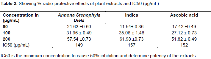
Radioprotective testing was done according to method described by Patel et al. (2012) with some modifications. The experimental sample was prepared by mixing 2 ml of extract, 5 ml of 0.9 % saline stock solution and 5 ml of RBC suspension. The negative control sample was prepared by replacing of fruit extract in experimental sample with 2 ml of 0.9% saline solution. The preparation of standard (positive control) was similar to that of test except that it had 2 ml of ascorbic acid instead of herbal extract. The concentration of herbal extracts and ascorbic acid ranged from 80-200 μg/ml. All the prepared mixtures were exposed immediately to UV (wavelength 365 nm) light for 30 min at room temperature. The mixtures were then centrifuged for 10 min at 3000 rpm. The absorbance of the supernatant was measured using UV visible spectrophotometer at 540 nm. Radioprotective efficacy of extracts was then determined by the extent of radiation induced haemolysis. Radioprotective efficacy was calculated as percentage inhibition of haemolysis using the following equation (Gunathilake et al., 2018).

Where: A 1 = Absorbance of isotonic buffered solution alone and RBC cell suspension after exposure to UV light; A 2 = Absorbance of isotonic buffered solution alone and RBC cell suspension test solution containing (Herbal extracts or ascorbic acid) exposed to UV light.
RESULTS AND DISCUSSION
DPPH assay
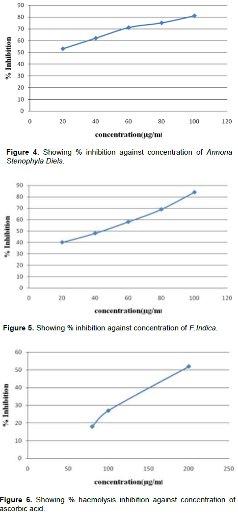
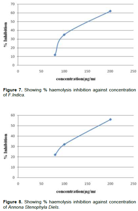
DPPH is a free radical compound with maximum absorption band ranging from 515 - 528 nm; therefore, it is a useful reagent for use in evaluation of antioxidant effects of phenolic compounds (Sánchez-Moreno, 2002). DPPH assay involves the reduction of DPPH by antioxidants to a yellow coloured compound called diphenylpicrylhydrazine. The reduction extent depends on hydrogen donating capacity of antioxidants. The hydrogen donating capacity of Annona Stenophyla Diels and F.Indica fruit extracts of concentrations ranging from 20 to 100 µg/ml were studied. The results obtained exhibited dose dependent DPPH radical scavenging activity as presented in Table 1. All the extracts and the positive control significantly decreased haemolysis (P ≤ 0.05). At low concentrations, F.Indica extract was less potent than ascorbic acid (standard) (P ≤ 0.05). There was no significant difference between % inhibition of Annona Stenophyla Diels and ascorbic acid (positive standard) (P ≤ 0.05). This implies that ascorbic acid and Annona Stenophyla Diels have equal antioxidant potency. However % IC50 which is also an indicator for potency shows that the extracts were more potent than ascorbic acid (standard) as depicted in Table 1. The results obtained in this study suggest that the fruit extracts are good radioprotectors and radical quenchers; therefore, eating these fruits can protect or postpone development of degenerative diseases such as cancer (Figures 1 to 8).


RBC haemolysis
Red blood cells are the most abundant cells in the human body, and they have their unique physiological and morphological characteristics (Hamidi and Tajerzadeh, 2003). Their membranes are rich in polyunsaturated fatty acids and envelopes haemoglobin carrying high oxygen concentrations; hence red blood cells are extremely prone to oxidative damage (Arbos et al., 2008). Oxidative damage of red blood cell membrane was implicated in haemolysis by (Arbos et al., 2008). In view of this, it is reasonable to use red blood cells in the study of oxidative stress and radio-protective effects of fruit plant extracts by exposing them to UV light, a free radical initiator. The antioxidant effect of plant fruit extracts was confirmed in sheep erythrocytes exposed to concentrations ranging from 100 to 200 µg/ml. All extracts showed concentration dependent inhibition of haemolysis as shown in Table 2. The extracts of both plant fruits and the positive control significantly decreased haemolysis (P ≤ 0.05). At higher concentrations F.Indica extract was more potent than ascorbic acid, the positive standard (P ≤ 0.05). There was no significant difference between % haemolysis inhibition of A. stenophyla diels and ascorbic acid (positive standard) (P ≤ 0.05). This implies that ascorbic acid and A. Stenophyla Diels have equal radio-protective potents. These results obtained show that if plant extracts can protect red blood cells from UV induced membrane peroxidation mediated haemolysis, they have the capacity to protect all other cells from UV induced mutations leading to cancer. UV light is a radical initiator; the plasma membrane forms peroxyl radicals upon UV light exposure. These radicals then undergo self-perpetuating reactions, forming more radicals leading to damage of important biomolecules such as proteins and DNA.
Phenol and flavanoid content
Studies have uncovered that free radicals such as hydroxyl, peroxide and superoxides play an important role in the pathogenesis of cancer, and plants are the main sources of antioxidants. Hence, plant derived supplements can be useful in maintenance of good health and combating degenerative diseases including cancer (Demiray et al., 2009). Results obtained in this study indicate that the phenol content of Annona Stenophyla Diels and F.Indica was 306 and 384 mg/gGAE (gallic acid equivalent) respectively, and the flavanoid content was 157 and 170 mg/gQE (quercetin equivalent) respectively.
CONCLUSION
The main aim of this research was to investigate the cytoprotective properties of plant fruit extracts using DPPH assay and UV induced haemolysis assay. The results obtain shows that the plant extracts inhibited DPPH absorption and can be used to protect cells from damaging reactive oxygen species. The plant extracts also suppressed UV light induced haemolysis. These findings suggest that fruit extracts can be used as armoury for protection against radiation exposure which induces peroxidation of lipids, forming radicals which can potentially damage DNA leading to the development of cancer. The results obtained further suggest that the fruits of A. Stenophyla Diels and F.Indica can be used in the formulation of nutraceuticals for prevention of cancer. These fruit extracts are alternative radio-protectors of plant origin which may provide radioprotection to other normal tissues during radiotherapy since radiation genotoxicity is largely free radical mediated and chemotherapy treatments whose mechanism involves free radical attack. The study was carried out in vitro, hence there is need to carry out in vivo studies to enable the inclusion of fruit extracts in herbal cancer prevention formulations. Further research should also focus on the potential use of fruit extracts to reduce the side effects of radiotherapy and its role in recovery of patients after a chemotherapy session. Further research should also focus on studies which include use of in-vitro cell lines as well as in-vivo studies.
AVAILABILITY OF DATA AND MATERIALS
The datasets used and/or analysed during the current study are available from the corresponding author on reasonable request.
ETHICS APPROVAL
The study was approved by Harare Institute of Technology.
CONFLICT OF INTERESTS
The author has not declared any conflict of interests.
REFERENCES
|
Adeboye PT, Bettiga M, Olsson L (2014). The chemical nature of phenolic compounds determines their toxicity and induces distinct physiological responses in Saccharomyces cerevisiae in lignocellulose hydrolysates. AMB Express 4(1). |
|
|
Arbos KA, Claro LM, Borges L, Santos CAM, Weffort-Santos AM (2008). Human erythrocytes as a system for evaluating the antioxidant capacity of vegetable extracts. Nutrition Research 28:457-463. |
|
|
Bhalodia N, Nariya P, Acharya R, Shukla V (2011). Evaluation of in vitro antioxidant activity of flowers of Cassia fistula Linn. International Journal of PharmTech Research (3)1: 589-599. |
|
|
Demiray S, Pintado ME, Castro PML (2009). Evaluation of phenolic profiles and antioxidant activities of Turkish medicinal plants: Tilia argentea, Crataegi folium leaves and Polygonum bistorta roots. World Academy of Science, Engineering and Technology 54:312- 317. |
|
|
Floyd RA (1990). The role of 8-hydroxydeoxyguanosine in carcinogenesis. Carcinogenesis (11):1447-1450. |
|
|
Fulgentius NL, Snyder AL, Shaikh K (2013). Determination of radical scavenging activity and total phenols of wine and spices. A randomized study. Antioxidants 2(3):110-121. |
|
|
Ganesh CJ (2007). Radioprotective potential of plants and herbs against the effects of ionizing radiation. Journal of Clinical Biochemistry and Nutrition 40(2):74-81. |
|
|
Gopalakrishnan A, Kong AN (2008). Anticarcinogenesis by dietary phytochemicals: cytoprotection by Nrf2 in normal cells and cytotoxicity by modulation of transcription factors NF-kappa B and AP-1 in abnormal cancer cells. Food and Chemical Toxicology 46(4):1257-1270. |
|
|
Gunathilake KDPP, Ranaweera KKDS, Vasantha RHP (2018). In vitro anti-inflammatory properties of selected green leafy vegetables. Biomedicines 6(4):107. |
|
|
Hamidi H, Tajerzadeh H (2003). Carrier erythrocytes: an overview. Drug Delivery 10(1):9-20. |
|
|
Kamtekar S, Keer V, Patil V (2014). Estimation of phenolic content, flavonoid content, antioxidant and alpha amylase inhibitory activity of marketed polyherbal formulation. Journal of Applied Pharmaceutical Science (4)09:061-065. |
|
|
Kwon KH, Barve A, Yu S, Huang MT, Kong AN (2007). Cancer by phytochemicals: potential molecular targets, biomarkers and animal models. Acta Pharmacologica Sinica 28(9):1409-1421. |
|
|
Lattanzio V (2013). Phenolic compounds: Introduction. In Handbook of Natural Products; Ramawat, K.G., Merillon, J.M., Eds.; Springer-Verlag: Berlin Heidelberg, Germany 1543-1580. |
|
|
Markham KR (1982). Techniques of flavonoid identification. Chap1 nd2 ed (pp. 113). London Academic Press. |
|
|
Maroyi A (2011). Ethnobotanical study of medicinal plants used by people in Nhema communal area, Zimbabwe. Journal of Ethnopharmacology 136(2):347-354. |
|
|
Martin-Moreno JM, Soerjomataram I, Magnusson G (2008). Cancer causes and prevention a condensed appraisal in Europe in 2008. European Journal of Cancer 44(10):1390-1403. |
|
|
Min K, Freeman C, Kang H, Choi SU (2015). The regulation by phenolic compounds of soil organic matter dynamics under a changing environment. BioMed Research International. |
|
|
Mut-Salud N, Álvarez PJ, Garrido JM, Carrasco E, Aránega A, Rodríguez-Serrano F (2016). Antioxidant intake and antitumor therapy: toward nutritional recommendations for optimal results. Oxidative Medicine and Cellular Longevity. |
|
|
Orwa C, Mutua A, Kindt R, Jamnadass R, Simons A (2009). Agroforestry database: A tree reference and selection guide version 4.0 (http://www.worldagroforestry.org/af/treedb/) |
|
|
Patel A, Patel A, Patel A, Patel NM (2010). Estimation of flavonoid, polyphenolic content and in vitro antioxidant capacity of leaves of Tephrosia purpurea Linn. (Leguminosae). International Journal of Pharma Sciences and Research 1(1):66-77. |
|
|
Patel S, Patel J, Patel RK (2012). To study proximate analysis & biological evaluation of Triphala Guggulu formulation. International Journal of Pharmaceutical Sciences and Research 4:1520-1526. |
|
|
Prakash V, Rana S, Sagar A (2017). Studies on analysis of antioxidant and enzyme inhibitory activity of Vitex negundo Linn. International Journal of Pharmacognosy and Phytochemical Research (9)6:833. |
|
|
Prasad S, Amit KT (2017). Reactive oxygen species (ROS) and cancer: Role of antioxidative nutraceuticals. Cancer Letters 387:95-105. |
|
|
Roya K, Fatemeh G (2013). Screening of total phenol and flavonoid content, antioxidant and antibacterial activities of the methanolic extracts of three Silene species from Iran. International Journal of Agriculture and Crop Sciences 5(3):305. |
|
|
Sánchez-Moreno C (2002). Methods used to evaluate the free radical scavenging activity in foods and biological systems. Food Science and Technology International 8:121-137. |
|
|
Shukla SK, Gupta ML (2010). Approach towards development of a radioprotector using herbal source against lethal irradiation. International Research Journal of Plant Science 1(5):118-125. |
|
|
Smith TA, Kirkpatrick DR, Smith S, Smith TK, Pearson T, Kailasam A, Agrawal DK (2017). Radioprotective agents to prevent cellular damage due to ionizing radiation. Journal of Translational Medicine 15(1):232. |
|
|
Soto ML, Falqué E, Domínguez H (2015). Relevance of natural phenolics from grape and derivative products in the formulation of cosmetics. Cosmetics 2(3):259-276. |
|
|
Taderera T, Gomo E, Chagonda LS (2015). The Antidiabetic activity of an aqueous root extract of Annona stenophylla engl. and diels in non-diabetic control and alloxan-induced diabetic rats. Journal of Biologically Active Products from Nature 6(4):315-322. |
|
|
Yu X, Kensler T (2005). Nrf2 as a target for cancer chemoprevention. Mutation Research/Fundamental and Molecular Mechanisms of Mutagenesis 591(1-2):93-102. |
|
Copyright © 2024 Author(s) retain the copyright of this article.
This article is published under the terms of the Creative Commons Attribution License 4.0


