Full Length Research Paper

ABSTRACT
Diabetes mellitus is the fourth killer disease globally. The available management strategies are quite expensive and sometimes unsafe. This necessitates the need for bio-active drugs from medicinal plants. Although Salvia officinalis (sage) is used in herbal medicine, the scientific validation for anti-diabetic effects of various extracts has been elusive. The present study aimed to determine and compare the anti-hyperglycaemic efficacy of methanolic, hexane, ethyl acetate, and aqueous leaf extracts of Salvia officinalis in alloxan-induced diabetic mice. Phytochemical screening of the extracts revealed presence of flavanone, sterols, saponins, tannins, alkaloids, and triterpenes. The extracts were subjected to preliminary in vivo bio-assays at dosage levels of 400 mg/kg for 7 days through oral administration. The aqueous extract demonstrated significant hypoglycaemic effect, pË‚0.05 hence subjected to further hypoglycaemic studies for 15 days. There was a significant decrease in blood sugar levels of groups treated with aqueous extract at 400 mg/kg and 600 mg/kg doses from 452.00 ± 11.13 mg/dL and 431.00 ± 10.65 mg/dL to 256.33 ± 5.12 mg/dL and 256.67 ± 8.74 mg/dL. Weight gain improved significantly from 28.05 ± 0.39 g and 27.38 ± 0.52 g to 29.32 ± 0.42 g and 28.55 ± 0.38 g respectively compared to controls, pË‚0.05. Histopathological studies revealed no significant changes in liver and kidney tissues. Besides, no significant cytotoxic effect was reported. Results from this study indicate that aqueous extract of Salvia officinalis is a potential anti-hyperglycaemic and can be used in modulating blood glucose levels.
Key words: Diabetes mellitus, Salvia officinalis, aqueous extract, hypoglycaemic effect, phytochemicals.
INTRODUCTION
Diabetes mellitus is a global non-communicable disease with rapidly increasing prevalence in the world (Mendenhall et al., 2017). It is a major healthcare problem experienced by many people due to defect of the endocrine system as well as a complex metabolic disorder that leads to syndromes such as stroke, heart attack, and peripheral vascular disease (Patel et al., 2011). High glucose levels in the blood (hyperglycemia) which is an indicator of diabetes is as a result of reduced/lack of insulin secretion by the pancreas, decreased sensitivity of the target tissues to the hormone insulin, or due to a combination of these two factors (International Diabetes Federation, 2013).
Diabetes can lead to numerous complications that are classified into acute, sub-acute or chronic. Complications associated with the acute form of diabetes mellitus include hypoglycemia, hyperosmolarite, hyperglycemia, diabetic ketoacidosis, and non-ketotic syndrome (Kitabchi et al., 2009). Sub-acute complications are polyuria, polydipsia, visual blurriness, weight loss, and lack of energy (Kuchake and Upasani, 2013). Chronic complications are associated with long-term damages like nephropathy, neuropathy, hypertension, hepatopathy, cardiomyopathy, diabetic foot ulcers, retinopathy, and reproductive damage (Lotfy et al., 2017). Chronic hyperglycemia leads to increase in production of oxygen free radicals due to autoxidation of glucose and glycation of the body proteins (Yaribeygi et al., 2019). This, generates oxidative stress resulting to secondary complications affecting various body organs such as the kidneys, eyes, arteries, and nerves (Henriksen et al., 2011).
Although diabetes was a rare disease in the past, it has become a big problem in recent years. In 2013, diabetes caused 5.1 million deaths, an increase of 0.5 million compared to 2011. It is anticipated that over 592 million people with diabetes by the year 2035 may die. In the year 2000, about 177 million people suffered from diabetes globally and by 2015 the number had risen to 415 million which is predicted to increase to about 642 million by the year 2040. In Africa, 14.2 million people are diabetic, and they are estimated to rise to 34.2 million by 2040 (International Diabetes Federation, 2015). Due to the increasing mortality incidence, search for newer, affordable, and safer bioactive anti-hyperglycaemic agents from medicinal plants has become an area of interest to scientists.
Salvia officinalis commonly known as sage, is an evergreen, perennial shrub in the Lamiaceae family (Sharma and Schaefer, 2019). It has grayish-green leaves, woody stems and blue to purplish flowers. It is native to the Mediterranean region but has naturalized in many parts of the world. Sage has been used in herbal medicine for a long time in the treatment of various illnesses. It is reported to have anti-inflammatory, antibacterial, and anti-fungal properties (Ghorbani and Esmaeilizadeh, 2017). It is also known for lipid profiling and antioxidant properties responsible for hypoglycaemic effects (Bommer et al., 2009; Schapowal et al., 2009). The aim of this study was to determine and compare the anti-hyperglycaemic efficacy of methanolic, hexane, ethyl acetate, and aqueous extracts of S. officinalis in alloxan-induced diabetic mice.
MATERIALS AND METHODS
Study area
Fresh leaves were collected from Egerton University’s Botanic Garden which is at an altitude of 2127 m and 1° 37″ south of Equator. The plant was authenticated by a qualified taxonomist, Prof Samuel Kariuki at Egerton University and the voucher specimen deposited at the Department of Biological Sciences, Egerton University with an assigned voucher specimen number (NB 238).
Extraction
The collected leaves were dried under shade to constant weight and ground to a fine powder using a blending machine (Thomas-Wiley Laboratory Mill Model 4). The powdered material (500 g) was soaked in 1.5 L of distilled methanol for 72 h at room temperature with intermittent shaking. The extract was decanted and filtered using Whatman No. 1 filter paper. The obtained filtrate was concentrated to dryness using a rotary Evaporator machine (BUCHI-R 205) under reduced pressure. The concentrated methanolic crude extract was divided into two parts with one portion stored in a vial at 4ºC awaiting bio-assays. The remaining portion was suspended in distilled water, extracted with hexane and thereafter ethyl acetate to give hexane, ethyl acetate, and aqueous crude fractions respectively (Kosgei et al., 2014).
Phytochemical screening
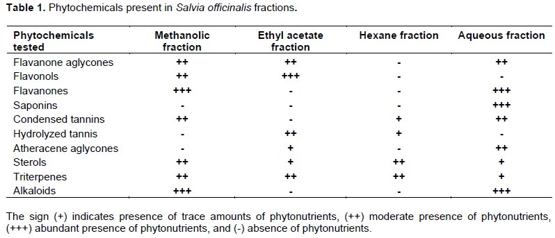
Phytonutrients present in various extracts were determined according to standard procedures by Harborne (1998). Tests were performed for alkaloids, saponins, triterpenes, steroid glycosides, sterols, anthracene aglycone, flavonone aglycones, flavonols, flavanone, and tannins.
Experimental animals
Two-month old healthy male Swiss albino mice weighing (25-30 g) were procured from Kenya Medical Research Institute (KEMRI), Nairobi, Kenya. The animals were housed in polypropylene cages, six mice in each with wood shaving as beddings. They were fed with standard mice pellets obtained from Unga Ltd, Nairobi, Kenya (crude protein 18.1%, crude fibre 7%, calcium 0.8%, phosphorous 0.8%, and fat 8%). The feed and water were available ad libitum except during the day of blood sampling when animals were fasted overnight with access to water only. The mice were acclimatized to standard laboratory environmental conditions, temperature (23 ± 2°C) dark and light cycles (12:12 h) for 2 weeks. The studies were performed with the approval of Institute of Primate Research (IPR) Animal Care and Ethics Committee [Ref: ISERC/10/2017].
Experimental design
Preliminary screening of the extracts was carried out using six mice per extract. The extracts were fed orally by use of an intra-gastric gavage for a period of 7 days. Each animal received 0.3 mL of the prepared extract (400 mg/kg). The aqueous extract was selected for extensive study as it lowered blood sugar levels significantly compared to other extracts. The mice were randomized into 5 groups of 6 mice each.
Group I: (Control- Non-diabetic- Untreated mice)
Group II: (Diabetic control- Diabetic treated group)
Group III: (Diabetic mice treated with 400 mg/kg of the aqueous extract)
Group IV: (Diabetic mice treated with 600 mg/kg of the aqueous extract) and
Group V: (Control-Diabetic mice treated with 2 mg/kg glibenclamide drug).
Induction of diabetes in mice
The animals were marked with wet picric acid for ease of identification. They were fasted overnight before injection of the alloxan® monohydrate chemical intraperitoneally as a single dose of 200 mg/kg bwt. After five (5) days (with access to food and water), the experimental animals were screened for hyperglycemia by determining their fasting blood sugar level using the glucose oxidase kit (SoftStyle Glucometer and SoftStyle Blood Glucose Test Strips from Chem-labs Limited, Nairobi Kenya). Blood glucose levels above 200 mg/dL was considered diabetic and used in the study.
Blood sugar and body weight determination
The animal’s tails were first sterilized with 10% alcohol. Blood was withdrawn by tail snipping from each animal for blood sugar analysis. Baseline blood sugar levels of all the animals were taken before extract administration and fasting blood sugar determined at intervals of 72 h (3 days) for 360 h (15 days). Baseline body weights of the various treatment groups were measured before and during the study period at intervals of 72 h using an electronic beam balance (model type: BL-220H).
Histopathology tests
Kidney and liver tissues were examined for histopathology using standard procedures according to Scott (1999). The organs were fixed and preserved in 10% (v/v) formalin and then processed for paraffin embedding. Paraffin embedded sections (7 μm thick) were stained with haematoxylin and eosin dye (H&E), and then examined under light microscopy (HumaScope AdvancedLED Binocular digital microscope) for pathology. Comparison of the various tissues was made between different treatment groups.
Data analysis
The obtained data was expressed as mean ± standard error of the mean (mean ± S.E.M). Data was subjected to one-way analysis of variance (ANOVA) followed by Turkey post hoc test using Statistical Package for Social Sciences (SPSS) software [version 23]. PË‚0.05 was considered statistically significant.
RESULTS
Phytochemical tests
S. officinalis leaves showed presence of various phytochemicals as shown in Table 1. Flavanones, saponins, and alkaloids were abundant in aqueous fraction.
Preliminary screening of the crude extracts
The effect of S. officinalis extracts on Fasting Blood Sugar (FBS) is indicated in Table 2. The aqueous extract lowered fasting blood sugar levels significantly from 487.67 ± 2.58 to 426.33 ± 2.95 mg/dL (pË‚0.05). Hexane and methanolic extracts as well indicated a significant decrease in blood sugar levels from 490.67 ± 3.01 and 497.00 ± 2.78 mg/dL to 473.00 ± 2.73 and 488.00 ± 2.82 mg/dL, respectively. The ethyl acetate extract, on the other hand, indicated an increase in blood sugar levels from 482.33 ± 0.42 to 519.00 ± 10.30 mg/dL.

Hypoglycaemic activity of aqueous extract
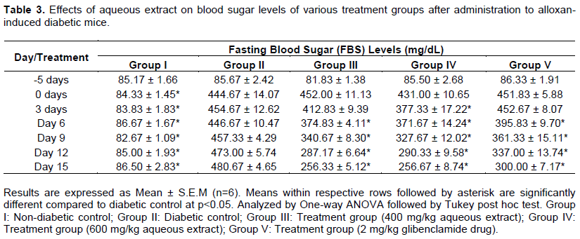
Oral administration of aqueous extract at doses of 400 and 600 mg/kg respectively indicated a significant decrease (p˂0.05) in fasting blood sugar levels compared to the controls (Table 3). Groups administered with aqueous extract indicated a greater decrease in blood sugar levels compared to glibenclamide treated group.
Body weight
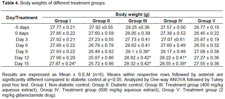
Aqueous extract at 400 and 600 mg/kg indicated a significant (pË‚0.05) improvement in body weight compared to the controls. Nevertheless, weight gained by Group V (administered with glibenclamide drug) was not significantly different (pË‚0.05) from the diabetic control (Table 4). Group III treated with 400 mg/kg recorded the highest weight gain from 27.57 ± 0.50 to 28.55 ± 0.38 g. On the other hand, Group V treated with glibenclamide drug recorded the least weight gain from 26.77 ± 0.19 to 27.55 ± 0.36 g.
Histopathological tests
Liver histopathology
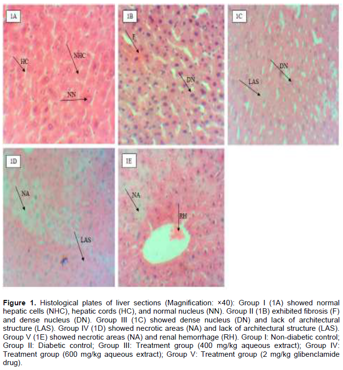
Liver cells of healthy mice showed normal cellular architecture characterized by normal hepatic cells with distinct nuclei and hepatic cords. Severe cellular degenerative changes were observed in diabetic control group. The general architectural structure of the liver cell was lost with severe necrosis and fibrosis noted. Tissues treated with the aqueous extract and glibenclamide drug also showed similar degenerative changes. Several necrotic areas were observed and normal cellular architecture destroyed (Figure 1).
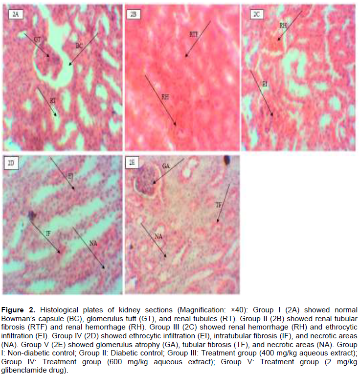
Kidney histopathology
The kidney sections of Group I showed normal renal architecture with distinct glomeruli, Bowman’s capsule and tubules. The kidney sections of Group II indicated tubular cellular necrosis and fibrosis. Tissues treated with the aqueous extract showed tubular cellular necrosis with intratubular fibrosis, renal hemorrhage, and erythrocytic infiltration. Similarly, group treated with glibenclamide drug indicated tubular and glomerulus atrophy, fibrosis and necrosis (Figure 2).
DISCUSSION
Alloxan is a diabetogenic agent that causes destruction of the pancreatic β-cells of the islets of Langerhans, thereby affecting insulin production (King, 2012). Insulin deficiency leads to increased glucose levels in the blood. Results from this study showed that oral administration of methanolic, hexane, and aqueous extracts at a dose of 400 mg/kg decreased fasting blood sugar of alloxan- induced diabetic mice significantly for a period of 7 days.
The glucose lowering effects of these extracts might be linked to presence of flavanones, flavonols, tannins, triterpenes, alkaloids, saponins, and sterols (Aghajanyan and Trchounian, 2018). Flavonoids and terpenes have been documented to possess anti-diabetic effect through insulin like-effect (Balasubashini et al., 2004). Ferulic acid and quercertin are flavonoids which have an effect on pancreatic β-cells of alloxan-induced diabetic rats. They cause cell proliferation hence secretion of more insulin (Mahesh and Menon, 2004). Flavonols and flavanones are classes of flavonoids identified in methanolic, ethyl acetate, and aqueous crude extracts. They possess anti-oxidant activity which scavenges free radicals generated during the progression of diabetes mellitus, thereby offering protection against possible damage to various tissues (Kawser et al., 2016).
Nevertheless, other classes of phytochemicals identified could also be responsible for hypoglycaemic effect. For instance, the hexane extract did not reveal any presence of flavanones and flavonols, but lowered blood sugar levels of diabetic mice significantly. Phenolic acids and tannins are polyphenols known to possess antioxidant activities hence it is responsible for free radical scavenging effect (de Almeida et al., 2005). Studies by Pan et al. (2003) and Rao and Gurfinkel (2000) have also demonstrated the hypoglycaemic activity exhibited by tannins. Since tannins were detected in the hexane extract in this study, they could have contributed to its hypoglycaemic effect.
A hyperglycaemic effect was noted in ethyl acetate extract where it demonstrated an increase in blood sugar levels. It showed presence of flavanone aglycones, flavonols, hydrolyzed tannis, atheracene aglycones, sterols, and triterpenes. These metabolites could be working in an antagonistic manner, thereby inhibiting the activity of each other (Njeru et al., 2015). This could probably be linked to the reason why ethyl acetate extract demonstrated lack of hypoglycaemic effect.
Administration of the aqueous extract at doses of 400 and 600 mg/kg for 15 days caused a significant drop in blood sugar levels and a gradual increase in weight gain (p˂0.05). The results obtained were consistent with those recorded by Eidi and Eidi (2009) who demonstrated the hypoglycaemic and weight improvement effect of S. officinalis aqueous extract. Similar results were reported by Salah et al. (2016) who documented the hypoglycaemic character of S. officinalis aqueous extract. Weight gain for the group administered with glibenclamide drug, however, was not statistically significant compared to the diabetic control. This demonstrates the potency of S. officinalis aqueous extract as compared to the glibenclamide drug. Diabetic control group also showed signs of polyphagia, polydipsia, and polyuria. Weight loss observed in a diabetic state is usually as a result of muscle wasting due to poor carbohydrate utilization. Consequently, it stimulates protein breakdown in order to provide amino acids necessary for gluconeogenesis to occur (Barazzoni et al., 2017).
Although the exact mode of action through which these extracts lower blood sugar levels has not yet been elucidated, it is thought to act through various mechanisms. Lima et al. (2006) reported that plant extracts cause regeneration of the destroyed pancreatic β-cells, protect the intact functional beta cells from further damage, increase plasma membrane permeability, and stimulate insulin secretion. Another study by Bnouham et al. (2006) also noted that plant extracts could possess insulin-like activity, increase peripheral utilization of glucose, decrease the rate of glycogenesis, increase synthesis of hepatic glycogen, or inhibit intestinal glucose absorption (Mahdizadeh et al., 2018).
Garcia-Compean et al. (2009) demonstrated that mitochondria oxidative stress generated due to diabetes leads to generation of free radicals that induces inflammation, necrosis, and fibrosis to the cells. Administration of aqueous extract on the diabetic mice at the dosage levels of 400 and 600 mg/kg did not indicate any significant histopathological changes compared to controls. In contrast, Essawy et al. (2018) documented protective effects of the aqueous extract of S. officinalis on damaged liver cells of mice. However, in this study, the hepatoprotective and nephroprotective effect of the aqueous extract is implicated. This could be attributed to the fact that the cells were already destroyed beyond repair or the treatment period was too short to give an effect. Nevertheless, the extract was not cytotoxic to the liver and kidney cells as no further damage was observed and no deaths of the animals was recorded during the study period. The extract could have, therefore, played a huge role in causing attenuation of the oxidative stress and also enhancing the antioxidant defense system (Ashour et al., 2017).
CONCLUSION
The study indicates that methanolic, hexane, and aqueous extracts of S. officinalis possess anti-diabetic potential at a dose of 400 mg/kg. At the same concentration, however, ethyl acetate extract proved to be hyperglycaemic. The aqueous extract at 400 and 600 mg/kg produced better effects compared to glibenclamide. Thus, it can be further explored for the development of phytomedicines for the management of diabetes mellitus. The histopathological studies and tissue examination of kidney and liver organs proved that aqueous extract is not toxic. Future research should involve active compounds identification, determination of the mode/mechanism of action, and possible synergistic interactions with other extracts.
CONFLICT OF INTERESTS
The authors have not declared any conflict of interests.
ACKNOWLEDGMENTS
The authors appreciate Biochemistry and Chemistry Departments of Egerton University for allowing the use of their facilities in achieving the objectives of this study. Dr. Nyaga and Mr. Areba of the Department of Veterinary Pathology, Microbiology and Parasitology of Egerton University are also recognized for their assistance during animal experiments, Prof. Makanya of Chiromo Campus, Nairobi University for helping in the preparation of histological slides, and Prof. S.T Kariuki of the Department of Biological Sciences, Egerton University for helping in identification of botanical materials.
REFERENCES
| Aghajanyan A, Trchounian A (2018). Antihyperglycemic properties of the herbal mixture composed of extracts from Salvia officinalis L., Calendula officinalis Linn., Glycyrrhizae radix L. And Echinacea purpurea L. on hyperglycemia induced by immobilization stress in rabbits. Chemistry and Biology 52(3):180-186. | |||
| Ashour MB, Ahmed OM, Abd AA, Ali MA (2017). Assessment of the Preventive Effects of Salvia officinalis and Ruta graveolens Ethanolic Leaf Extracts on Chlorpyrifos-and Methomyl-induced Renal Toxicity and Oxidative Stress in Albino Rats. International Journal of Prevention and Treatment 6:34-44. | |||
|
Balasubashini MS, Rukkumani R, Viswanathan P, Menon VP (2004). Ferulic acid alleviates lipid peroxidation in diabetic rats. Phytotherapy Research: An International Journal Devoted to Pharmacological and Toxicological Evaluation of Natural Product Derivatives 18(4):310-314. Crossref |
|||
|
Barazzoni R, Deutz NEP, Biolo G, Bischoff S, Boirie Y, Cederholm T, Cuerda C, Delzenne N, Sanz ML, Ljungqvist O, Muscaritoli M (2017). Carbohydrates and insulin resistance in clinical nutrition: Recommendations from the ESPEN expert group. Clinical Nutrition 36(2):355-363. Crossref |
|||
|
Bnouham M, Ziyyat A, Mekhfi H, Tahri A, Legssyer A (2006). Medicinal plants with potential antidiabetic activity-A review of ten years of herbal medicine research (1990-2000). International Journal of Diabetes and Metabolism 14(1):1-17. Crossref |
|||
|
Bommer S, Klein P, Suter A (2009). A multicenter open clinical trial to assess the tolerability and efficacy of sage tablets in menopausal patients with hot flushes. Planta Medica 75(9):159-181. Crossref |
|||
|
de Almeida ME, Mancini FJ, Guerra NB (2005). Characterization of antioxidant compounds in aqueous coriander extract (Coriandrum sativum L.). LWT-Food Science and Technology 38(1):15-19. Crossref |
|||
|
Eidi A, Eidi M (2009). Antidiabetic effects of sage (Salvia officinalis L.) leaves in normal and streptozotocin-induced diabetic rats. Diabetes and Metabolic Syndrome: Clinical Research and Reviews 3(1):40-44. Crossref |
|||
| Essawy AE, Lamfon HA, Al Harbi AB, Ali AM, Lamfon NA (2018). The Effect of Salvia officinalis Extract on Alleviating Oxidative Stress and Hepatic Dysfunction Induced by Carbon Tetrachloride in Mice. Jordan Journal of Biological Sciences 12(4):403-408. | |||
|
Garcia-Compean D, Jaquez-Quintana JO, Gonzalez-Gonzalez JA, Maldonado GH (2009). Liver cirrhosis and diabetes: risk factors, pathophysiology, clinical implications and management. World Journal of Gastroenterology 15(3):280-295. Crossref |
|||
|
Ghorbani A, Esmaeilizadeh M (2017). Pharmacological properties of Salvia officinalis and its components. Journal of Traditional and Complementary Medicine 7(4):433-440. Crossref |
|||
|
Harborne AJ (1998). Phytochemical methods a guide to modern techniques of plant analysis. In phytochemical methods. Chapman & Hall, London, UK 15:1-36. Crossref |
|||
|
Henriksen EJ, Diamond-Stanic MK, Marchionne EM (2011). Oxidative stress and the etiology of insulin resistance and type 2 diabetes. Free Radical Biology and Medicine 51(5):993-999. Crossref |
|||
| International Diabetes Federation (2013). IDF Diabetes Atlas, 6th ed. International Diabetes Federation 128:40-50. | |||
| International Diabetes Federation (2015). International diabetes federation. IDF Diabetes Atlas, 7th edn. Brussels, Belgium: International Diabetes Federation. | |||
|
Kawser HM, Abdal DA, Han J, Yin Y, Kim K, Kumar SS, Yang GM, Choi H, Cho SG (2016). Molecular mechanisms of the anti-obesity and anti-diabetic properties of flavonoids. International Journal of Molecular Sciences 17(4):569. Crossref |
|||
|
King AJ (2012). The use of animal models in diabetes research. British Journal of Pharmacology 166(3):877-894. Crossref |
|||
|
Kitabchi AE, Umpierrez GE, Miles JM, Fisher JN (2009). Hyperglycaemic crises in adult patients with diabetes. Diabetes Care 32(7):1335-343. Crossref |
|||
|
Kosgei CJ, Mwendia CM, Mwaniki CG, Matasyoh JC (2014). Phytochemical screening, cytotoxicity studies and larvicidal activity of hexane extract of Lippia kituiensis against Rhipicephalus appendiculatus. Journal of Biomedical and Pharmaceutical Research 3(4):105-110. Crossref |
|||
| Kuchake VG, Upasani CD (2013). Effect of Vitamin E and C Plus Reduced Glutathione in Treatment of Diabetic Nephropathy. International Journal of Pharmacy Research and Review 2:1-5. | |||
| Lima CF, Azevedo MF, Araujo R, Fernandes-Ferreira M, Pereira-Wilson C (2006). Metformin-like effect of Salvia officinalis (common sage): is it useful in diabetes prevention? British Journal of Nutrition 96(2):326-333. | |||
|
Lotfy M, Adeghate J, Kalasz H, Singh J, Adeghate E (2017). Chronic complications of diabetes mellitus: a mini review. Current Diabetes Reviews 13(1):3-10. Crossref |
|||
| Mahdizadeh R., Moein S, Soltani N, Malekzadeh K, Mahmoodreza M (2018). Study the molecular mechanism of Salvia species in prevention of diabetes. International Journal of Pharmaceutical Sciences and Research 9(11):4512-4521. | |||
| Mahesh T, Menon PV (2004). Quercetin alleviates oxidative stress in streptozoticin induced diabetic rats. The Journal of nutrition 135:2555-2565. | |||
|
Mendenhall E, Kohrt BA, Norris SA, Ndetei D, Prabhakaran D (2017). Non-communicable disease syndemics: poverty, depression, and diabetes among low-income populations. The Lancet 389(10072):951-963. Crossref |
|||
|
Njeru SN, Obonyo MA, Nyambati SO, Ngari SM (2015). Antimicrobial and cytotoxicity properties of the crude extracts and fractions of Premna resinosa (Hochst) Schauer (Compositae): Kenyan traditional medicinal plant. BMC complementary and alternative medicine 15(1):295-311. Crossref |
|||
|
Pan GY, Huang ZJ, Wang GJ, Fawcett JP, Liu XD, Zhao XC, Sun JG, Xie YY (2003). The antihyperglycaemic activity of berberine arises from a decrease of glucose absorption. Planta Medica 69(7):632-636. Crossref |
|||
|
Patel DK, Kumar R, Prasad SK, Sairam K, Hemalatha S (2011). Antidiabetic and in vitro antioxidant potential of Hybanthus enneaspermus (Linn) F. Muell in streptozotocin-induced diabetic rats. Asian Pacific Journal of Tropical Biomedicine 1(4):316-322. Crossref |
|||
|
Rao ΑV, Gurfinkel DM (2000). The bioactivity of saponins: triterpenoid and steroidal glycosides. Drug Metabolism and Drug Interactions 17(1-4):211-236. Crossref |
|||
| Salah A, Al-Chalabi HMM, Shukri RM, Mahmood LI, Al-Anbar BK (2016). Effect of Salvia officinalis L.(Sage) aqueous extract on liver and testicular function of diabetic albino male rats. Journal of Babylon University/Pure and Applied Sciences 24(4):83-90. | |||
|
Schapowal A, Berger D, Klein P, Suter A (2009). Echinacea/sage or chlorhexidine/lidocaine for treating acute sore throats: a randomized double-blind trial. European Journal of Medical Research 14(9):406-417. Crossref |
|||
| Scott T (1999). Introduction to Medical Laboratory Technology. British Journal of Biomedical Science 56(2):154. | |||
| Sharma Y, Schaefer JFJ (2019). Ethnobotany, phytochemistry, cultivation and medicinal properties of Garden sage (Salvia officinalis L.). Journal of Pharmacognosy and Phytochemistry 8(3):3139-3148. | |||
|
Yaribeygi H, Atkin SL, Sahebkar A (2019). A review of the molecular mechanisms of hyperglycemiaâ€induced free radical generation leading to oxidative stress. Journal of Cellular Physiology 234(2):1300-1312. Crossref |
|||
Copyright © 2024 Author(s) retain the copyright of this article.
This article is published under the terms of the Creative Commons Attribution License 4.0


