Full Length Research Paper
ABSTRACT
Advanced glycation end-products (AGEs) have been shown to be implicated in many degenerative disorders as well as diabetic pathology. Of interest, several natural compounds including phenolic constituents are capable of inhibiting the formation of AGEs. Bio-guided fractionation of a methanol extract from Mangifera indica leaves led to the characterization of six antiglycative constituents including gallic acid (1), iriflophenone-3-C-β-D-glucoside (2), mangiferin (3), iriflophenone-3-C-(2'-O-galloyl)-β-D-glucoside (4), hyperoside (5) and isoquercitrin (6). With IC50 values lower than that of aminoguanidine, the six identified phenolic compounds can be regarded as remarkable inhibitors of advanced glycation end products formation and M. indica leaves should be considered as a potential nutraceutical resource to prevent carbonyl stress-related diseases.
Key words: Mangifera indica, antiglycation, radical scavenging, phenolics, benzophenone.
INTRODUCTION
Mangifera indica L. (family: Anacardiaceae) is widely growing in many parts of the world particularly in tropical countries (Rodeiro et al., 2006). In traditional medicine, leaves of M. indica are employed to treat a wide range of diseases including malaria, diabetes, diarrhea, dysentery and so on (Yakubu et al., 2015). It has been reported that its bioactive compounds are ascorbic acid, carotenoids as well as phenolic derivatives including gallotannins, flavonoids, xanthones and benzophenones (Shaheen et al., 2017). These constituents have been shown to exert several interesting pharmacological activities such as antimicrobial, anti-inflammatory, immunomodulatory, analgesic and antidiabetic effects (Fernández-Ponce et al., 2015; Vasil’ev et al., 2006).
Of interest, all these properties can be, at least in part, related to positive action on oxidative and/or carbonyl stress (Halake et al., 2016; Mildner-Szkudlarz et al., 2017). It has to be noted that the potent antioxidant activity of mango leaves has already been reported by different studies (Pereira et al., 2007; Fernández-Ponce et al., 2012; Ling et al., 2009). By contrast, very limited investigations have been done regarding inhibitory effects on the formation of advanced glycation end products (AGEs) (Itoh et al., 2017). AGEs are the final products of a nonenzymatic reaction, also known as Maillard reaction, occurring between reducing sugars and nucleic acids, lipids or free amino groups of proteins. Numerous studies have demonstrated that the formation and accumulation of AGEs can accelerate the ageing process (Kim et al., 2017).
Additionally, AGEs are involved in the development of numerous pathologies such as atherosclerosis, neuro-degenerative diseases as well as diabetic complications (Yang et al., 2018). Inhibition of the formation of these harmful glycotoxins is thus considered as a promising preventive or therapeutic target for a wide range of disorders (Brings et al., 2017) and increasing consideration is recently being given to the evaluation of phytoconstituents as antiglycating agents.
The present study aimed thus at performing a bio-guided fractionation of a methanolic extract from mango leaves in order to identify potential AGEs inhibition constituents. In vitro bovine serum albumin/D-ribose fluorimetric assay was chosen to evaluate the AGEs inhibition properties of the fractions and compounds. Knowing that scavenging reactive oxygen species is also considered as a mechanism of AGEs inhibition (Reddy et al., 2006), antioxidant activity was also assessed by using colorimetric DPPH assay.
MATERIALS AND METHODS
Reagents
Methanol, dichloromethane, ethyl acetate, butanol, trifluoroacetic acid, acetonitrile, D-ribose, bovine serum albumin (BSA), aminoguanidine hydrochloride, gallic acid, 1,1-diphenyl-2-picryl-hydrazyl (DPPH), Trolox, fluorescein, 2,2′-azobis(2-methylpropionamidine) dihydrochloride, Iron (II) chloride and ferrozine were bought from Sigma-Aldrich (Saint-Quentin Fallavier, France). Ethylenediaminetetraacetic acid disodium salt (EDTA) was purchased from Fisher Chemical (Illkirch, France).
Instrumentation
Proton and carbon nuclear magnetic resonance spectra were recorded in acetone-d6 on a Bruker DRX 500 spectrometer (1H:500 MHz, 13C:125 MHz) (Bruker Biospin SAS, Wissembourg, France). HPLC analyses were performed with a reverse phase column (Uptisphere® C18, 250 mm × 4.6 mm, 3.5 μm particle size) using a UHPLC Ultimate 3000 RSLC apparatus with a DAD UV/VIS 3000 RS detector (Thermo-Fisher Scientific, Germany) and mass spectral analysis was carried out on a Orbitrap Q-Exactive (Thermo-Fisher Scientific, Germany).
Plant material
M. indica leaves (500 g) were collected from cultivated trees at Diender, Senegal, in July 2016 and identified by Dr. William Diatta. A voucher specimen (N°1166 M. indica L.) has been deposited in a laboratory herbarium (Laboratory of Pharmacognosy, Faculty of Medicine, Pharmacy and Odontology from University Cheikh Anta Diop of Dakar, Senegal).
Extraction and isolation of bioactive compounds
The air-dried plant material was powdered and macerated twice for 48 h with methanol at room temperature. The extract was then filtered and methanol was completely removed under reduced pressure on a rotary evaporator.
Crude methanol extract (20 g) was dissolved in distilled water and the aqueous solution was successively partitioned with dichloromethane, ethyl acetate and butanol. Each resulting solvent fraction was concentrated as described earlier to yield a dichloromethane fraction (DF, 1.68 g), an ethyl acetate fraction (EAF, 5.32 g), a butanol fraction (BF, 6.81 g) and an aqueous fraction (AF, 5.93 g). A portion of BF (1.5 g) was fractionated using steric exclusion column chromatography (Sephadex® LH-20, 90 g) eluted with a MeOH and water gradient system (60:40 to 80:20), yielding a total of 12 combined fractions. Fraction 5 afforded directly compound 2 (220 mg). Besides, compound 4 was detected as the major constituent of fraction 7 (150 mg). Further purification was performed by mean of semi-preparative HPLC (Nucleosil® RP-18, 250 mm × 100 mm, 5 μm particle size) eluted with a mobile phase consisting of water containing 0.1% trifluoroacetic acid and acetonitrile to yield 15 mg of compound 4. Compounds 2 and 4 were unambiguously characterized on the basis of their MS and NMR data as well as through comparison with published reports (Barreto et al., 2008; Pranakhon et al., 2015).
Iriflophenone-3-C-β-D-glucoside (2): ESI-MS m/z 407.0980 [M – H]− (calcd for C19H19O10, 407,0978). 1H NMR (acetone-d6) δ: 3.40-3.70 (4H, m), 3.85 (2H, br s), 4.93 (1H, d, J = 9.75 Hz), 5.99 (1H, s), 6.85 (2H, d, J = 8.75 Hz), 7.64 (2H, d, J = 8.75 Hz).
Iriflophenone 3-C-(2'-O-galloyl)-β-D-glucoside (4): ESI-MS m/z 559.1091 ([M – H]− (calcd for C26H23O14, 559,1088). 1H NMR (acetone-d6) δ: 3.50-3.85 (3H, m), 3.89 (2H, br s), 5.18 (1H, d, J = 10 Hz), 5.43 (1H, t, J = 10 Hz), 5.89 (1H, s), 6.83 (2H, d, J = 7.5 Hz), 7.06 (2H, s), 7.61 (2H, br s).
Evaluation of antiglycation activity
Inhibition of AGEs formation was conducted according to Derbré et al. (2010). Briefly, 40 μL of bovine serum albumin (BSA, 25 mg/mL), 40 μL of D-ribose (120 mM) and 20 µL of sample solutions were incubated at 37°C in a phosphate buffer, 50 mM, pH 7.4. Sample solutions (extract, fractions and pure compounds) were prepared in phosphate buffer (50 mM, pH = 7.4) and DMSO (final concentration 1% (v/v)). Extract and fractions were evaluated at final concentrations of 0.05 to 1 mg/mL while pure compounds were assessed at concentrations of 25 to 500 µM. Aminoguanidine, a widely used inhibitor of AGEs formation (Abdallah et al., 2016; Yang et al., 2018), was employed as positive control (0.5 to 10 mM). Complete reaction mixture including BSA and ribose in PBS was used as negative control (100% of AGEs formation). After 24 h of incubation, AGEs fluorescence was measured with a microplate reader (TECAN infinite F200 PRO) using 370 and 440 nm as the excitation and emission wavelengths, respectively. The IC50 value was calculated from the linear regression analysis of dose-response curve plotting extract or compounds concentration versus inhibition percentage of AGEs formation. All analyses were performed in triplicate and results were expressed as mean ± standard error of mean (SEM).
Evaluation of radical scavenging activity
The DPPH radical scavenging activity was evaluated according to a previously published method (Meda et al., 2017). Briefly, 10 µL of extract or fractions (0.1-2.5 mg/mL in methanol) or pure compounds (1-40 mM in methanol) were added to 2.5 mL of fresh DPPH solution (25 µg/mL in methanol). After 30 min of incubation, the decrease in DPPH absorbance was recorded at 515 nm. A standard curve of Trolox in the range of 0.1 to 6 mM was constructed (R2 = 0.9978, y = 1101.1x + 1.5196) and results were indicated as µmol of Trolox Equivalents per g (µmol TE/g) for fractions and as IC50 for pure compounds. Each assay was conducted in triplicate and results were expressed as mean ± SEM.
RESULTS AND DISCUSSION
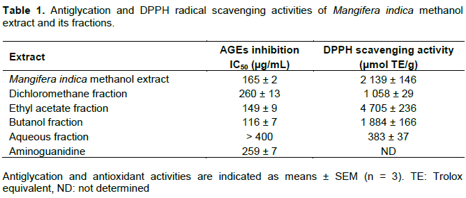
M. indica methanolic extract (MIME) was first evaluated for its antiglycation and antioxidant activities. As illustrated in Table 1, results indicated that the studied extract exerted very potent anti-AGEs and radical scavenging effects. Indeed, with an IC50 value of 165 ± 2 μg/mL, MIME was shown to possess a stronger AGEs inhibition activity than the positive control aminoguanidine (259 ± 7 μg/mL). Consistently with previous investigations of M. indica leaves (Ling et al., 2009), a substantial DPPH radical scavenging effect was also highlighted (2139 ± 146 μmol Trolox equivalent (TE)/gram of dry extract). The whole extract was thus submitted to liquid-liquid fractionation using solvents of increasing polarities, yielding dichloromethane (DF), ethyl acetate (EAF), butanol (BF) and aqueous (AF) fractions. As indicated in Table 1, BF was shown to exert the highest AGEs inhibition activity as demonstrated by its very low IC50 value of 116 ± 7 µg/mL. By contrast, the best DPPH radical scavenging activity was obtained for EAF, highlighting the fact that the most potent anti-AGEs compounds of M. indica leaves were not the best DPPH radical scavengers. Taking these data into consideration, we thus embarked upon an investigation of BF chemical composition, seeking for new anti-AGEs natural compounds.
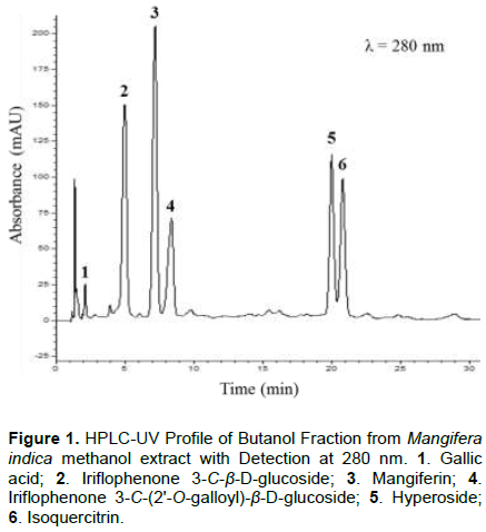
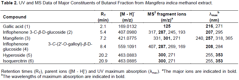
HPLC-DAD/MS analyses were performed to identify the main constituents of BF. As shown in Figure 1, six major compounds were detected at a wavelength of 280 nm. Compounds 1, 3, 5 and 6 were respectively identified as gallic acid, mangiferin, hyperoside and isoquercitrin by comparing their retention times, UV spectra and MS data (Table 2) with those of reference commercial standards. Conversely, compounds 2 and 4 did not match with any available standards and purification of these two constituents was achieved to unequivocally determine their structures. A portion of BF was then submitted to steric exclusion column chromatography to afford directly compound 2. This constituent was unambiguously characterized as iriflophenone-3-C-β-D-glucoside on the basis of its MS (Barreto et al., 2008) and NMR data (Pranakhon et al., 2015). Further purification by mean of semi-preparative HPLC led to the isolation of compound 4. Analysis of its 1H and 13C NMR data allowed the characterization of a benzophenone glycoside acylated with a galloyl unit. Deeper investigation of 2D NMR spectra led to the unambiguous identification of iriflophenone 3-C-(2'-O-galloyl)-β-D-glucoside. Of note, spectral data of this very unusual compound were in agreement with a previous chemical study of M. indica leaves (Barreto et al., 2008) which constituted the sole description of this constituent in plant kingdom.
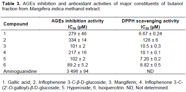
As indicated in Table 3, the six identified constituents were all shown to exert substantial inhibitory activity against in vitro protein glycation, as attested by their IC50 values lower than that of aminoguanidine. With IC50 values ranging from about 100 µM, compounds 3, 5 and 6 were determined as the most potent inhibitors of the studied fraction. Of note, these results are consistent with previous published studies highlighting the strong anti-AGEs capacity of xanthone derivatives and flavonoid glycosides (Abdallah et al., 2017; Derbré et al., 2010). More precisely, the potent antiglycation activities of compounds 3, 5 and 6 have been previously reported by two distinct studies (Itoh et al., 2017; Kim et al., 2011) using glucose or fructose as reducing sugars. By contrast, data regarding benzophenone constituents are scarcer and the noticeable antiglycation activity of compound 4 is firstly reported. As illustrated in Table 3, compounds 3, 4, 5 and 6 were also identified as potent DPPH radical scavengers indicating that their potent anti-AGEs activities might be, at least in part, related to antioxidant action. It has however been noted that, in the same way as for fractions evaluation, no perfect correlation occurs between antioxidant and antiglycation activities of the studied constituents (R2 = 0.4592). For instance, compound 1 was identified as the most potent DPPH scavenger of the fraction but was only inducing moderate antiglycation activity compared to compounds 3, 4, 5 and 6. It suggests thus that other mechanisms than antioxidant effect might account for the antiglycation properties of the most potent inhibitors constituents of M. indica leaves.
The formation of AGEs is indeed considered as a multifactorial process (Abdallah et al., 2016). Generated through several different pathways including Maillard reaction as well as glucose autoxidation, lipid peroxidation or the polyol pathway, α-dicarbonyl species such as glyoxal (GO) and methylglyoxal (MGO) have also been identified as crucial intermediates in the formation of AGEs (Nowotny et al., 2015). It is now well established that scavenging these highly reactive carbonyl species constitute an effective strategy for limiting AGEs formation (Li et al., 2014). Of interest, several flavonoid derivatives including quercetin have been shown to efficiently trap GO and MGO under physiological conditions (Li et al., 2014). It is worth mentioning that A ring of quercetin was identified as a crucial structural feature for activity. Indeed, characterization of its MGO adducts proved that positions 6 and 8 of quercetin were the active sites for trapping dicarbonyl derivatives. To the best of our knowledge, no data are available concerning hyperoside (5) and isoquercitrin (6). However, these two quercetin-3-O-glycosides possess the exact same A ring than quercetin and might also be capable of reacting with carbonyl species. By contrast, gallic acid (1) has been previously shown to exert almost no trapping effect of MGO (Shao et al., 2014). The lack of reactivity of that phenolic acid towards reactive dicarbonyl species could thus explain, at least in part, the modest antiglycation activity of compound 1 compared to flavonoid derivatives of the studied extract.
CONCLUSION
The present results attest that the leaves of M. indica exert strong and promising in vitro antiglycative activity. Further investigations using in vivo models would now be of major interest to confirm the potential of M. indica leaves to prevent carbonyl stress-related diseases. By using a bio-guided approach, this study also identified six potent antiglycative constituents including gallic acid, mangiferin, quercitrin, hyperoside, iriflophenone 3-C-β-D-glucoside and iriflophenone 3-C-(2'-O-galloyl)-β-D-glucoside. With IC50 values lower than that of aminoguanidine, these six phenolic compounds can be regarded as remarkable inhibitors of AGEs formation as well as suitable markers for quality control of mango leaves.
CONFLICT OF INTERESTS
The authors have not declared any conflict of interests.
REFERENCES
|
Abdallah HM, El-Bassossy H, Mohamed GA, El-Halawany AM, Alshali KZ, Banjar ZM (2016). Phenolics from Garcinia mangostana inhibit advanced glycation endproducts formation: effect on Amadori products, cross-linked structures and protein thiols. Molecules 21(2):251. |
|
|
Abdallah HM, El-Bassossy HM, Mohamed GA, El Halawany AM, Alshali KZ, Banjar ZM (2017). Mangostana xanthones III and IV: advanced glycation end-product inhibitors from the pericarp of Garcinia mangostana. Journal of Natural Medicines 71(1):216-226. |
|
|
Barreto JC, Trevisan MTS, Hull WE, Erben G, DE Brito ES, Pfundstein B, Würtele G, Spiegelhalder B, Owen RW (2008). Characterization and Quantitation of Polyphenolic Compounds in Bark, Kernel, Leaves, and Peel of Mango (Mangifera indica L.). Journal of Agricultural and Food Chemistry 56(14):5599-5610. |
|
|
Brings S, Fleming T, Freichel M, Muckenthaler MU, Herzig S, Nawroth PP (2017). Dicarbonyls and advanced glycation end-products in the development of diabetic complications and targets for intervention. International Journal of Molecular Sciences 18(1):1-24. |
|
|
Derbré S, Gatto J, Pelleray A, Coulon L, Séraphin D, Richomme P (2010). Automating a 96-well microtiter plate assay for identification of AGEs inhibitors or inducers: application to the screening of a small natural compounds library. Analytical and bioanalytical chemistry 398(4):1747-1758. |
|
|
Fernández-Ponce MT, Casas L, Mantell C, Rodríguez M, De la Ossa EM (2012). Extraction of antioxidant compounds from different varieties of Mangifera indica leaves using green technologies. The Journal of Supercritical Fluids 72:168-175. |
|
|
Fernández-Ponce MT, Casas L, Mantell C, De la Ossa EM (2015). Use of high pressure techniques to produce Mangifera indica L. leaf extracts enriched in potent antioxidant phenolic compounds. Innovative Food Science and Emerging Technologies 29:94-106. |
|
|
Halake K, Birajdar M, Lee J (2016). Structural implications of polyphenolic antioxidants. Journal of Industrial and Engineering Chemistry 35:1-7. |
|
|
Itoh K, Murata K, Sakaguchi N, Akai K, Yamaji T, Shimizu K, Isaki K, Matsukawa T, Kajiyama S, Fumuro M, Iijima M, Matsuda H (2017). Inhibition of advanced glycation end products formation by Mangifera indica leaf extract. Journal of Plant Studies 6(2):102-107. |
|
|
Kim CS, Park S, Kim J (2017). The role of glycation in the pathogenesis of aging and its prevention through herbal products and physical exercise. Journal of Exercise Nutrition and Biochemistry 21:55-61. |
|
|
Kim HK, Lee JM, Yokozawa T, Sakata K, Lee S (2011). Protective activity of flavonoid and flavonoid glycosides against glucose-mediated protein damage. Food Chemistry 126:892-895. |
|
|
Li X, Zheng T, Sang S, Lv L (2014). Quercetin inhibits advanced glycation end product formation by trapping methylglyoxal and glyoxal. Journal of Agricultural and Food Chemistry 62:12152-12158. |
|
|
Ling LT, Yap S, Radhakrishnan AK, Subramaniam T, Cheng HM, Palanisamy UD (2009). Standardised Mangifera indica extract is an ideal antioxidant. Food Chemistry 113:1154-1159. |
|
|
Meda NR, Fraisse D, Gnoula C, Vivier M, Felgines C, Senejoux F (2017). Characterization of antioxidants from Detarium microcarpum Guill. et Perr. leaves using HPLC-DAD coupled with pre-column DPPH assay. European Food Research and Technology 243:1659-1666. |
|
|
Mildner-Szkudlarz S, Siger A, Szwengiel A, Przygonski K, Wojtowicz E, Zawirska-Wojtasiak R (2017). Phenolic compounds reduce formation of Nε-(carboxymethyl)lysine and pyrazines formed by Maillard reactions in a model bread system. Food Chemistry 231:175-184. |
|
|
Nowotny K, Jung T, Höhn A, Weber D, Grune T (2015). Advanced glycation end products and oxidative stress in type 2 diabetes mellitus. Biomolecules 5:194-222. |
|
|
Pereira CG, Meireles MAA (2007). Evaluation of global yield, composition, antioxidant activity and cost of manufacturing of extracts from lemon verbena (Aloysia triphylla [l'hérit.] britton) and mango (Mangifera indica l.) leaves. Journal of Food Process Engineering 30:150-173. |
|
|
Pranakhon R, Aromdee C, Pannangpetch P (2015). Effects of iriflophenone 3-C-β-glucoside on fasting blood glucose level and glucose uptake. Pharmacognosy Magazine 11:82-89. |
|
|
Reddy VP, Beyaz A (2006). Inhibitors of the Maillard reaction and AGE breakers as therapeutics for multiple diseases. Drug Discovery Today 11:646-654. |
|
|
Rodeiro I, Cancino L, González JE, Morffi J, Garrido G, González RM, Nu-ez A, Delgado R (2006). Evaluation of the genotoxic potential of Mangifera indica L. extract (Vimang), a new natural product with antioxidant activity. Food and Chemical Toxicology 44:1707-1713. |
|
|
Shaheen N, Lu Y, Geng P, Shao Q, Wei Y (2017). Isolation of four phenolic compounds from Mangifera indica L. flowers by using normal phase combined with elution extrusion two-step high speed countercurrent chromatography. Journal of Chromatography B 1046:211-217. |
|
|
Shao X, Chen H, Zhu Y, Sedighi R, Ho CT, Sang S (2014). Essential structural requirements and additive effects for flavonoids to scavenge methylglyoxal. Journal of Agricultural and Food Chemistry 62:3202-3210. |
|
|
Vasil'ev SA, Garazd MM, Khilya VP (2006). 3-Phenoxychromones: Natural distribution, synthetic and modification methods, biological properties. Chemistry of Natural Compounds 42:241-253. |
|
|
Yakubu MT, Salimon SS (2015). Antidiarrhoeal activity of aqueous extract of Mangifera indica L. leaves in female albino rats. Journal of Ethnopharmacology 163:135-141. |
|
|
Yang R, Wang WX, Chen HJ, He ZC, Jia AQ (2018). The inhibition of advanced glycation end-products by five fractions and three main flavonoids from Camellia nitidissima Chi flowers. Journal of Food and Drug Analysis 26:252-259. |
|
Copyright © 2024 Author(s) retain the copyright of this article.
This article is published under the terms of the Creative Commons Attribution License 4.0


