Full Length Research Paper
ABSTRACT
The investigation was led from February-November 2014 longitudinally to recognize aerobic bacterial isolates, estimate incidence rate, identify the associated risk factors and antimicrobial sensitivity patterns of heifer and cow mastitis in and around Debre-Libanos district. An aggregate of 31 Jersey and Holstein-Fresian cross heifers that were left for less than a month to calve were sampled and pursued for the initial two lactation stages after calving. Clinical heifer mastitis was distinguished by physical examination of the udder and milk while sub-clinical one was recognized by California mastitis test. The incidence rate of heifer mastitis per gland month at risk was observed to be 37.4%. The event of new contamination in heifers and cows was not altogether influenced by gland position and udder cleanliness (P>0.05), yet by the management system, lactation stage and dry cow therapy (P<0.01). From 231 isolates, Staphylococcus aureus (25.1%) was the most prevalent pathogen pursued by coagulase negative Staphylococcus and Streptococcus species (each 14.7%). Other bacterial isolates included Micrococcus species (10.38%), Klebsiela pneumoniea (9.95%), Esherishia coli (12.98%), Corynebacterium species (5.62%), Enterobacter aerogens (4.32%) and Bacillus species (4.32%). Antimicrobial sensitivity test demonstrated that tetracycline (71.7%) was observed to be increasingly successful antibiotic among the whole tried antibiotics against all bacterial isolates while the least effective antibiotic was observed to be penicillin G (24.2%). The present investigation has demonstrated that heifer and cow mastitis specifically; sub-clinical mastitis is a critical sickness affecting heifers' milk production.
Key words: heifer mastitis, incidence rate, risk factors, Ethiopia, antimicrobial sensitivity test
INTRODUCTION
Mastitis is the irritation of the parenchyma of the mammary gland paying little heed to the reason. There is swelling, warmth or agony in the mammary gland in clinical cases. Be that as it may, considerable extents of mastitic organs are not promptly discernible by manual palpation nor by visual examination of the milk utilizing a strip cub; these quarters speak to sub-clinical form of mastitis. Bovine mastitis is related with a wide range of infectious agents, ordinarily isolated into those causing contagious mastitis; which spread from contaminated quarters to different quarters and cows and inhabit normal teat skin and those causing environmental mastitis, which are generally present in the cow's environment and achieve the nipple from that source (Biffa et al., 2005). Pathogens causing mastitis in cows are additionally divided into major pathogens (those that reason clinical mastitis) and minor pathogens (those that ordinarily cause sub-clinical mastitis and less every now and again cause clinical mastitis) (Radostits et al., 2006).
Generally, heifers have been thought of as a gathering free of intramammary infections (Aarestrup and Jensen, 1997). Be that as it may, intramammary infection (IMI) before calving and new cases of mastitis promptly following calving have been reported (Barkema et al., 1998). Mastitis affects the production performance of heifers and cows and it is a critical concern to dairy producers. Heifer and cow mastitis causes monetary misfortunes because of a reduction in milk output, treatment costs and culling costs (Huijps et al., 2007). Nielsen et al. (2009) assessed a milk loss of 155 kg on 305 days lactation and Huijps et al. (2007) evaluated the expense of mastitis at 55/heifer present on a farm.
The prevalence, incidence and types of mastitis causing pathogens differ among herds (Oliver et al., 2005). The incidence of clinical mastitis in the peripartum period is moderately higher in heifers than in cows (Biffa et al., 2005) and there is a positive relationship among peripartum and postpartum infections (Aarestrup and Jensen, 1997).
There are several works about the prevalence, antimicrobial resistance profile and risk factors associated with bovine mastitis in Ethiopia (Almaw et al., 2009). However, information on the incidence and risk factors of heifer and cow mastitis is scarce. Knowledge about the types of heifer and cow mastitis causing pathogens, incidence and risk factors related with it are essential for setting up control methodologies. Moreover, knowledge regarding the persistence of IMI with various pathogens is important to settle on choices in regards to antibiotic treatment.
Along these lines, the objectives of this investigation were to survey the incidence rate of heifer and mastitis and to recognize the associated risk factors. Also, we isolate and identify bacteria causing heifer and cow mastitis and assess their affectability to various antibacterial agents.
MATERIALS AND METHODS
Study area
This study was conducted in and around Debre-Libanos district in North Showa zone of Oromia, Ethiopia. Debre-Libanos district is located 9° 48’ N and 38° 44’ E at about 89 km north of Addis Ababa. The altitude of the district is between 1500-2700 m above sea level, its minimum and maximum temperature varies from 19 to 23°C. It gets bimodal rain fall that ranges from 800-1200 mm and the predominant soil type are black soil (56%) and red soil (38%). Two agro ecologies are found in the area and mixed agricultural activities are performed. There are about 80,796 head of cattle, 84,507 goats, 23,723 sheep, 10,899 equines and 75,305 poultry in the districts. All of these livestock species are reared mainly by smallholder farmers under intensive, semi intensive and extensive production system. The district is divided into 11 administrative peasant associations and 13,000 L of milk is collected per day from Debre-tsege town only (Debre-Libanos district Agricultural and Rural development office, personal communication).
Study animals and husbandry practices
The studied animals were 31 pregnant heifers that were left for less than a month to calve and were followed for the first two lactations stages after calving. Two breeds of heifers from smallholder and commercial dairy farms (Holistein-Fresian and Jersey crosses) in and around Debre-Libanos district were included in the study. 20 of them were from Debre-Libanos district while 6 and 5 heifers were from Wuchale and Grar-Jarso district, respectively. The heifers were managed intensively and semi intensively by the smallholder dairy farmers while the commercial farm managed them semi-intensively.
Sample size and sampling method
Purposeful sampling technique was used in this study and 31 pregnant heifers that were left with less than four weeks to calve were included in the study. Numbered ear tags or local names were used to identify the pregnant heifers in each herd. The heifers were immobilized and secretion samples for microbiological tests were taken from open udder quarters. The assessment of open or closed udder quarters was done using the method described by Quinn et al. (1994). It involved applying light pressure to the teat sinus by using a gentle milking action with the thumb and index finger, allowing the contents to slip upward within the teat. When the pressure applied by this action resulted in a drop of secretion at the orifice, the teat was classified as being open, otherwise as closed. Only open quarters were sampled. Due to the small volume of secretion presented in most glands, foremilk secretion was not discarded strictly before collection of the sample ante partum. After calving, two samples were taken representing the first and the second stage of lactation. And here, foremilk secretion was discarded before taking the samples.
Data collection
Questionnaire survey
A semi-structured questionnaire was developed and pre-tested, and all information relating to the study objectives were recorded. Data were collected on potential risk factors for the occurrence of mastitis in heifers and cows based on observation and by interviewing the farm owners. The animal level factors such as herd size, presence of teat lesion, teat blindness, body condition and breed difference were recorded. The farm level factors such as housing types, farm hygiene, barn floor status, type of milking method, use of towels, milking sequences and hygiene were also recorded. Udder and milk abnormalities (injuries, blindness, tick infestation and indurations, swelling, milk clots, abnormal secretion, etc.) were also recorded.
Physical examination of the udder
Cows with clinical mastitis were identified first by examining the udder visually and then through palpation to detect possible fibrosis, inflammatory swellings, visible injury, tick infestation, atrophy of the tissue, heat, pain and/or hardness and swelling of supramammary lymph nodes (Biffa et al., 2005). Viscosity and appearance of milk secreted from each mammary quarter were examined for the presence of clots, flakes, blood and watery secretions. Rectal temperature of those cows with clinical mastitis was taken to check systemic involvement (Aarestrup and Jensen, 1997).
California mastitis test
The California mastitis test (CMT) was carried out as screening test for subclinical mastitis and for selection of samples for culture. Subclinical mastitis was diagnosed based on CMT results and the nature of coagulation and viscosity of the mixture (milk and CMT reagent), which show the presence and severity of the infection, respectively. In case of sub-clinical mastitis identification, a squirt of milk sample was placed in each of the cups on the CMT paddle from each quarter of the udder and an equal amount of 3% CMT reagent was added to each cup and mixed well. Reactions were then graded as 0 and trace for negative samples, or +1, +2 and +3 for positive ones. Quarters with CMT score +1 or above was judged as positive (Quinn et al., 1999). Cows were considered positive, when at least one quarter was tested positive using CMT. But during the first sampling, CMT was found to be not effective method to detect mastitis as the sampling volume from pregnant heifers was very minute and the content (keratin like) was not milk and all the samples were directly cultured and culture positive samples were recorded as CMT positive.
Milk sample collection
Milk sample were collected according to the procedure recommended by Quinn et al. (1994). Strict aseptic procedures were followed when collecting milk samples in order to prevent contamination with microorganisms present on the skin udder and teats, on the hands of samplers and on the barn environment. Teat ends were cleaned and disinfected with ethanol (70%) before sampling. Strict foremilk (first jets) were discarded to reduce contamination from the teat canal (Quinn et al., 1999). Sterile universal bottle with tight fitting cups were used to collect milk samples. To reduce contamination of teat ends during sample collection, the near teats were sampled first and then followed by the far ones. The collecting universal bottle was held as horizontal as possible and by turning the teat to the near horizontal position; 10 ml of milk sample were collected from each quarter of the udder into the universal bottle. After samples were collected, they were properly packed and transported in an ice box to the microbiology laboratory, Faculty of Veterinary Medicine, Addis Ababa University. Samples that were not immediately processed were refrigerated at +4°C for 24-72 h.
Bacteriological isolation and characterization
Milk samples were bacteriologically examined according to the procedures employed by Quinn et al. (1999). In refrigerated milk samples, bacteria may be concentrated in the cream layer and held with clumps of fat globules (Quinn et al., 1994). Hence, dispersion of fat and bacteria was accomplished by warming the samples at 25ºC for 15 min and shaken before plating on a standard bacteriological media. A loopful of milk sample collected each infected quarter was inoculated separately on to MacConkey agar and blood agar base enriched with 7% defibrinated sheep blood. The inoculated plates were then incubated aerobically at 37ºC for 24 to 48 h. When growth was not observed after incubation for 24 to 48 h, the quarter’s milk sample was inoculated on an enriched tryptone-soya broth to amplify the bacterial growth. Identification of the bacteria on primary culture was made based on colony morphology, hemolytic characteristics, Gram stain reaction including shape and arrangements of the bacteria, catalase and oxidation-fermentation tests. Staphylococci were identified based on Catalase test, growth characteristics on Mannitol salt agar and purple agar and tube coagulase test. Identification of Streptococci was made according to growth characteristics on blood agar, Gram reaction and arrangement, catalase test. Gram negative isolates grown on MacConkey agar were identified based on growth characteristics on MacConkey agar, oxidase reaction, catalase test, triple sugar iron (TSI) agar, the IMViC (indole, methyl red, Voges-Proskaur,and citrate) test (Quinn et al., 1999).
Antimicrobial sensitivity testing
The antimicrobial resistance profiles of the bacterial isolates were determined using Kirby-Bauer-disk diffusion method (Quinn et al., 1999). The antibiotic impregnated disks that were used for the test were; trimethoprim 5 µg, ampicillin 10 µg, chloramphenicol 30 µg, cefoxitin 30 µg, kanamycin 30µg, penicillin 6 µg, gentamycin 30 µg and tetracycline 30 µg. Well isolated bacterial colonies of the same morphologic type were inoculated into 7 ml of tryptophan soya broth and incubated at 37ºC for 8 h until turbidity was seen and was compared to a 0.5 McFarland standards. Mueller-Hinton Agar for less fastidious bacterial isolates and 5% sheep blood added Mueller-Hinton Agar for Streptococcus species isolates were used as planting medium. Fifteen minutes after inoculation of the plates, the antibiotic impregnated disks were applied on the surface of inoculated plates with sterile forceps. All the disks were gently pressed down onto the agar with forceps to ensure complete contact with the agar surface. The plates were inverted and then incubated aerobically for 18 h at 37ºC. The diameters of the zone inhibition were measured to the nearest whole millimeter using the transparent rule. Zone of inhibition for individual antimicrobial agents were translated into susceptible, intermediate and resistant categories by referring the recommended CLSI.org interpretative standard.
Data storage and analysis
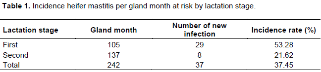
All the research findings were stored in Microsoft Excel (version 2007) and prepared for analysis. The incidence rates were estimated at gland level and expressed in terms of gland month at risk according to Thrusfield (2005). Chi-square test and relative risk (RR) with confidence interval (CI) of 95% were used to evaluate risk factors. The data were analyzed using Epi InfoTM (version 3.5.1., Center for Disease Control, USA). Statistical significance was set at a P≤0.05.
RESULTS
Study area
This study was conducted in and around Debre-Libanos district in North Showa zone of Oromia, Ethiopia. Debre-Libanos district is located 9° 48’ N and 38° 44’ E at about 89 km north of Addis Ababa. The altitude of the district is between 1500-2700 m above sea level, its minimum and maximum temperature varies from 19 to 23°C. It gets bimodal rain fall that ranges from 800-1200 mm and the predominant soil type are black soil (56%) and red soil (38%). Two agro ecologies are found in the area and mixed agricultural activities are performed. There are about 80,796 head of cattle, 84,507 goats, 23,723 sheep, 10,899 equines and 75,305 poultry in the districts. All of these livestock species are reared mainly by smallholder farmers under intensive, semi intensive and extensive production system. The district is divided into 11 administrative peasant associations and 13,000 L of milk is collected per day from Debre-tsege town only (Debre-Libanos district Agricultural and Rural development office, personal communication).
Study animals and husbandry practices
The studied animals were 31 pregnant heifers that were left for less than a month to calve and were followed for the first two lactations stages after calving. Two breeds of heifers from smallholder and commercial dairy farms (Holistein-Fresian and Jersey crosses) in and around Debre-Libanos district were included in the study. 20 of them were from Debre-Libanos district while 6 and 5 heifers were from Wuchale and Grar-Jarso district, respectively. The heifers were managed intensively and semi intensively by the smallholder dairy farmers while the commercial farm managed them semi-intensively.
Sample size and sampling method
Purposeful sampling technique was used in this study and 31 pregnant heifers that were left with less than four weeks to calve were included in the study. Numbered ear tags or local names were used to identify the pregnant heifers in each herd. The heifers were immobilized and secretion samples for microbiological tests were taken from open udder quarters. The assessment of open or closed udder quarters was done using the method described by Quinn et al. (1994). It involved applying light pressure to the teat sinus by using a gentle milking action with the thumb and index finger, allowing the contents to slip upward within the teat. When the pressure applied by this action resulted in a drop of secretion at the orifice, the teat was classified as being open, otherwise as closed. Only open quarters were sampled. Due to the small volume of secretion presented in most glands, foremilk secretion was not discarded strictly before collection of the sample ante partum. After calving, two samples were taken representing the first and the second stage of lactation. And here, foremilk secretion was discarded before taking the samples.
Data collection
Questionnaire survey
A semi-structured questionnaire was developed and pre-tested, and all information relating to the study objectives were recorded. Data were collected on potential risk factors for the occurrence of mastitis in heifers and cows based on observation and by interviewing the farm owners. The animal level factors such as herd size, presence of teat lesion, teat blindness, body condition and breed difference were recorded. The farm level factors such as housing types, farm hygiene, barn floor status, type of milking method, use of towels, milking sequences and hygiene were also recorded. Udder and milk abnormalities (injuries, blindness, tick infestation and indurations, swelling, milk clots, abnormal secretion, etc.) were also recorded.
Physical examination of the udder
Cows with clinical mastitis were identified first by examining the udder visually and then through palpation to detect possible fibrosis, inflammatory swellings, visible injury, tick infestation, atrophy of the tissue, heat, pain and/or hardness and swelling of supramammary lymph nodes (Biffa et al., 2005). Viscosity and appearance of milk secreted from each mammary quarter were examined for the presence of clots, flakes, blood and watery secretions. Rectal temperature of those cows with clinical mastitis was taken to check systemic involvement (Aarestrup and Jensen, 1997).
California mastitis test
The California mastitis test (CMT) was carried out as screening test for subclinical mastitis and for selection of samples for culture. Subclinical mastitis was diagnosed based on CMT results and the nature of coagulation and viscosity of the mixture (milk and CMT reagent), which show the presence and severity of the infection, respectively. In case of sub-clinical mastitis identification, a squirt of milk sample was placed in each of the cups on the CMT paddle from each quarter of the udder and an equal amount of 3% CMT reagent was added to each cup and mixed well. Reactions were then graded as 0 and trace for negative samples, or +1, +2 and +3 for positive ones. Quarters with CMT score +1 or above was judged as positive (Quinn et al., 1999). Cows were considered positive, when at least one quarter was tested positive using CMT. But during the first sampling, CMT was found to be not effective method to detect mastitis as the sampling volume from pregnant heifers was very minute and the content (keratin like) was not milk and all the samples were directly cultured and culture positive samples were recorded as CMT positive.
Milk sample collection
Milk sample were collected according to the procedure recommended by Quinn et al. (1994). Strict aseptic procedures were followed when collecting milk samples in order to prevent contamination with microorganisms present on the skin udder and teats, on the hands of samplers and on the barn environment. Teat ends were cleaned and disinfected with ethanol (70%) before sampling. Strict foremilk (first jets) were discarded to reduce contamination from the teat canal (Quinn et al., 1999). Sterile universal bottle with tight fitting cups were used to collect milk samples. To reduce contamination of teat ends during sample collection, the near teats were sampled first and then followed by the far ones. The collecting universal bottle was held as horizontal as possible and by turning the teat to the near horizontal position; 10 ml of milk sample were collected from each quarter of the udder into the universal bottle. After samples were collected, they were properly packed and transported in an ice box to the microbiology laboratory, Faculty of Veterinary Medicine, Addis Ababa University. Samples that were not immediately processed were refrigerated at +4°C for 24-72 h.
Bacteriological isolation and characterization
Milk samples were bacteriologically examined according to the procedures employed by Quinn et al. (1999). In refrigerated milk samples, bacteria may be concentrated in the cream layer and held with clumps of fat globules (Quinn et al., 1994). Hence, dispersion of fat and bacteria was accomplished by warming the samples at 25ºC for 15 min and shaken before plating on a standard bacteriological media. A loopful of milk sample collected each infected quarter was inoculated separately on to MacConkey agar and blood agar base enriched with 7% defibrinated sheep blood. The inoculated plates were then incubated aerobically at 37ºC for 24 to 48 h. When growth was not observed after incubation for 24 to 48 h, the quarter’s milk sample was inoculated on an enriched tryptone-soya broth to amplify the bacterial growth. Identification of the bacteria on primary culture was made based on colony morphology, hemolytic characteristics, Gram stain reaction including shape and arrangements of the bacteria, catalase and oxidation-fermentation tests. Staphylococci were identified based on Catalase test, growth characteristics on Mannitol salt agar and purple agar and tube coagulase test. Identification of Streptococci was made according to growth characteristics on blood agar, Gram reaction and arrangement, catalase test. Gram negative isolates grown on MacConkey agar were identified based on growth characteristics on MacConkey agar, oxidase reaction, catalase test, triple sugar iron (TSI) agar, the IMViC (indole, methyl red, Voges-Proskaur,and citrate) test (Quinn et al., 1999).
Antimicrobial sensitivity testing
The antimicrobial resistance profiles of the bacterial isolates were determined using Kirby-Bauer-disk diffusion method (Quinn et al., 1999). The antibiotic impregnated disks that were used for the test were; trimethoprim 5 µg, ampicillin 10 µg, chloramphenicol 30 µg, cefoxitin 30 µg, kanamycin 30µg, penicillin 6 µg, gentamycin 30 µg and tetracycline 30 µg. Well isolated bacterial colonies of the same morphologic type were inoculated into 7 ml of tryptophan soya broth and incubated at 37ºC for 8 h until turbidity was seen and was compared to a 0.5 McFarland standards. Mueller-Hinton Agar for less fastidious bacterial isolates and 5% sheep blood added Mueller-Hinton Agar for Streptococcus species isolates were used as planting medium. Fifteen minutes after inoculation of the plates, the antibiotic impregnated disks were applied on the surface of inoculated plates with sterile forceps. All the disks were gently pressed down onto the agar with forceps to ensure complete contact with the agar surface. The plates were inverted and then incubated aerobically for 18 h at 37ºC. The diameters of the zone inhibition were measured to the nearest whole millimeter using the transparent rule. Zone of inhibition for individual antimicrobial agents were translated into susceptible, intermediate and resistant categories by referring the recommended CLSI.org interpretative standard.
Data storage and analysis
All the research findings were stored in Microsoft Excel (version 2007) and prepared for analysis. The incidence rates were estimated at gland level and expressed in terms of gland month at risk according to Thrusfield (2005). Chi-square test and relative risk (RR) with confidence interval (CI) of 95% were used to evaluate risk factors. The data were analyzed using Epi InfoTM (version 3.5.1., Center for Disease Control, USA). Statistical significance was set at a P≤0.05.
Bacterial isolates
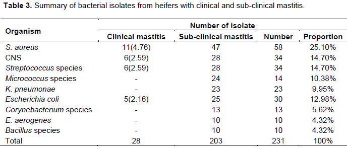
A total of 231 bacterial isolates were identified from milk samples. Pre-calving prevalence of quarters with IMI of any pathogen was 46% (Table 3). S. aureus was the most prevalent pathogen isolated (10%), and in decreasing proportion were CNS (8%), Streptococcus species (7%), Micrococcus species (5.6%), K. pneumonae (4.8%), E. coli (4%), Corynebacterium species (3.2%), Bacillus species and Entrobacter aerogens (each 1.6%). In the first lactation stage prevalence of IMI with any of the listed pathogens was 63%. S. aureus (16%) was again the most prevalent pathogen and in decreasing proportion were Streptococcus species (9.6%), CNS (8.1%), E. coli (7.2%), K. pneumonae (5.6%), Micrococcus species (4%) and Bacillus species, E. aerogens and Corynebacterium species (3.2% each). In the second lactation stage prevalence of IMI was 79.8%. S. aureus were again the most prevalent pathogen isolated (20.9%) and in decreasing proportion were E.coli (12.9%), CNS (11.2%), Streptococcus species (10.4%), K. pneumonae (8%), Micrococcus species (5.6%), Corynebacterium species (4%) and E. aerogens and Bacillus species (each 3.2%). Clinical mastitis was diagnosed in 5.4% of the quarters. Only S. aureus, CNS, Streptococcus species and E. coli were isolated from clinical mastitis.
Antimicrobial susceptibility test
The antibiotic susceptibility test revealed that highest number of S. aureus was susceptible to tetracycline 76.92% and the least number of S. aureus were susceptible to penicillin G 1.92% (Table 4).The antibiotic susceptibility test showed a highest number of E. coli was susceptible to tetracycline 76.92% and least susceptible to penicillin G 38.46%.
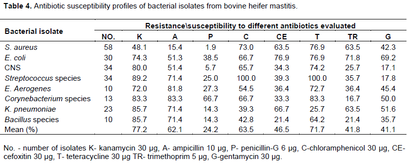
The antibiotic susceptibility test revealed that highest and the least number of coagulase negative Staphylococcuswas susceptible to Kanamycin 80% and penicillin G 5.71% respectively. The antibiotic susceptibility test suggested that highest number of Streptococcus species were susceptible to Tetracycline and Chloramphenicol 100% and least susceptible to Gentamycine 17.80%. The antibiotic susceptibility test indicated that highest number of Bacillus species were susceptible to Kanamycin 85.71% and least susceptible to penicillin G 14.28%.
DISCUSSION
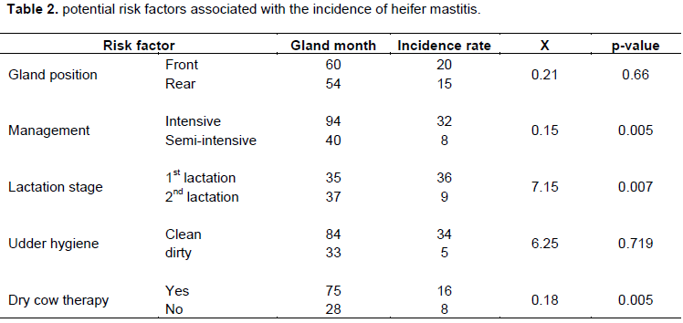
The incidence rate of mastitis was higher in heifers and cows held under intensive management system contrasted with heifers and cows under semi intensive management system. This could be because of limited exercise of animals and the transmission of pathogens from infected cows to primiparous as animals were housed together. Higher risks of mastitis with restricted exercise (Ginn et al., 1982; Gillespie et al., 1999) and in combined housing management of heifers with multiparous cows (Barkema et al., 1998) were reported. Incidence rate of mastitis was likewise higher in cows at first lactation stage than in cows at mid lactation stage. First lactation stage had higher incidence rate of mastitis (53.28%) than mid lactation stage (21.62%); the difference is statistically significant (P<0.05). This outcome was in close arrangement with a report made somewhere else (Smith et al., 1994). Nonattendance of dry dairy animals treatment routine could be the main consideration adding to high prevalence at early lactation and early infection related with delayed diapedesis of neutrophils into the mammary gland (Schalm 1942). Cows that were not treated amid dry period were more affected than those treated and there was factual contrast among treated and untreated groups (P<0.05). This distinction could be related with low bactericidal and bacteriostatic nature of milks and mammary gland 15 days before and after calving. Also, the limit of the quarters to give phagocytic and bactericidal action by and large decreases amid this period (Nordhaug et al., 1994).
In this investigation, subclinical mastitis has been observed to be higher in heifers and cows than clinical mastitis. This could be ascribed to the little consideration given to subclinical mastitis while treating clinical cases. Also, ranchers in Ethiopia are not all around educated about the quiet instances of sub-clinical mastitis (Almaw et al., 2009). Among the farms visited during this study, only one farm managed its cows semi-intensively did use CMT arbitrarily to detect sub-clinical mastitis.
Quarter prevalence of mastitis (47.04%) found in this investigation was tantamount with the finding of Trinidad et al. (1990a) who announced quarter prevalence rate of 53.1%, yet higher than the report made by Trindad et al. (1990b) and Nickerson et al. (2000) who revealed 35.25 and 27.57%, respectively. When contrasted with the others, the right back quarter were affected with the most noteworthy infection rate (48.9%) while the left back quarters were the second with an infection rate of 48.11%. This may be because of the high production capacity of the hind quarters (Radostits et al., 2006) and the high possibility of getting fecal and environmental defilement (Almaw et al., 2009).
The outcome gotten from bacteriological examination of the samples uncovered that from an aggregate of 372 quarter tested, 175 (47.04%) were bacteriologically positive which is higher than Alemaw et al. (2009) who revealed extents of 18%. This might be because of bactericidal properties of inflammatory udder secretions. Mastitis is the aftereffect of cooperation between three components like bacteria, dairy animals and environment. In this investigation, the transcendent life forms isolated from clinical and sub-clinical mastitis were found to be Staphylococcus aureus followed by CNS and Streptococcus species. The predominance and primary role of S. aureus isolate in bovine mastitis have additionally been accounted for in different investigations (Harmon et al., 1986). Radostits et al. (2006) stated that S. aureus is very much adjusted to get by in the udder and more often establishes a mild sub-clinical disease of long term from which it shed in milks encouraging transmission to sound animals for the most part amid milking. The least distinguished isolate was Bacillus species as was seen by Fox et al. (1995).
The predominance of Staphylococcus aureus may likewise be clarified by inadequate milking and particularly when it is related with excruciating sores or any injuries on the surface of the udder (Alemaw et al., 2009). Staphylococcus is an opportunistic pathogenic bacterium which survives on the skin of the udder and can infect the udder via teat canal or any wound. Further, the prevalence of E. aerogens in the present investigation might be because of poor sterile conditions in the herds and this infection is ending up increasingly incessant and will in general, pursue the contamination of Staphylococcus species (Harmon et al., 1986). The prevalence of Streptococcus species might be because of poor dairy practicing techniques, which is a contagious organism that infects other healthy animals in the herd. The prevalence of Bacillus species in the present investigation might be because of natural variables like soil, water and feaces; these are the fundamental wellspring of microorganisms when animals are presented to the above ecological elements, and microscopic organisms like Bacillus enter by means of teat canals. Comparable finding demonstrated that higher prevalence of E. coli might be because of poor sterile conditions as E. coli originates from the heifer’s and cow’s environment and infect the udder via teat canal (Nickerson et al., 2000).
Further in the present investigation, antibiotic susceptibility tests were likewise directed for the detaches by utilizing eight antibiotics which were utilized much of the time in the investigation zones for treatment of mastitis and tetracycline was observed to be the most effective antibiotics among all the tried antibiotics against every one of the bacterial isolates pursued by kanamycin, chloramphinicol, ampicillin, gentamycin, cefoxitin, trimethoprim and penicillin G.
The susceptibility bacterial isolates in the present investigation was similar to the existing reports by Alemaw et al. (2009), whereas Oliver et al. (2005) have reported that ciprofloxacin was the best antibiotic against bacteria isolated from cattle and buffaloes. Gillespie et al. (1999) have discovered that most noteworthy affectability of bacteria was appeared to enrofloxacin and gentamycin; comparative discoveries have been accounted for by Alemaw et al. (2009) in which gentamicin was observed to be best antibiotics. Rather than these discoveries, it has been accounted for that 64% of the isolates, from mastitis milk were sensitive to tetracycline and 52.8% to ampicillin (Oliver et al., 2005). As of late, it has been accounted for that the most noteworthy number of S. aureus and CNS isolated from bovine mastitis were susceptible to ceftriaxone (Gillespie et al., 1999).
The present study has demonstrated that heifer and cow mastitis exceptionally; sub-clinical one is a vital illness influencing heifer's milk production due to dispose of milk, blind teat and culling. Absence of execution of routine mastitis prevention and control rehearses by every one of the breeders was observed and preponderance of risk factors noted are the primary explanations behind the observed high incidence rate of heifer mastitis. In like manner, cows found at their first lactation stage, managed intensively and that did not get dry cow therapy, were at higher danger of contracting mastitis. The present investigation has additionally uncovered that S. aureus is an imperative reason for heifer and cow mastitis and higher public health risk because of utilization of crude milk and its products. Besides, the investigation plainly demonstrated that heifer’s mastitis etiological organisms are the same as cows. Antimicrobial sensitivity test obviously demonstrated that tetracycline is the best medication for the majority of the pathogens while penicillin G is the least one.
Accordingly, the present investigation warrants the requirement for applying possible heifer and cow mastitis intercession procedure with unique accentuation on sub-clinical mastitis including solid dairy extension service focused on awareness creation and applying dry cow therapy.
The animal health delivery framework need to concentrate on standard screening of dairy cows; including heifers, for sub-clinical mastitis and treating of the cases both in lactation and dry period. At long last, the balanced utilization of antibiotics and regular antibiogram surveillance ought to be made an integral part of the methodology.
CONFLICT OF INTERESTS
The authors have not declared any conflict of interests.
REFERENCES
|
Aarestrup F, Jensen N (1997). Prevalence and duration of intramammary infection in Danish heifers during the peripartum period. Journal of Dairy Science, pp. 80:307-312. |
|
|
Alemaw G, Molla W, Melaku A (2009). Prevalence of bovine subclinical mastitis and antimicrobial resistance pattern in Gondar town and surrounding areas, Ethiopia. Livestock Research for Rural Development 21(7). |
|
|
Barkema HW, Schukken YH, Lam TJ, Beiboer ML, Wilmink H, Benedictus G, Brand A (1998). Incidence of clinical mastitis in dairy herds grouped in three categories by bulk milk somatic cell counts. Journal of Dairy Science 81:41-49. |
|
|
Biffa E, Debela F, Beyene, K (2005). Prevalence and Risk factors of mastitis in lactating dry cows in southern Ethiopia. The International Journal of Applied Research in Veterinary Medicine 3(3):189-198. |
|
|
Fox LK, Chester ST, Hallberg JW, Nickerson SC, Pankey JW, Weaver LD (1995). Survey of intramammary infections in dairy heifers at breeding age and first parturition. Journal of Dairy Science 78(7):1619-1628. |
|
|
Gillespie BE, Owens WE, Nickerson SC, Oliver SP (1999). Deoxyribonucleic acid finger printing of Staphylococcus aureus from heifer mammary secretions and from horn flies. Journal of Dairy Science 82(7):1581-1585. |
|
|
Ginn R, Case VS, Packard H, Tatini G (1982). Quantitative assay of beta lactam residues in raw milk using a disc assay method. Journal of Food Protection 45(6):571-57. |
|
|
Harmon WL, Crist RW, Hemken BE, Langlois T (1986). Prevalence of minor udder pathogens after intramammary dry treatment. Journal of Dairy Science 69(3):843-49. |
|
|
Huijps KS, de Vliegher M, Hogeveen H (2007). Heifer mastitis: it takes money. In the Proceedings of the 2007 Heifer Mastitis Conference, pp. 88-89. |
|
|
Nickerson WE, Owens HG, Boddie RL (2000). Efficacy of a Staph. aureus mastitis vaccine in dairy heifers. In Proceedings of the Symposium on Immunology and the Ruminant Mammary Gland, pp. 426-431. |
|
|
Nielsen U, Emanuelson B, Berglund N, Strandberg E (2009). Relationship between somatic cell count and milk yield in different stages of lactation. Journal Dairy Science 92(7):3124-3133. |
|
|
Nordhaug LL, Nesse NL, Norcross H, Gudding R (1994). A field trial with an experimental vaccine against Staphylococcus aureus mastitis in cattle. Clinical parameters. Journal of Dairy of Science 77(5):1267-1275. |
|
|
Oliver BE (1988). Frequency of isolation of environmental mastitis causing pathogens and incidence of new intramammary infection during the non lactating period. American Journal of Veterinary Research 49(11):1789-93. |
|
|
Oliver BE, Gillespie SJ, Headrick MJ, Lewis HH (2005). Prevalence, risk factors and strategies for controlling mastitis in heifers during the periparturient period. International Journal of Applied Research in Veterinary Medicine 3(2):150-162. |
|
|
Quinn PJ, Carter ME, Markey BK,. Carter GR (1994). Mastitis. Clinical Veterinary Microbiology. Wolfe Publishing, London, pp. 327-344. |
|
|
Quinn PJ, Markey BK, Carter GR (1999). Clinical Veterinary Microbiology, Mosby international Limited, London pp. 21-66. |
|
|
Radostits CC, Gay CD, Hinchcliff KW, Constable PD (2006). Diseases of mammary gland. In: Veterinary Medicine. A text book of the diseases of Cattle, Sheep, Goats, Pigs and Horses. 10th edition. W.B. Saunders Company, London P 674. |
|
|
Schalm OW (1994). Streptococcus agalactiae in the udder of heifers at parturition traced to suckling among calves. Cornell Veterinary 32:49-60. |
|
|
Smith JS, Hogan DA, Todhunter WP, Weiss PS (1994). Intramammary infection and clinical mastitis in heifers at calving and dynamics over a 14 year period in a dairy herd. Journal of Dairy Science 77(Suppl. 1):197. (Abstr). |
|
|
Trinidad P, Nickerson SC, Alley TK (1990a). Prevalence of intramammary infection and teat canal colonization in unbred and primigravid dairy heifers. Journal of Dairy Science 73(1):107-114. |
|
|
Trinidad P, Nickerson SC, Luther DG (1990b). Antimicrobial susceptibilities of staphylococcal species isolated from mammary glands of unbred and primigravid dairy heifers. Journal of Dairy Science 73(2):357-62. |
|
Copyright © 2024 Author(s) retain the copyright of this article.
This article is published under the terms of the Creative Commons Attribution License 4.0


