ABSTRACT
Bovine coccidiosis is a protozoan infection caused by different species of Eimeria which has a worldwide distribution. The disease which mainly affects calves belongs to large herd size where hygiene is not well managed and is associated with poor body condition. A cross-sectional study was conducted from November 2016 to April 2017 to determine the prevalence of bovine coccidiosis and identify the associated risk factors in semi intensive and extensive dairy farms in and around Holeta town, Finfine Zuria Liyu Zone, Oromia Regional State, Ethiopia. Fecal samples were randomly collected from three hundred and eighty four calves belonging to dairy farms and examined for the presence of the oocyts of Eimeria by floatation technique using saline solution. The study revealed that the overall prevalence of coccidiosis was 26.04%. The risk factors considered were age, sex, breed, production system, herd size, fecal consistency, body condition and hygienic status of the house. The prevalence of coccidiosis was higher within calves in poor hygienic (58.6%) dairy farms than calves from better hygienic farms (14.6%). There was also significant difference (P<0.05) in the prevalence of coccidiosis between different herd sizes with higher prevalence in herd size >10 animals (39.3%). The highest prevalence of coccidiosis was recorded in calves with diarrheic faeces (91.7%) than calves with soft, constipated and normal fecal consistency (P<0.05). Appropriate monitoring and control of the disease is advisable in the study farms.
Key words: Calf, coccidiosis, dairy farms, Eimeria, Holeta, prevalence.
Ethiopia is endowed with abundant livestock resources of varied and diversified genetic roles with specific adaption to its wide range of agro ecologies (CSA, 2015). This great livestock potential is not properly exploited due to many prevailing socio economic values and attitudes, traditional management methods, limited genetic potential and rampant disease. Gastrointestinal parasite infections are a problem for both small and large scale farms; however, their impact is greater in sub-Saharan Africa. The prevalence ofgastrointestinal parasites and the severity of infection vary considerably depending on the genera of helminth and protozoan parasites involved, animal species, local environmental conditions such as humidity, temperature, rainfall, vegetation and management practices (Debela, 2002; Sandhu and Singla, 2005; Wadhawa et al., 2011 ).The most important disease problems in the young calf are pneumonia and diarrhea. The pathogens associated with calf diarrhea are rotavirus, corona virus, Salmonella species, protozoan parasites, Eimeria and Cryptosporidium species (Bhat et al., 2012; Brar et al., 2017). Bovine coccidiosis is a protozoan disease of the intestinal tract caused by microscopic organisms called coccidia; and is one of the most common and important disease of cattle worldwide(The Merck Veterinary Manual, 2005). This disease is usually the most known and devastating protozoan disease in calves under age of one year (Ernst et al., 1987). Previous works which were conducted in different parts of Ethiopia showed that coccidiosis is a paramount important protozoan disease in younger calves (<12 months) which were kept in poor hygienic status as well as improperly nourished with colostrum (Abebe et al., 2008; Mehreteab et al., 2012; Alemayehu et al., 2013; Temesgen, 2016). Although, coccidiosis is an important cause of calf morbidity and mortality in Ethiopia in general, and in the study area in particular, there is no previous detail information on its prevalence coccidiosis in the study area. Therefore, this study was conducted to determine the prevalence and associated risk factors of calf coccidiosis in farms in and around Holeta, Finfine Zuria Liyu zone, Ethiopia.
Study area
The present study was conducted in extensive and semi intensive farms found in and around Holeta town located in Finfine Zuria Liyu Zone, Oromia Regional State, Ethiopia during the period between November 2016 and April 2017 to determine calf coccidiosis and its putative risk factors. Holeta is located 45 km west of Addis Ababa at altitude 2400 m above sea level. It is geographically located between 9° 3´N latitude and 38° 30´ E longitudes. The area experienced bimodal rainfall pattern with a short rainy season from February to April and the long rainy season from the middle of June to end of September. The remaining months are dry periods. The area gets an annual rain fall of 1000 to 1100 mm and the annual temperature ranges between 18 and 24°C. The total cattle population of the study area is estimated to be 175,741, out of which 172,769 (98.3%) heads of cattle are local breeds and 2972 (1.7%) are crosses kept under extensive and semi intensive management systems (WoWAHA, 2015).
Study animals
The study was carried out on 384 calves within the age of 1 month to 1 year old. The samples were randomly collected from calves reared under semi intensive and extensive management systems. Examined calves were categorized based on their age and grouped into three as: group I (1 to 4 months age), group II (5 to 8 months) and group III (9 to 12 months of age), based on house hygiene grouped into three (good, moderate and poor) and also based on size of herd grouped into three (<10, 11-20 and >20 head of calves).
Study design and sample size determination
The type of study was cross sectional with simple random sampling technique conducted between November 2016 and April 2017 to determine the prevalence and associated risk factors of calf coccidiosis in and around Holeta town. The desired sample size for this study was determined by using the single population proportion formula according to Thrusfield (2005). Since there is no previous report on calf coccidiosis in the study area, the sample size was established based on the 50% expected prevalence, 5% desire absolute precision and 95% confidence level (Cl).
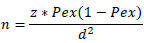
Where, n = required sample size; z=1.962; pex = expected prevalence; d = desired absolute precision. Thus, the desire sample size for Pex = 0.5 is n= 384 calves included in this study. While collecting faecal samples, data related to age, sex, bred, herd size management system, body condition, fecal consistency and hygienic status of barn were properly recorded.
Faecal sample collection and examination
A total of 384 faecal samples were collected directly from the rectum of each sampled animal with strict sanitation, and placed in air and water tight sample vials. After collection, the samples were transported in ice box to Holeta Agricultural Research Center (HARC) Parasitological Laboratory for fecal examination using simple floatation technique with saline solution (Yu et al., 2011; Gupta and Singla 2012).
Data management and analysis
All data collected were entered and managed in MS-Excel software program and analyzed using SPSS 20.0 statistical software version. Descriptive statistics such as percentage was used to approximate the prevalence of calf coccidiosis in the study area. Prevalence was calculated as number of positive calves harboring Eimeria oocytes divided by the total calves examined. Chi-square (χ2) statistics were used to test the association between variables. At p <0.05 was taken as statistically significant.
Overall prevalence
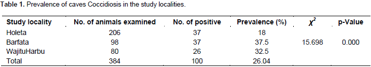
In the present study, out of total 384 samples tested, 100 (26.04%) were positive for the presence of Eimeria oocyts. The prevalence was higher in Barfata (37.5%) as compared to other study localities (p <0.05) as shown in Table 1.
Potential risk factors
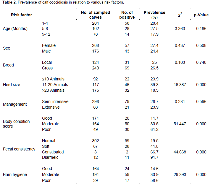
The analysis of putative risk factors was made by Chi-square analysis. The result showed strong significant associations between coccidian infection and herd size (χ2 =16.387, P = 0.000), different body condition scores (χ2 = 51.447, P = 0.000), faecal consistency (χ2 = 44.668, P = 0.000) and hygienic status of the house (χ2 = 29.393, P= 0.000). However, no significant difference was observed between the prevalence of coccidiosis and different age groups (χ2=3.363, P =0.186), sexes (χ2 = 0.437, p = 0.508), breeds (0.103, P =0.748) and different management systems (χ2 = 0.281, P =0.596) as shown in Table 2.

Calf coccidiosis causes a significant economic loss through morbidity and mortality worldwide. The result of the present study conducted in three localities of Holota town and its environs showed an overall prevalence of 26.04% coccidian infection in calves. The 26.04% prevalence of coccidiosis in this study is in line with previous studies of Bekele et al. (2012) [22.7%] in Dire Dawa, Temesgen (2016) [24.3%] in South Wollo Zone Amhara region, Ethiopia, Toaleb et al. (2011) [24.2%] in Egypt. However, the prevalence result of the present study is much lower than many of the previous reports in Ethiopia, namely, 68.1% in Addis Ababa and Debre Zeit Dairy Farms (Abebe et al., 2008), 31.9% in Kombolcha, south Wollo (Alemayehu et al., 2013), 62.5% in Asella town (Ibrahim, 2016) and 38.9% in and around Asella town dairy farms (Tsegaye, 2016). The findings of this study are also much lower than that of other countries of the world. For instance, a prevalence of 47.1% was reported in China (Dong et al., 2012). The results of the present study, however, is higher than previous reports by Gillhuber et al. (2014) (13.3%) in Southern Germany, Hussin (2016) (9.5%) in Iraq and Das et al. (2015) (11.9%) in India. Such inconsistency in the prevalence rate of coccidiosis may be due to the variation in diagnostic tests, age of the animals, susceptibility of different breeds to the disease, stress level, handling, climatic and other factors of agro-ecology, variation in the study season, number and target group of the study animals and husbandry practices(Radostitis et al., 2007a; Abebe et al., 2008; Heidari et al., 2014). This study showed variation in the prevalence of calf coccidiosis between different study localities (18% to 37.5%). Similar results were obtained in different parts of Zimbabwe (17.4 to 32.6%) (Pfukenyi et al., 2007), Al-Baha, Saudi Arabia (29.84 to 32.51%) (Ibrahim et al., 2015) and in Addis Aababa and DebreZeit, Ethiopia (57.2 to 76.4%) (Abebe et al., 2008). This geographical difference in distribution of positive cases could be explained by the management practices and the bio-security followed by the farm owners.
Analysis of risk factor with regard to the age of the calves revealed that there is no statistically significant association (χ2= 3.363, P= 0.186) between the age of the calves and coccidian infection. This result agrees with the reports of Abebe et al. (2008), Tsegaye (2016), Alemayehu et al. (2013), Bekele et al. (2012) and Gillhuber et al. (2014). This similarity could be due to the fact that animal husbandry practices in the study areas are identical, and also, different age groups were kept and housed together without separation. There was no statistically significant difference in prevalence of coccidian infection between male and female animals (χ2 = 0.437, p =0.508), which is in agreement with the reports of Abebe et al. (2008), Heidari and Gharekhani (2014), Alemayehu et al. (2013) and Ibrahim (2016). This might be associated with the fact that different sex groups kept in similar husbandry system might have equal chance of accessing the oocyts. Despite this, previous studies done on adult cattle showed higher prevalence of Eimeria in female animals than in males (Manya et al., 2008; Rehman et al., 2011). Nevertheless, this could be attributed to the physiological stress loaded on female animals in relation to pregnancies and giving birth as compared to males (Radostits et al., 2007a). The breed related prevalence of coccidian infection in the present study showed no statistical significant difference between breeds (χ2 = 0.103, p =0.748), which is in agreement with the findings of Abebe et al. (2008) and Alemayehu et al. (2013). The possible explanation for this similarity could be the fact that calf rearing condition in the study areas was identical and different breeds were housed together without separation. In contrast to the current findings, susceptibility differences were reported between local and cross breeds (Ibrahim, 2016).
This breed susceptibility difference could be related to the dose of oocytes ingested and the species of Eimeria involved in the infection (Taylor et al., 2007). In the current study, coccidian infection was significantly higher in calves reared in large herd size than in small size (χ2 = 16.387, P = 0.000). This finding is in line with the report of Nasir et al. (2009). This similarity might be due to rapid spread of infection from calf to calf as well as greater contamination of feeding and watering troughs when animals are communally feed and overcrowded (Radostitis et al., 2007a). However, the current finding disagrees with that of Abebe et al. (2008). This variation might be due to the differences in the study seasons, husbandry practices and the treatment regime given to the calves. There was no significant difference (χ2 = 0.281, p = 0.596) in the prevalence of coccidiosis and management systems. This result agrees with the report of Abebe et al. (2008), Alemayehu et al. (2013) and Temesgen (2016). This similarity might be due to equal chance of accessing the oocysts when grazing from contaminated field. However, the current finding is in contrast with the previous report of Abisola (2004). This variation might be due to hygienic condition of the barn, nutritional status of the calves, contamination level of the feed, water, floor and treatment given to the animals. There was statistically significant (χ2=44.668, P=0.000) difference in prevalence rate between fecal consistency and coccidian infection which agrees with the findings of Pundit (2009) and Alemayehu et al. (2013).
However, this finding disagrees with the report of Abebe et al. (2008). In the present study, 91.7% diarrheic calves were found tobe positive to Eimeria. However, there were no apparent clinical signs in most of the animals sampled for the study. A strong significant association (χ2= 51.447, P= 0.000) was recorded between body condition score and coccidian infection in the current study. Similarly, Mehreteab et al. (2012) reported a higher infection rate in calves with poor body condition score than in calves with good and moderate body condition score. This might be due to the weak immune status of the calves with poor body condition score. As a result, malnutrition and other parasitic infections result in immune compromised calves. This condition produces a higher infection rate in poor state animals than in good-state animals (Radostits et al., 2007b). In the current study, association of Eimeria infection in relation to the hygienic status of calf house was verified (p < 0.05). This result agrees with the report of Bekele et al. (2012) Mundt et al. (2005a, b) and Dawid et al. (2012). The similarity implies that poor sanitation in the calving and calf housing areas as well as poor management of housing favors infection of coccidiosis. Obviously, poor ventilation, droughts, poor calf nutrition, group pens, heavy stocking, cows present with calves, soiled bedding are regarded as risk factors for coccidiosis (Radostits et al., 2007b).
CONCLUSION AND RECOMMENDATIONS
In conclusion, this study provides proof of a coccidian infection in dairy farms in Holeta and its environs. Hygiene of calf’s house can be considered as a risk factor for the occurrence of coccidia. Large herd size, diarrheic faeces and poor body condition of the calves also increased the risk of infection with coccidia. However, sex, age, breed and management system of calves did not show any difference with the occurrence of protozoan parasite infection. In general, different risk factors were considered to affect the rate of infection of calves with this protozoan parasite. Based on these findings, it was recommended that calves with severe diarrhea be isolated and treated with appropriate drugs; any possibility of fecal contamination of the farm and the calves be minimized. Further epidemiological investigations are required to determine the protozoan parasite species composition and their economic impact.
The authors declare that there is no conflict of interest.
REFERENCES
|
Abebe R, Wossene A, Kumssa B (2008). Epidemiology of Eimeri infections in calves in Addis Ababa and DebreZeit Dairy Farms, Ethiopia. Int. J. Appl. Res. Vet. Med. 6(1):24-30.
|
|
|
|
Abisola TO (2004). Studies on Bovine coccidian. [Apicomplexia: eimeriidae] In: Parts of Plateau State, Nigeria. Available at:
View
|
|
|
|
|
Alemayehu A, Nuru M, Belina T (2013). Prevalence of bovine coccidia in Kombolcha district of South Wollo, Ethiopia. J. Vet. Med. Anim. Health 5(2):41-45.
|
|
|
|
|
Bekele M, Ferid D, Yeshitila A (2012). Calf coccidiosis in selected dairy farms of Dire Dawa, Eastern Ethiopia. Glob. Vet. 9(4):460-464.
|
|
|
|
|
Bhat SA, Juyal PD, Singla LD (2012). Prevalence of cryptosporidiosis in neonatal buffalo calves in Ludhiana district of Punjab, India. Asian J. Anim. Vet. Adv. 7(6):512-520.
Crossref
|
|
|
|
|
Brar APS, Sood NK, Kaur P, Singla LD, Sandhu BS, Gupta K, Narang D, Singh CK and Chandra M (2017). Periurban outbreaks of bovine calf scour in Northern India caused by Cryptosporidium in association with other enteropathogens. Epidemiol. Infect. 145(13):2717-2726.
Crossref
|
|
|
|
|
Central Statistical Agency (CSA) (2015). Federal Democratic Republic of Ethiopia: Central Statical Agency Agricultural sample survey, Volume II, report on Livestock and Livestock chracterstics, Bulletin No.578. Addis Ababa. pp. 12-15.
|
|
|
|
|
Das M, Deka DK, Sarmah PC, Islam S, Sarma S (2015). Diversity of Eimeria spp. in dairy cattle of Guwahati, Assam, India. Vet. World 8(8):941-945.
Crossref
|
|
|
|
|
Dawid F, Amede Y, Bekele M (2012). Calf coccidiosis in selected dairy farms of Dire Dawa, Eastern Ethiopia. Glob. Vet. 9 (4):460-464.
|
|
|
|
|
Debela E (2002). Epidemiology of gastro-intestinal helminthiasis of Rift Valley goats under traditional husbandry system in Adami Tulu district, Ethiopia. Ethiop. J. Sci. 25: 35-44.
|
|
|
|
|
Dong H, Zhao Q, Han H, Jiang L, Zhu S, Li T, Kong C, Huang B (2012). Prevalence of coccidian infection in dairy cattle in shanghais, China. J. Parasitol. 98(5):963-966.
Crossref
|
|
|
|
|
Ernst JV, Stewart TB, Witlock DR (1987). Quantitative determination of coccidian oocysts in beef calves from the coastal plain area of Georgia (USA). Vet. Parasitol. 23(1-2):1-10.
Crossref
|
|
|
|
|
Gillhuber J, Rügamer D, Pfister K, Scheuerle MC (2014). Giardiosis and other enteropathogenic infections: A study on diarrhoeic calves in Southern Germany. BMC Res. Notes 7:112-120.
Crossref
|
|
|
|
|
Gupta SK, Singla LD (2012). Diagnostic trends in parasitic diseases of animals. In: Veterinary Diagnostics: Current Trends. Gupta RP, Garg SR, Nehra V and Lather D (Eds), Satish Serial Publishing House, Delhi. pp. 81-112.
|
|
|
|
|
Heidari H, Gharekhani J (2014). Detection of Eimeria species in Iranian native cattle. Int. J. Adv. Res. 2(7):731-734.
|
|
|
|
|
Heidari H, Sadeghi-Dehkordi Z, Moayedi R, Gharekhani J (2014). Occurrence and diversity of Eimeria species in cattle in Hamedan province, Iran. Vet. Med. 59(6):271-275.
Crossref
|
|
|
|
|
Hussin AG (2016). Prevalence and Associated Risk Factors of Eimeria spp. in cattle of Baghdad, Iraq. J. Appl. Anim. Sci. 9(1):37-44.
|
|
|
|
|
Ibrahim DAYDN (2016). Prevalence and associated risk factors of calf coccidiosis in and around Asela Town, Southeast Ethiopia. Prevalence 6(3).
|
|
|
|
|
Ibrahim MM, Soliman MF, Alghamdi AO (2015). Subclinical bovine coccidiosis in Al-Baha Area, Saudi Arabia. Int. J. Vet. Sci. Res. 1(1):023-028.
|
|
|
|
|
Manya P, Sinha SR, Sinha S, Verma SB, Sharma SK, Mandal KG (2008). Prevalence of bovine coccidiosis at patna. J. Vet. Parasitol. 22:5-12.
|
|
|
|
|
Mehreteab B, Ferid D, Yeshitila A (2012). Calf coccidiosis in selected dairy farms of Diredawa, eastern Ethiopia. Glob. Vet. 9:460-464.
|
|
|
|
|
Mundt HC, Bangoura B, Rinke M, Rosenbruch M, Daugschies A (2005a). Pathology and treatment of Eimeria zuernii coccidiosis in calves: investigations in an infection model. Parasitol. Int. 54(4):223-230.
Crossref
|
|
|
|
|
Mundt HC, Bangaura B, Mengel H, Keidel J, Daugschies A (2005b). Control of clinical coccidiosis of caslves due to E. bovis and E.zuerni with toltrazuril under field conditions. Parasitol. Res. 97:134-142.
Crossref
|
|
|
|
|
Nasir A, Avais M, Khan MS, Ahmad N (2009). Prevalence of Cryptosporidium parvum infection in Lahore (Pakistan) and its association with diarrhea in dairy calves. Int. J. Agric. Biol. 11(2):221-224.
|
|
|
|
|
Pfukenyi DM, Willingham AL, Mukaratirwa S, Monrad J (2007). Epidemiological studies of parasitic gastrointestinal nematodes, cestodes and coccidia infections in cattle in the highveld and lowveld communal grazing areas of Zimbabwe. Onderstepoort J. Vet. Res. 74(2):129-142.
Crossref
|
|
|
|
|
Radostits OM, Gay CC, Blood DC, Hinchcliff KW (2007a). Veterinary Medicine - A Textbook of the Diseases of Cattle, Horses, Sheep, Pigs and Goats, 10th edition reviewed by Sameeh M. Abutarbush, BVSc, MVetSc, Diplomate ABVP, Diplomate ACVIM Saunders, USA Available at:
View
|
|
|
|
|
Radostits OM, Gay CC, Hinchcliff KW, Constable PD (2007b). Diseases associated with protozoa. Veterinary Medicine: A Textbook of the Diseases of Cattle, Sheep, Goats, Pigs and Horses. 10th ed. Philadelphia, Pennsylvania: Saunders Elsevier. pp. 1498-1515.
|
|
|
|
|
Rehman TU, Khan MN, Sajid MS, Abbas RZ, Arshad M, Iqbal Z, Iqbal A (2011). Epidemiology of Eimeria and associated risk factors in cattle of district Toba Tek Singh, Pakistan. Parasitol. Res. 108(5):1171-1177.
Crossref
|
|
|
|
|
Sandhu BS, Singla LD (2005) Coccidiosis in bovines: an increasing problem. In: Compendium of Winter School on Novel Approaches for Diagnosis and Control of Parasitic Diseases of Domestic and Wild Animals held from 07-27 October, 2005 at PAU, Ludhiana. pp. 190-194.
|
|
|
|
|
Taylor MA, Coop RL, Wall RL (2007). Veterinary Parasitology. 3rd ed. Oxford, UK: Blackwell Publishing. Available at:
View
|
|
|
|
|
Temesgen GK (2016). Epidemiological studies on calf coccidiosis in dairy farms in South Wollo Zone Amhara Region, Ethiopia. J. Vet. Sci. Technol. 7(392):2.
|
|
|
|
|
The Merck Veterinary Manual (2005). Merck & Co. Inc. and Merial Limited introduce the Ninth Edition of the Merck Veterinary Manual. Available at:
View
|
|
|
|
|
Toaleb NI, El-Moghazy FM, Hassan SE (2011). Diagnosis of eimeriosis in cattle by ELISA using partially purified antigen. World Appl. Sci. J. 12:33-38.
|
|
|
|
|
Thrusfield M (2005).Veterinary Epidemiology. 3rd Ed., Blackwell Science Ltd., Oxford, UK. pp.233-261.
|
|
|
|
|
Tsegaye E (2016). Occurrence of coccidiosis in diarrheic calves in and around Asella town dairy farms. World J. Biol. Med. Sci. 3(3):48-54.
|
|
|
|
|
Wadhawa A, Tanwar RK, Singla LD, Eda S, Kumar N and Kumar Y (2011). Prevalence of gastrointestinal helminths in cattle and buffaloes in Bikaner, Rajasthan, India. Vet. World 4(9):417-419.
Crossref
|
|
|
|
|
WoWAHA(2015).WolmeraWereda Animal Health Agency. Statistical Abstract. Wolmera, FinfineLiyuZuria Zone, Oromia, Ethiopia.
|
|
|
|
|
Yu SK, Gao M, Huang N, Jia YQ, Lin Q (2011). Prevalence of coccidian infection in cattle in Shaanxi province, Northwestern China. J. Anim. Vet. Adv. 10:2716-2719.
|
|