Seablite (Suaeda maritima) is a salt marsh plant growing in mangrove forest. Its young leaves can be used as fresh vegetable or cooked. The cooked seablite is quite salty, so it should be cooked with other types of vegetable to reduce salty taste (Tanaka, 1976). Local people in Samut Songkram province use seablite for different types of cooking such as traditional seablite salad, seablite curry with crabs, or scalded seablite with chili paste. The edible part is the young leaves which should be scalded for about 10 to 15 min and then knocked with cold water to make them crispier (Pornpitakdamrong and Sudjaroen, 2014). In the South of India, seablite is pickled in vinegar or used for cooking as well as domestic animal food (Bandaranayke, 2002). Sudjaroen (2012) studied the nutritional values of seablite reporting that the amounts in seablite were of water, protein (3.46 ± 0.04 %w/w), fat (0.15 ± 0.01 %w/w), carbohydrate (2.18 ± 0.02 %w/w), fibers (6.21 ± 0.01 %w/w), calcium (2,471.37 ± 0.054 mg/100 g), beta-carotene (3,545.16 ± 0.093 mg/100 g). It can be seen that seablite is an interesting vegetable with high nutritional values. It has been studied that the seablite leaves can be used to prevent hepatitis (Bandaranayke, 2002; Ravikumar et al., 2011) and for antivirus. Such biological activity is related to triterpenoids and sterols. Patra et al. (2011) determined the antioxidant and antimicrobial activity of seablite’s leaf and stem extracted by organic solvent (acetone, ethanol and methanol) and water. The finding was seablite extract from leaves and stems in all solvent can perform 70 to 92% of antioxidant from total antioxidant capacity, total phenolic content, and 2,2-diphenyl-1-picrylhydrazyl (DPPH) radical scavenging. From other ways to test the antioxidant of seablite, the result was also satisfying. The antimicrobial activities showed that seablite acetone extract was inhibited four types of pathogenic bacteria: Vibrio cholera, Staphylococcus epidermidis, Bacillus subtilis and Escherichia coli.
Seablite can grow naturally, so it is the low cost vegetable with high nutritional value. There should be a promotion of seablite as ready-made products for the convenience of the consumers in other regions. Moreover, it can be a new source of income of the people in the community. Pornpitakdamrong and Sudjaroen (2014) were developed dried seablite in lower scale production by using hot air blower (abc electro, Kirchheim-Teck, Germany), which can dried fresh seablite 200 to 300 g and yielded dried seablite only 30 to 50 g. On this point, seablite should be drying on larger scale and use simple technique, such as solar stove. Solar stove (size 60 x 120 cm with 4 sieves) can dry 2.5 to 3.0 kg that is10 time when compared with hot air blower and will be save energy supply, which is appropriate to people who living in rural area. The present study was aimed to produce dried seablite (Suaeda maritima) by solar stove, evaluate nutritive values and antioxidant activity of dried seablite during production and storage.
Seablite collection and dried seablite production
The basis data of seablite, such as, cultivated area, last annual yield of production and local expert’s interview, which were supported by Samut Songkram agricultural extension office. The seablite was harvested during December 2014 to February 2015, which scalded in boiling water added with 0.5% sugar boiling water for 5 min and after that it was soaked in cool water (Pornpitakdamrong and Sudjaroen, 2014). Scalded seabilte (2.5 to 3 kg) was blowing by electrical fan 1 to 2 h before dried by solar stove. Scalded seabilte was dried by solar stove (size 60 x 120 cm with 4 sieves) during 1 to 2 days. Internal temperature of solar stove and time of drying were recorded. The processed seablite must be dried in constant weigh prior to package in plastic bag and removed air by vacuum sealer (Karabada Sealer DZ-300A, Japan). Maximum of sunshine was optimal time for seablite drying during 10.00 a.m. to 4.00 p.m., which yielded 45 to 60°C in internal temperature of solar stove. Scalded seablite was dried within 1 to 2 days and remained 250 to 300 g of dried seablite. Preserved dried seablite in vacuumed plastic bags was100 g/each bag. Packaged dried seablite was further evaluated for nutritive values (Table 1) and biological assays, which were done in duplicate.
Evaluation of the nutritive values
Proximate analysis
The proximate analysis was carried out according to the methods to be described, or based on the Official Methods of Analysis of AOAC International, 16th ed (AOAC, 1995). The fresh samples were used for the water content determination. The remaining samples were dried at 105°C for 3 h, ground, and then stored in air-tight containers in a cool, dry place for other analyses.
Water content determination
Three to five grams of each sample was dried at 105°C for 3 h. The dried sample was then weighed. The water content was calculated as the percentage on the wet weight basis.
Determination of crude protein
Crude protein was determined by Kjeldahl method (AOAC, 1995), using Buchi Digestion Unit (B-435) and Distillation Unit (B-323) (Buchi, Switzerland). Dried sample (0.2 g) was digested with 20 ml of conc.H2SO4, using 3 g of the selenium and copper sulfate mixture as the catalyst. The digestion was continued for half an hour after the digestion mixture turned clear green. Then 60 ml of 32% sodium hydroxide solution was added, and the mixture was distilled for 3 min. The distillate was collected in a flask containing 60 ml of 2% (w/w) boric acid solution, with methylene blue and methyl red as the indicators. The distillate was then titrated with 0.1 N (w/w) H2SO4 solution; the end point was purple. Crude protein was calculated as the percentage on the wet weight basis (N × 6.25).
Determination of crude fat
One gram of the dried sample was extracted with 25 ml of petroleum ether in a Goldfisch apparatus (Labconco, U.S.A.) for 3 to 4 h. The petroleum ether extract was evaporated to dryness at 105°C. The residue was weighed and then calculated as the percentage of crude fat on the wet weight basis.
Determination of dietary fiber
Insoluble dietary fiber content was determined according to the AOAC Official Method 991.42 (AOAC, 1995). Amyloglucosidase (conc.) in the amount of 0.1 ml was used instead of 0.3 ml of the normal strength enzyme. Soluble dietary fiber content was determined according to the AOAC Official method by modified as in insoluble dietary fiber determination. The sum of both values was recorded as the total dietary fiber content of each sample.
Determination of total ash content
One gram of each sample was ignited in a muffle furnace at 525°C until ash was obtained. The residue was weighed and expressed as total ash on the wet weight basis.
Determination of carbohydrate
The carbohydrate content was obtained by difference, subtracting the water content, crude protein, crude fat, total dietary fiber, and total ash contents from 100% w/w.
Determination of β-carotene, Vitamin E and vitamin C
a) Measure β-carotene was applied from the method of Munzuroglu et al. (2003). Sample (50 g) was mashed in a homogenizer and 2g homogenate paste per sample was taken for extraction of β-carotene. To the above homogenates, 4 ml of ethanol were added, vortexed and the mixture centrifuged (MistralÓ 2000) at 2000 rpm for 3 min at 4°C. The supernatant was also filtered through a Whatman No.1 paper, and to the filtrate 0.15 ml n-hexane was added and mixed. β-carotene was extracted twice in the hexane phase and the collected extract was dried under a stream of liquid nitrogen. Dried extract was solubilized in 0.2 ml methanol and then filtered through a 0.45 μm membrane filter before high performance liquid chromatography (HPLC) injection. Injections were made in duplicate for each sample. The quantification utilized absorption spectra of 436 nm for β-carotene. HPLC separations were accomplished at room temperature with a Perkin-Elmer liquid chromatograph system (Series 1100), consisting of a sample injection valve (Cotati 7125) with a 20 μl sample loop, an ultraviolet (UV) spectrophotometric detector (Cecil 68174), integrator (HP 3395) and a Techsphere ODS-2 packed (5 mm particle and 80 A ? pore size) column (250 ´ 4.6 i.d. mm) with a methanol: acetonitrile: chloroform (47:42:11, v/v) mobile phase at 1 ml/min flow rate.
b) Measure vitamin E was applied from the method of Qian et al. (1998). An initial extraction procedure was developed as follows. Sample was ground in a warring blender and screened through an 80 mesh sieve. One g of the sample was precisely weighed and transferred to a 10 ml screw-capped extraction tube. Four ml of n-hexane was added to the tube and the tube was flushed with a steam of N2 to protect vitamins from air exposure before capping. The mixture was shaken on a vortex mixer for 0.5 min, rested for 5 min, and shaken another half minute. After centrifugation at 4000 rpm for 5 min, 1 ml of supernatant was transferred to a 1.5 ml vial and evaporated under nitrogen to remove the solvent. The residue was re-dissolved in 0.3 ml n-butanol and filtered through a 0.45 μm membrane filter before being injected into the HPLC system.
Chromatographic separations were performed on a 150 ´ 3.9 mm Novapak C column (Waters). Methanol was used as mobile phase at a flow-rate of 1.5 ml/min and a pressure of 1000 p.s.i. (1 p.s.i. = 6894.76 Pa) All injections were 50 μl loop injections on a M710B autosampler (Waters). A Model M510 Waters pump and a Model M490 Waters variable Wavelength UV–visible detector set at 290 nm were used. All quantitation was by peak area using a Waters M740 integrator. Based on the established chromatographic conditions, repeated injections of 0.1, 0.5, 1, 5 and 10 mg/L of the standard vitamin E was made duplicated onto the HPLC system. The retention time for vitamins E was 4.1 min. A Shimadzu MPS-2000 universal spectrophotometric scanner was used to determine the spectrograms of vitamin E in n-butanol.
c) Measure vitamin C was applied from the method of Sanchez-Moreno et al. (2003). Total vitamin C (ascorbic acid plus dehydroascorbic acid) were determined by HPLC. The procedure employed to determine total vitamin C was the reduction of dehydroascorbic acid to ascorbic acid, using DL-dithiothreitol as reductant reagent. 50 mg of each dried seablite was homogenized with 40 ml of an extraction solution (3% metaphosphoric acid plus 8% acetic acid).The resulting mixture was centrifuged, filtered, and adjusted to 100 ml with distilled water. Samples were filtered through a 0.45-μm membrane filter, and duplicates of 20 μl for each extract were analyzed by HPLC. Results were expressed as milligrams of ascorbic acid per 100 ml. An aliquot of the mixture was taken to react with 2.0 ml of a solution 20 mg/ml DL-dithiothreitol for 2 h at room temperature and in darkness. During this time the reduction of dehydroascorbic acid to ascorbic acid has been placed. Samples were filtered through a 0.45 μm membrane filter, and duplicates of 20 μl for each extract were analyzed by HPLC. Results were expressed as milligrams of total vitamin C per 100 ml. A Hewlett-Packard model 1050 quaternary solvent delivery controller pump was used for analysis. Samples was introduced onto the column via a manual injector (Rheodyne) equipped with a sample loop (20 μl). Separation of ascorbic acid was performed by HPLC using a reversed-phase C18 Hypersil ODS (5 μm) stainless steel column (250 ´ 4.6 i.d. mm) (Technochroma). The solvent system used was an isocratic elution of a 0.01% solution of H2SO4, adjusted to pH 2.5-2.6. The flow rate was fixed at 1.0 ml/min. A Hewlett-Packard 1040A UV-visible photodiode array detector was set at 245 nm; chromatographic data and UV-visible spectra were collected, stored, and integrated using a Hewlett-Packard Chem Station and related software. Identification of the ascorbic acid was carried out by HPLC by comparing the retention time and UV-visible absorption spectrum with those of the standard previously referred to. Calibration curves were built with 10, 25, 50 and 100 mg/100 ml of ascorbic acid standard.
Determination of calcium, iron and sodium
The microwave-assisted treatment was adapted from that employed by us for the determination of the mineral profile of diets (Mir-Marqués, et al., 2015). Calcium, iron and sodium determinations were done by inductively coupled plasma optical emission spectrometry (ICP-OES) techniques. ICP -OES Optima 5300 DV inductively coupled plasma optical emission spectrometer Perkin Elmer (Norwalk, CT, USA) equipped with an auto sampler AS 93-plus, and a ultrasonic nebulizer U6000AT+ Cetac (Nebraska EEUU) were used for all mineral determination. The operating conditions of the ICP-OES equipment were as follow 15 L/min of argon plasma gas flow rate, 0.2 L/min of auxiliary gas flow rate, 0.8 L/min nebulizer gas flow rate, 1300W of radio frequency (RF) power, and 1.1 ml/min of sample flow rate. The calibration standards were prepared in 0.5% nitric acid. The calibration range for all elements was evaluated from 0.05 to 2 mg/L except calcium for which calibration curves was prepared from 2 to 10 mg/L. Ruthenium (1 mg/L) was used as internal standard and added to all samples, reagent blanks and standards.
Test of biological activities
It was used100 g of ground dried seablite for continuous extraction, then, extract with ethanol and water using Soxhlet apparatus. Finally, get the solvent evaporated through rotary evaporation apparatus under vacuum.
Total phenolic content (TPC)
Measurement using Folin-Ciocalteu reagent (Singleton et al., 1999) was done by comparing it with standard solvent, that is, gallic acid at 1 to 0.125 mg/ml concentration; then, calculating total phenolic content (TPC) of gallic acid in mg/g of the extracts.
Antioxidant activity measurement
a) DPPH radical scavenging assay to measure the decreasing light absorbance of a,a-diphenyl-b-picrylhydrzyl (DPPH) radical (Yen and Duh, 1994) using negative control by DPPH radical (6×10⁻⁵ M), promptly measured at 515 nm using spectrophotometer (Genesis 20, Thermo Fisher Scientific, USA). 50 ml of methanolic extract (1 to 20 mg/ml) was placed in a cuvette, and 2 ml of DPPH (6×10⁻⁵ M) was added and then decrease in absorbance was determined. Vitamin C (0.1 mg/ml) was used as positive controls and unit of antioxidant activity was according by mg of gallic acid equivalent, GAE.
b) ABTS cation radical scavenging assay similar to the 1st method but using2, 2-azinobis (3-ethyl-benzothiazoline-6-sulfonic acid) (ABTS) radical instead (Re et al., 1999) and also using Trolox (water-soluble vitamin E analogue) as standard substance to create standard graph (0.5 to 5.0 mg/ml concentration). The antioxidant activity of the dried seablite would be shown in Trolox equivalent antioxidant capacity (TEAC)/g of the dried seablite extracts.
c) Oxygen radical absorbance capacity (ORAC) to measure ability of extract to scavenge oxygen radical (Prior et al., 2003) and the florescent signal generated by fluorescene sodium salt (Sigma-aldrich, Inc.) was measured by FLUOstar OPTIMA microplate reader (BMG) on 1 h. The antioxidant activity of dried seablite would be also shown in Trolox equivalent antioxidant capacity (TEAC)/g of the dried seablite extracts.
Cytotoxic activity screening test
Test for cytotoxic activity on primate cell line (Vero cell) using green fluorescent protein (GFP)-based assay (Hunt, 1999) by ellipticine as a positive control and 0.5%DMSO as a negative control.
Dried seablite preparation
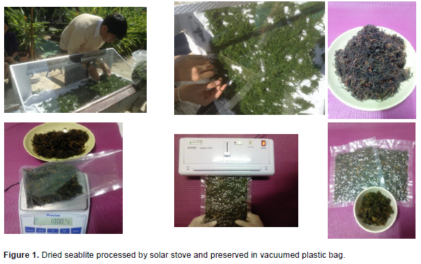
2.5 to 3 kg of scalded seablite was remained 250 to 300 g of dried seablite after processed by solar stove and yielded was approximately 10%. All pictures of dried seablite prepared in Figure 1.
Nutritional value of dried seablite
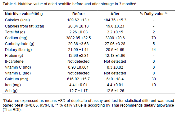
Nutritive values (mean ± SD) of dried seablite on beginning storage and after 3 months kept were shown in Table 1, however, there were no statistical different at p<0.05 when analyzed by paired t-test. β-carotene and vitamin E of dried seablite were vanished by scalding and drying processes on dried seablite production. Only vitamin C was still remained, however, it was decreased after 3 months storage.
Biological properties of dried seablite
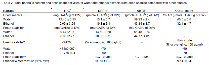
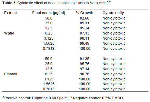
As results, it was found that dried seablite extracted with water exhibited the higher TPC and antioxidant activities (except DPPH) than ethanol extract (TPC of 13.46 ± 2.35mg GAE/ g of DW, DPPH values of 15.1 ± 5.7 μmole TEAC/ g of DW, ABTS+· values of 59.23 ± 2.4 μmole TEAC/ g of DW and ORAC values of 45.6 ± 5.6 μmole TEAC/ g of DW), which is shown in Table 2. The test about the cytotoxic activity on cell showed that hexane and ethanol extract yielded no toxic on Vero cell at 50 μg/ml of extract (Table 3). The antioxidant activity values from all methods were related to amount of total phenolic content.
Preserving of dried seablite in vacuumed plastic bags and evaluation for nutritive values, dried seablite was contained high dietary fiber (21.99 /100 g) and high calcium (616.02 mg/100 g). Dietary fiber and calcium in dried seablite were insignificant decreased (20.5 g/100 g and 610 mg/100 g, respectively) after 3 months storage. However, high amount of sodium in dried seablite should avoid intake in some risk group, such as, hypertension, cardiovascular diseases and renal diseases. Seablite was difficulty scalding for removing salty taste (Pornpitakdamrong and Sudjaroen, 2014) on summer in Thailand and unable to prepared for cooking. If seablite was prepared as dried seablite on other period, it can be useful for cooking on summer time. In addition, dried seablite is easier to transport and cook in other place where far from mangrove area that mean dried seablite can be value-added product.
These results of antioxidant tests were corresponded to the previous study (Pornpitakdamrong and Sudjaroen, 2014; Patra et al., 2011), which found water extract had higher antioxidant activities than ethanol extract. Our water extract had higher polarity, thus water extract may contain high amount of phenolic compounds that correlated to TPC as well, and however, this finding was contrasted with Patra et al. (2011) study. When comparing antioxidant activities of dried seablite to fresh seablite on previous studies (Pornpitakdamrong and Sudjaroen, 2014; Patra et al., 2011; Ravikumar et al., 2011), the antioxidant activities of dried seablite were decreased, which due to food processing, such as, scalding and drying, however, it still remained antioxidant effect in dried seablite. This results were contrasted with previous studies may depended on sample type (between dried and fresh samples), variation of plant, method of extraction, concentration, and solubility of extract and type of assay. The cytotoxic test of seablite was non-toxic (at concentration = 50 μg/ml), which was corresponded to fresh seablite on previous study (Sudjaroen, 2014) and natural antioxidants have been proved to inhibit tumor growth selectively, because of different redox status between normal cells and cancer cell (Nair et al., 2007).