Journal of
Pharmacognosy and Phytotherapy

  • Abbreviation: J. Pharmacognosy Phytother.
  • Language: English
  • ISSN: 2141-2502
  • DOI: 10.5897/JPP
  • Start Year: 2009
  • Published Articles: 234

Full Length Research Paper

Phytochemical screening of methanolic dried galls extract of Quercus infectoria using gas chromatography-mass spectrometry (GC-MS) and Fourier transform-infrared (FT-IR)

Ameera Omran Hussein
  • Ameera Omran Hussein
  • College of Ecology, Al-Qasim Green University, Iraq.
  • Google Scholar
Ghaidaa Jihadi Mohammed
  • Ghaidaa Jihadi Mohammed
  • College of Science, Al-Qadisia University, Iraq.
  • Google Scholar
Mohammed Yahya Hadi
  • Mohammed Yahya Hadi
  • College of Biotechnology, Al-Qasim Green University, Iraq.
  • Google Scholar
and Imad Hadi Hameed*
  • and Imad Hadi Hameed*
  • Department of Biology, Babylon University, Hilla City, Iraq
  • Google Scholar


  •  Received: 26 August 2015
  •  Accepted: 20 November 2015
  •  Published: 31 March 2016

 ABSTRACT

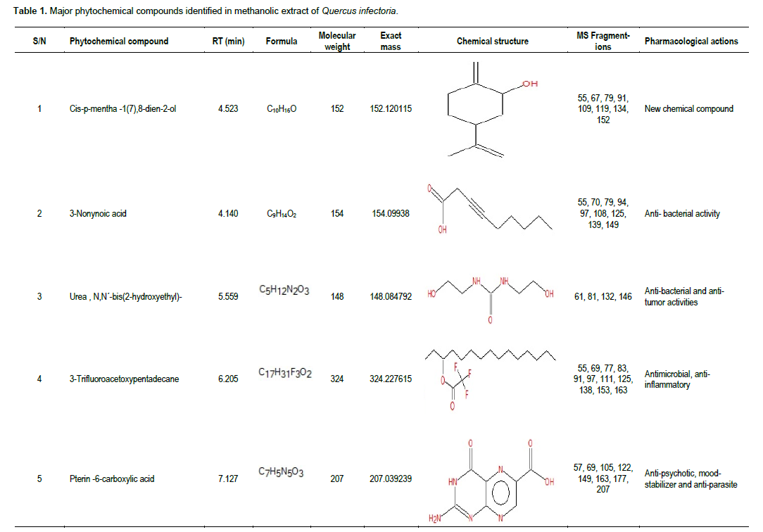
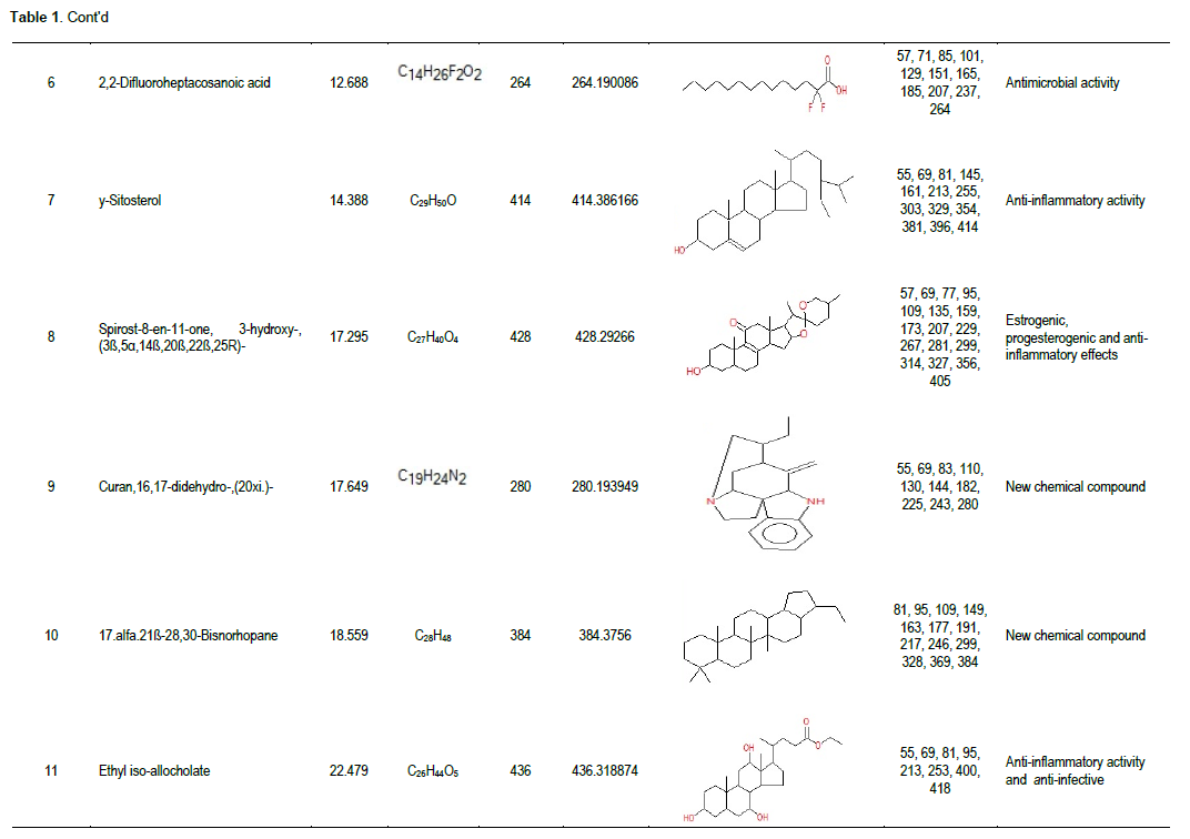
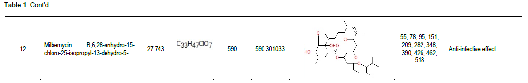
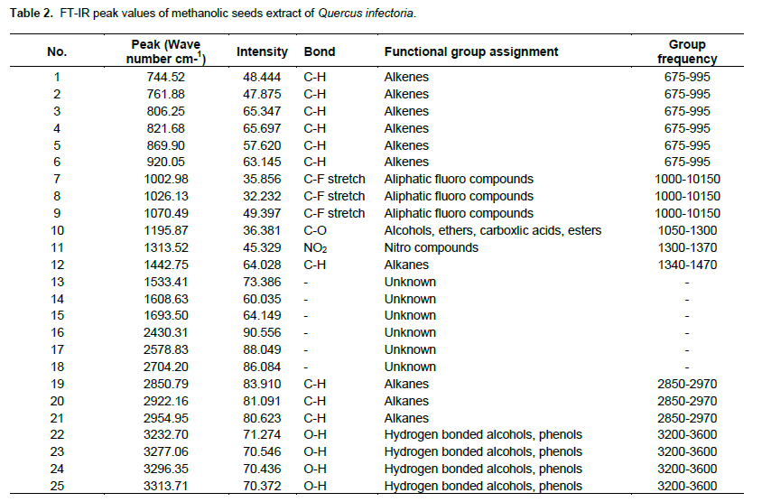
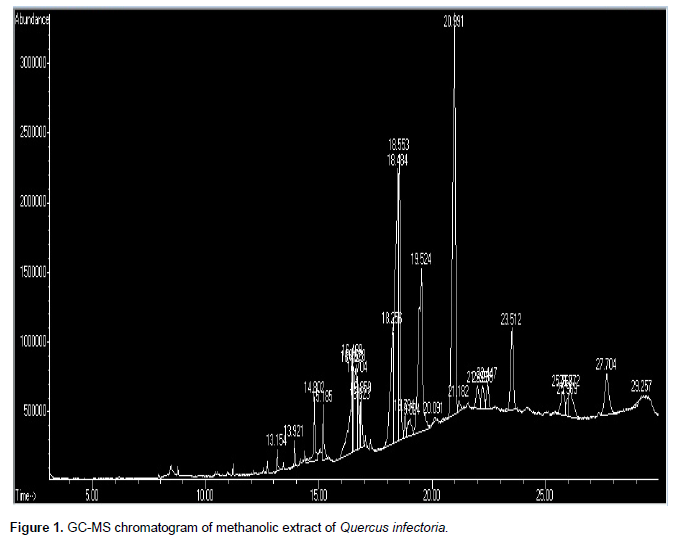
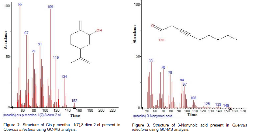
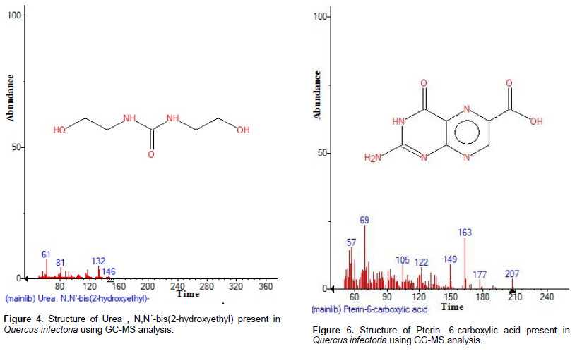
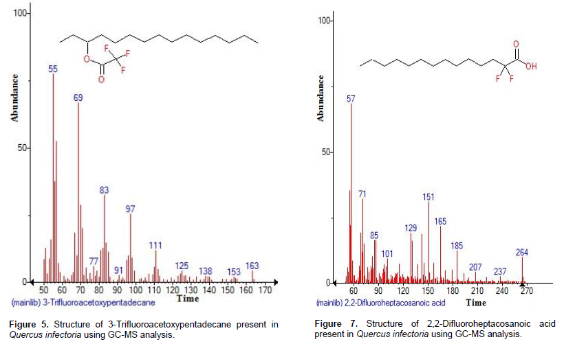
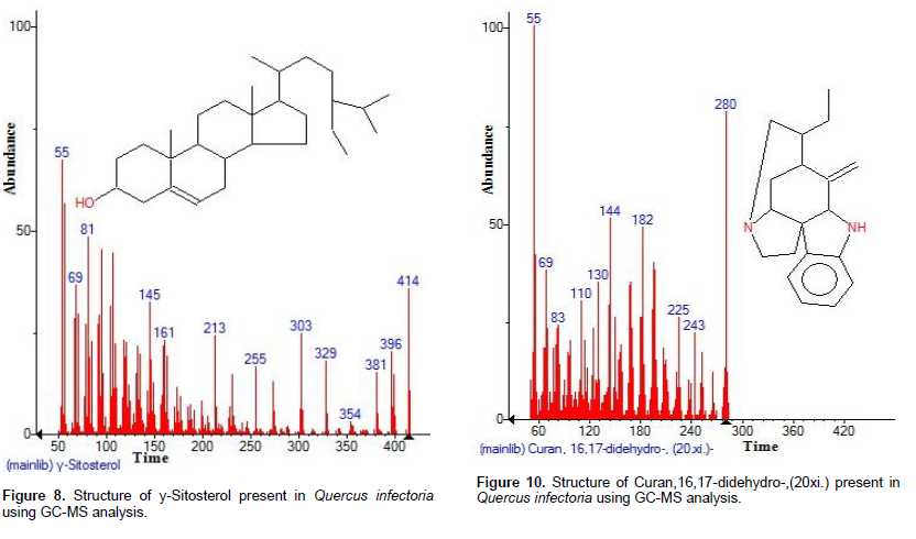
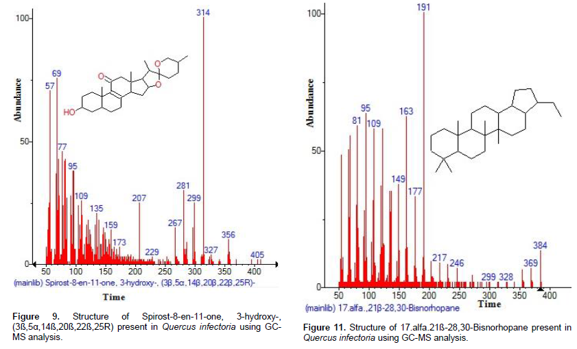
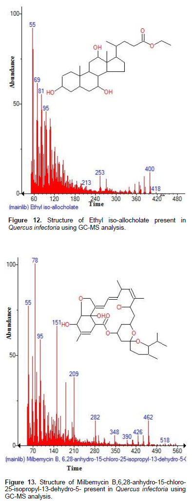
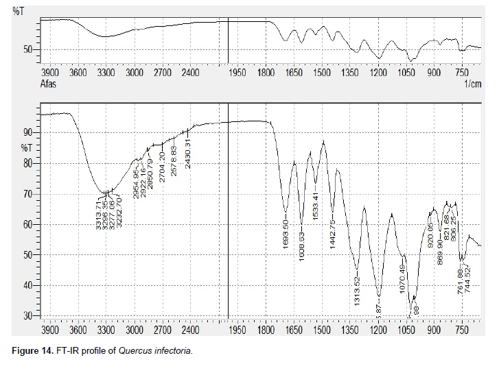
The main objective of this study was to determine the phytochemical composition from the dried galls of Quercus infectoria, using methanolic extraction and report the main functional components by using infrared (IR) technique. The phytochemical compound screened by gas chromatography-mass spectrometry (GC-MS) method. Twelve bioactive phytochemical compounds were identified in the methanolic extract of Q. infectoria. The identification of phytochemical compounds is based on the peak area, retention time molecular weight, and molecular formula. GC-MS analysis of Q. infectoria revealed the existence of the Cis-p-mentha -1(7),8-dien-2-ol, 3-Nonynoic acid, Urea, N,N´-bis(2-hydroxyethyl)-, 3-Trifluoroacetoxypentadecane, Pterin -6-carboxylic acid, 2,2-Difluoroheptacosanoic acid, y-Sitosterol, Spirost-8-en-11-one, 3-hydroxy-, (3ß,5α,14ß,20ß,22ß,25R)-, Curan,16,17-didehydro-,(20xi.)-, 17.alfa.21ß-28,30-Bisnorhopane, Ethyl iso-allocholate, Milbemycin B,6,28-anhydro-15-chloro-25-isopropyl-13-dehydro-5-. The Fourier transform-infrared (FTIR) analysis of Q. infectoria proved the presence of alkenes, aliphatic fluoro compounds, nitro compounds, alkanes, hydrogen bonded alcohols, and phenols.

Key words: Quercus infectoria, Fourier transform-infrared (FT-IR), gas chromatography-mass spectrometry (GC-MS) analysis, phytochemicals.


 INTRODUCTION

Quercus infectoria is an oak tree of the family Fagaceae in the Mediterranean  area,  especially  in  Greece,  Syria, Iran, and Asia Minor (Samuelsson, 1999). The galls arise on  young  branches  of  this  tree  as a result of attack by female gasp-wasp Adleria gallae-tinctoria and Cynips gallae tinctoria by deposition of the eggs (Greenish, 1999). Q. infectoria is a small tree widely distributed in Greece, Asia Minor, and Iran. It has been evaluated in terms of its pharmacological effects and it was found that it had antiparkinsonian, antitremorine, antiinflammatory, antidiabetic, and antioxidant effects (Aivazi and Vijayan, 2009; Altameme et al., 2015a).  Traditionally, galls are used in postpartum practice (Soon et al., 2007) and in the treatment of diarrhea, hemorrhage, and skin disease (Greenish, 1999; Hameed et al., 2015a). The galls of Q. infectoria were documented to possess antibacterial (Basri et al., 2005; Darogha, 2009), anti-MRSA (Chusri and Voravuthikunchai, 2009), antiviral (Hussein et al., 2000), antifungus (Yamunarani et al., 2005; Yoshikawa et al., 2007; Hameed et al., 2015b), and anti-inflammatory activities. Previous investigation revealed that the ethanol extract of the nutgalls consists of tannins, flavonoids, and steroidal compounds (Rukayadi et al., 2006; Chusri and Voravuthikunchai, 2009; Mekseepralard et al., 2010). The constituents of the galls of Q. infectoria comprise a large amount of tannins, gallic acid, syringic acid, ellagic acid, beta sitosterol, amentoflavone hexamethyl ether, isocryptomerin, methyl betulate, methyl olenate, and hexagalloyl glucose (Lodhi et al., 2012; Hameed et al., 2015c). Larvacidal activity of the gall extracts of Q. infectoria was initially reported against Anopheles stephensi (Aivazi and Vijayan, 2009). The main constituents of the galls are tannin (50 to 70%) with small amount of free gallic acid and starch. The present study aimed to analyze the methanol extract of Q. infectoria galls.


 MATERIALS AND METHODS

Collection and preparation of plant
 
The dried galls were purchased from local market in Hilla city, middle of Iraq. After thorough cleaning and removal of foreign materials, the dried galls were stored in airtight container to avoid the effect of humidity and then stored at room temperature until further use (Altameme et al., 2015b; Hameed et al., 2015d).
 
Preparation of sample
 
About 15 g of the plant sample powdered were soaked in 75 ml methanol for 14 h in a rotatory shaker. Whatman No.1 filter paper was used to separate the extract of the plant. The filtrates were used for further phytochemical analysis (Hussein et al., 2015; Jasim et al., 2015; Hamza et al., 2015). It was again filtered through sodium sulphate in order to remove the traces of moisture.
 
Gas chromatography-mass spectrum (GC-MS) analysis
 
GC-MS technique was used in this study to identify the components present in the extract which was carried out at Indian Institute of Science, Bangalore. The GC-MS analysis of the plant extract was made in a Agilent 7890 A instrument under computer control at 70 eV (Kareem et al.,  2015;  Imad  et  al.,  2014a).  About  1 μl  of  the methanol extract was injected into the GC-MS using a micro syringe and the scanning was done for 45 min. As the compounds were separated, they eluted from the column and entered a detector which was capable of creating an electronic signal whenever a compound was detected. The greater the concentration in the sample is, the bigger the signal obtained which was then processed by a computer. The time from when the injection was made (Initial time) to when elution occurred is referred to as the retention time (RT) (Imad et al., 2014b). While the instrument was run, the computer generated a graph from the signal called Chromatogram. Each of the peaks in the chromatogram represented the signal created when a compound eluted from the gas chromatography column into the detector. The X-axis showed the RT and the Y-axis measured the intensity of the signal to quantify the component in the sample injected. As individual compounds eluted from the gas chromatographic column, they entered the electron ionization (mass spectroscopy) detector, where they were bombarded with a stream of electrons causing them to break apart into fragments. The fragments obtained were actually charged ions with a certain mass. The Mass/Charge (M/Z) ratio obtained was calibrated from the graph obtained, which was called the mass spectrum graph which is the fingerprint of a molecule. Before analyzing the extract using gas chromatography and mass spectroscopy, the temperature of the oven, the flow rate of the gas used, and the electron gun were programmed initially. The temperature of the oven was maintained at 100°C. Helium gas was used as a carrier as well as an eluent. The flow rate of helium was set to 1 ml/min. The electron gun of mass detector liberated electrons having energy of about 70 eV. The column employed here for the separation of components was Elite 1 (100% dimethyl poly siloxane) (Mohammed and Imad, 2013; Imad et al., 2014c). The identity of the components in the extracts was assigned by comparison of their retention indices and mass spectra fragmentation patterns with those stored on the computer library and also with published literatures (Yang et al., 2010).


 RESULTS AND DISCUSSION

GC-MS analysis of compounds was carried out in methanolic dried galls extract of Q. infectoria and is shown in Table 1. The GC-MS chromatogram of the twenty peaks of the compounds detected as shown in Figure 1. Chromatogram GC-MS analysis of the methanol extract of Q. infectoria showed the presence of twenty major peaks and the components corresponding to the peaks were determined as follows. The first set up peak were determined to be Cis-p-mentha -1(7),8-dien-2-ol (Figure 2). The next peaks were considered to be 3-Nonynoic acid, Urea , N,N´-bis(2-hydroxyethyl)-, 3-Trifluoroacetoxypentadecane, Pterin -6-carboxylic acid, 2,2-Difluoroheptacosanoic acid, y-Sitosterol, Spirost-8-en-11-one, 3-hydroxy-, (3ß,5α,14ß,20ß,22ß,25R)-, Curan,16,17-didehydro-,(20xi.)-, 17.alfa.21ß-28,30-Bisnorhopane, Ethyl iso-allocholate, Milbemycin B,6,28-anhydro-15-chloro-25-isopropyl-13-dehydro-5 (Figures 3 to 13). Plant based antimicrobials have enormous therapeutic potential as they can serve the purpose with lesser side effects. Further continued exploration of plant derived antimicrobials is needed today. The FTIR analysis of Q. infectoria proved the presence of alkenes, aliphatic fluoro compounds, nitro compounds Table 2, alkanes, hydrogen bonded alcohols and phenols which show major peaks at  744.52,  806.25,  920.05,  1026.13, 1195.87, 1313.52, 1442.75, 2850.79, 2954.95, 3232.70, and 3313.71 (Figure 14).


 CONCLUSION

Q. infectoria is a native plant of Iraq. Thus, the GC-MS analysis of methanolic extract of Q. infectoria showed a highly complex profile containing approximately twelve components. This study may be useful to further explore the pharmacological and biosynthetic activity of the plants.


 ACKNOWLEDGEMENT

The authors thank Dr. Abdul-Kareem Al-Bermani, Lecturer, Department of Biology, for valuable suggestions and encouragement.


 REFERENCES

Aivazi AA, Vijayan VA (2009). Larvacidal activity of oak Quercus infectoria Oliv. Fagaceae) gall extracts against Anopheles stephensi Liston. Parasitol. Res. 104(6):1289-1293.
crossref


Altameme HJ, Hameed IH, Idan SA, Hadi MY (2015a). Biochemical analysis of Origanum vulgare seeds by Fourier-transform infrared (FT-IR) spectroscopy and gas chromatography-mass spectrometry (GC-MS). J. Pharmacogn. Phytother. 7(9):221-237.
crossref


Altameme HJ, Hameed IH, Kareem MA (2015b). Analysis of alkaloid phytochemical compounds in the ethanolic extract of Datura stramonium and evaluation of antimicrobial activity Afr. J. Biotechnol. 14(19):1668-1674.


Basri DF, Ha FS, Zin NM, Jantan I (2005). Antibacterial activity of the galls of Quercus infectoria. Malaysian J. Sci. 24:257–262.


Chusri S, Voravuthikunchai SP (2009). Detailed studies on Quercus infectoria Olivier (nutgalls) as an alternative treatment for methicillin-resistant Staphylococcus aureus infections. J. Appl. Microbiol. 106(1):89– 96.
crossref


Darogha SN (2009). Antibacterial activity of Quercus infectoriaextracts against bacterial isolated from wound infection. J. Kirkuk Univ. Sci. Stud. 4(1):20–30.


Greenish HG (1999). MateriaMedica, Scientific Publisher, Jodhpur, India, 3rd edition,


Hameed IH, Hussein HJ, Kareem MA, Hamad NS (2015a). Identification of five newly described bioactive chemical compounds in methanolic extract of Mentha viridis by using gas chromatography-mass spectrometry (GC-MS). J. Pharmacogn. Phytother. 7(7):107-125.
crossref


Hameed IH, Ibraheam IA, Kadhim HJ (2015b). Gas chromatography mass spectrum and Fourier-transform infrared spectroscopy analysis of methanolic extract of Rosmarinus oficinalis leaves. J. Pharmacogn. Phytother. 7(6):90-106.
crossref


Hameed IH, Jasim H, Kareem MA, Hussein AO (2015c). Alkaloid constitution of Nerium oleander using gas chromatography-mass spectroscopy (GC-MS). J. Med. Plants Res. 9(9):326-334.
crossref


Hameed IH, Hamza LF, Kamal SA (2015d). Analysis of bioactive chemical compounds of Aspergillus niger by using gas chromatography-mass spectrometry and Fourier-transform infrared spectroscopy. J. Pharmacogn. Phytother. 7(8):132-163.
crossref


Hamza LF, Kamal SA, Hameed IH (2015). Determination of metabolites products by Penicillium expansum and evaluating antimicobial activity. J. Pharmacogn. Phytother. 7(9):194-220.
crossref


Hussein AO, Hameed IH, Jasim H, Kareem MA (2015). Determination of alkaloid compounds of Ricinus communis by using gas chromatography-mass spectroscopy (GC-MS). J. Med. Plants Res. 9(10):349-359.
crossref


Hussein G, Miyashiro H, Nakamura N, Hattori M, Kakiuchi N, Shimotohno K (2000). Inhibitory effects of Sudanese medicinal plant extract on hepatitis C virus protease. Phytother. Res. 14(7):510–516.
crossref


Imad H, Mohammed A, Aamera J (2014a). Genetic variation and DNA markers in forensic analysis. Afr. J. Biotechnol. 13(31):3122-3136.
crossref


Imad H, Mohammed A, Cheah Y, Aamera J (2014b). Genetic variation of twenty autosomal STR loci and evaluate the importance of these loci for forensic genetic purposes. Afr. J. Biotechnol. 13:1-9.
crossref


Imad H, Muhanned A, Aamera J, Cheah Y (2014c). Analysis of eleven Y-chromosomal STR markers in middle and south of Iraq. Afr. J. Biotechnol. 13(38):3860-3871.
crossref


Jasim H, Hussein AO, Hameed IH, Kareem MA (2015). Characterization of alkaloid constitution and evaluation of antimicrobial activity of Solanum nigrum using gas chromatography mass spectrometry (GC-MS). J. Pharmacogn. Phytother. 7(4):56-72.


Kareem MA, Hussein AO, Hameed IH (2015). Y-chromosome short tandem repeat, typing technology, locus information and allele frequency in different population: A review. Afr. J. Biotechnol. 14(27):2175-2178.
crossref


Lodhi G, Singh HK, Pant KK, Rao C, Hussain Z (2012). Hepatoprotective effects of Quercus infectoria gall extract against carbon tetrachloride treated liver injury in rats. Int. J. Appl. Res. Nat. Prod. 5(3):17-22.


Mekseepralard C, Kamkaen N, Wilkinson JM (2010). Antimicrobial and antioxidant activities of traditional Thai herbal remedies for aphthous ulcers. Phytother. Res. 24:1514–1519.
crossref


Mohammed A, Imad H (2013). Autosomal STR: From locus information to next generation sequencing technology. Res. J. Biotechnol. 8(10):92-105.


Rukayadi Y, Yong D, Hwang JK (2006). In vitro anticandidal activity of xanthorrhizol isolated from Curcuma xanthorrhiza Roxb. J. Antimicrob. Chemother. 57:1231-1234.
crossref


Samuelsson G (1999). "Drug of natural origin," in A Textbook of Pharmacognosy, Swedish Pharmaceutical Press, Stockholm, Sweden, 4th edition.


Soon LK, Hasni E, Law KS, Waliullah SS, Farid CG (2007). Ultrasructural findings and elemental analysis of Quercus infectoria Oliv. Ann. Microsc. 7:32–37.


Yamunarani K, Jaganathan R, Bhaskaran R, Govindaraju P, Velazhahan R (2005). In vitro antifungal activity of a 29-kDa glycoprotein purified fromthe galls of Quercus infectoria. Acta Phytopathologica et Entomologica Hungarica 40(1-2):43–54.
crossref


Yang SA, Jeon SK, Lee EJ, Shim CH, Lee IS (2010). Comparative study of the chemical composition and antioxidant activity of six essential oils and their components. Nat. Prod. Res. 24: 140-151.
crossref


Yoshikawa M, Morikawa T, Kobayashi H, Nakamura A, Matsuhira K, Nakamura S, Matsuda H (2007). Bioactive saponins and glycosides. Structures of new cucurbitane-type triterpene glycosides and antiallergic constituents from Citrullus colocynthis. Chem. Pharm. Bull. l55:428-434.
crossref


          */?>